Review
. 2023 Aug 31;13(4):743-761.
doi: 10.21037/cdt-23-79. Epub 2023 Jul 21.

Opportunistic screening at chest computed tomography: literature review of cardiovascular significance of incidental findings

Affiliations
Review

Opportunistic screening at chest computed tomography: literature review of cardiovascular significance of incidental findings

Arzu Canan et al. Cardiovasc Diagn Ther. .

Abstract

Background and objective: Several incidental cardiovascular findings are present in a routine chest computed tomography (CT) scan, many of which do not make it to the final radiology report. However, these findings have important clinical implications, particularly providing prognosis and risk-stratification for future cardiovascular events. The purpose of this article is to review the literature on these incidental cardiovascular findings in a routine chest CT and inform the radiologist on their clinical relevance.

Methods: A time unlimited review of PubMed and Web of Science was performed by using relevant keywords. Articles in English that involved adults were included.

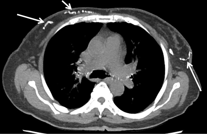
Key content and findings: Coronary artery calcification (CAC) is the most common incidental cardiac finding detected in a routine chest CT and is a significant predictor of cardiovascular events. Noncoronary vascular calcifications in chest CT include aortic valve, mitral annulus, and thoracic aortic calcifications (TAC). Among these, aortic valve calcification (AVC) has the strongest association with coronary artery disease and cardiovascular events. Additional cardiac findings such as myocardial scar and left ventricular size and noncardiac findings such as thoracic fat, bone density, hepatic steatosis, and breast artery calcifications can also help in risk stratification and patient management.

Conclusions: The radiologist interpreting a routine chest CT should be cognizant of the incidental cardiovascular findings, which helps in the diagnosis and risk-stratification of cardiovascular disease. This will guide appropriate referral and management.

Keywords: Chest computed tomography (CT); cardiac; cardiovascular; incidental.


Conflict of interest statement

Conflicts of Interest: All authors have completed the ICMJE uniform disclosure form (available at https://cdt.amegroups.com/article/view/10.21037/cdt-23-79/coif). AC and PSR report that they received royalties from Elsevier, unrelated to the content of this manuscript. PSR serves as an unpaid editorial board member of Cardiovascular Diagnosis and Therapy from September 2021 to August 2023. The other authors have no conflicts of interest to declare.

Figures

Figure 1
Figure 1
Axial non-contrast chest CT images show coronary artery calcification. (A) Mild coronary artery calcification in left circumflex artery (arrow); (B) moderate coronary artery calcifications in the left anterior descending and left circumflex coronary arteries (arrows); (C) severe coronary artery calcifications including left main, left anterior descending, and left circumflex artery (arrows).
Figure 2
Figure 2
Axial non-contrast chest CT images show aortic valve calcification. (A) Mild calcification of aortic valve leaflets (arrow); (B) moderate calcification of aortic valve leaflets (arrows); (C) severe calcification of aortic valve leaflets (arrow).
Figure 3
Figure 3
Axial non-contrast chest CT images show mitral annulus calcification. (A) Mild calcification of mitral annulus (arrow); (B) moderate calcification of mitral annulus (arrow); (C) severe calcification of mitral annulus (arrow).
Figure 4
Figure 4
Non-contrast chest CT images show thoracic aortic calcification. (A) Mild calcification of aortic arch (arrow); (B) moderate calcification of descending thoracic aorta (arrow); (C) sagittal oblique multiplanar reformatted image shows severe calcification of entire thoracic aorta. CT, computed tomography.
Figure 5
Figure 5
Non-contrast chest CT images show epicardial and paracardiac adipose tissue. (A) Axial non-contrast chest CT image shows the epicardial fat (asterisk) between the myocardium and pericardium (arrowheads); (B) color-coded image shows the borders of the epicardial (yellow areas) and paracardiac fat (red areas). Blue line represents the pericardium. CT, computed tomography.
Figure 6
Figure 6
Axial non-contrast chest CT images show epicardial fat within the pericardium between the anterior pericardium (arrowheads) and the myocardium (arrows). (A) Mild amount of epicardial fat; (B) moderate amount of epicardial fat; (C) large amount of epicardial fat. CT, computed tomography.
Figure 7
Figure 7
Axial non-contrast chest CT images show myocardial scar. (A) Subendocardial calcification and fatty replacement (arrow) of the left ventricle indicative of myocardial scar and fatty metaplasia from previous myocardial infarction; (B) moderate subendocardial calcification (arrow) of the left ventricle mid and apical septum, consistent with myocardial scar from chronic myocardial infarction. CT, computed tomography.
Figure 8
Figure 8
Axial non-contrast chest CT image shows fatty metaplasia of subendocardial layer of the lateral wall of the left ventricle (arrows), indicating remote myocardial infarction. CT, computed tomography.
Figure 9
Figure 9
Axial contrast enhanced chest CT image shows left ventricular enlargement with a diameter of 70.2 mm. CT, computed tomography.
Figure 10
Figure 10
Hepatic steatosis on non-contrast chest CT. (A) Axial upper abdominal image from a non-contrast chest CT shows diffuse hepatic steatosis. Note the decreased attenuation of the liver compared to surrounding hemidiaphragm (arrows) and the spleen (asterisks); (B) the HU of these areas in the liver are less than <40 HU and the ratio of liver/spleen parenchymal attenuation is <1, consistent with hepatic steatosis. CT, computed tomography; HU, Hounsfield unit.
Figure 11
Figure 11
Non-contrast chest CT images show decreased mineral density of the vertebral body with increased trabeculation (arrows) on axial (A) and sagittal (B) reconstructions. CT, computed tomography.
Figure 12
Figure 12
Axial maximal intensity projection reconstruct from axial non-contrast chest CT images shows bilateral breast arterial calcifications (arrows). CT, computed tomography.

Similar articles

Cited by

References

    1. Choy G, Kröpil P, Scherer A, et al. Pertinent reportable incidental cardiac findings on chest CT without electrocardiography gating: review of 268 consecutive cases. Acta Radiol 2013;54:396-400. 10.1177/0284185113475918 - DOI - PubMed
    1. Sverzellati N, Arcadi T, Salvolini L, et al. Under-reporting of cardiovascular findings on chest CT. Radiol Med 2016;121:190-9. 10.1007/s11547-015-0595-0 - DOI - PubMed
    1. Kuetting D, Müller A, Feisst A, et al. Incidental Cardiac Findings in Non-Electrocardiogram-gated Thoracic Computed Tomography of Intensive Care Unit Patients: Assessment of Prevalence and Underreporting. J Thorac Imaging 2018;33:168-75. 10.1097/RTI.0000000000000309 - DOI - PubMed
    1. Obisesan OH, Osei AD, Uddin SMI, et al. An Update on Coronary Artery Calcium Interpretation at Chest and Cardiac CT. Radiol Cardiothorac Imaging 2021;3:e200484. 10.1148/ryct.2021200484 - DOI - PMC - PubMed
    1. LaMonte MJ, FitzGerald SJ, Church TS, et al. Coronary artery calcium score and coronary heart disease events in a large cohort of asymptomatic men and women. Am J Epidemiol 2005;162:421-9. 10.1093/aje/kwi228 - DOI - PubMed