. 2023 Aug 22:14:1222339.
doi: 10.3389/fimmu.2023.1222339. eCollection 2023.

The aging kidney is characterized by tubuloinflammaging, a phenotype associated with MHC-II gene expression

Affiliations

The aging kidney is characterized by tubuloinflammaging, a phenotype associated with MHC-II gene expression

Julius Sinning et al. Front Immunol. .

Abstract

Introduction: Even during physiologic aging, the kidney experiences a loss of mass and a progressive functional decline. This is clinically relevant as it leads to an increased risk of acute and chronic kidney disease. The kidney tubular system plays an important role in the underlying aging process, but the involved cellular mechanisms remain largely elusive.

Methods: Kidneys of 3-, 12- and 24-month-old male C57BL/6J mice were used for RNA sequencing, histological examination, immunostaining and RNA-in-situ-hybridization. Single cell RNA sequencing data of differentially aged murine and human kidneys was analyzed to identify age-dependent expression patterns in tubular epithelial cells. Senescent and non-senescent primary tubular epithelial cells from mouse kidney were used for in vitro experiments.

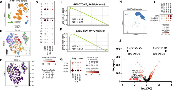
Results: During normal kidney aging, tubular cells adopt an inflammatory phenotype, characterized by the expression of MHC class II related genes. In our analysis of bulk and single cell transcriptional data we found that subsets of tubular cells show an age-related expression of Cd74, H2-Eb1 and H2-Ab1 in mice and CD74, HLA-DQB1 and HLADRB1 in humans. Expression of MHC class II related genes was associated with a phenotype of tubular cell senescence, and the selective elimination of senescent cells reversed the phenotype. Exposure to the Cd74 ligand MIF promoted a prosenescent phenotype in tubular cell cultures.

Discussion: Together, these data suggest that during normal renal aging tubular cells activate a program of 'tubuloinflammaging', which might contribute to age-related phenotypical changes and to increased disease susceptibility.

Keywords: CD74; MHC-II; aging kidney; epithelial cell; inflammation; senescence; tubular cell.


Conflict of interest statement

RS received honoraria for scientific lectures from Otsuka Pharmaceutical, AstraZeneca, Bayer, Fresenius Medical Care. The remaining authors declare that the research was conducted in the absence of any commercial or financial relationships that could be construed as a potential conflict of interest.

Figures

Figure 1
Figure 1
(A) Schematic of the generation of RNAseq data sets from senescent (sen) and non-senescent (ns) primary tubular epithelial cells (PTEC) (n=4). (B, C) Quantification of transcripts of senescence marker genes Cdkn1a (p21) and Cdkn2a (p16INK4a) by RNAseq data from PTEC. (D–F) Quantification of transcripts of MHC-II related genes Cd74, H2-Ab1 and H2-Eb1 by RNAseq data from PTEC. (G) Schematic of the generation of RNAseq data sets from kidneys of three, 12 and 24 months (m) old mice (each group n=3). (H) Bar diagram showing upregulation of immune-related pathways by GSEA. (I) Venn diagram showing upregulation of 109 overlapping differentially expressed genes (DEGs) in both data sets generated by RNAseq. (J) Bar diagram showing enriched pathways of overlapping genes by GSEA. (K–M) Quantification of transcripts of MHC-II related genes Cd74, H2-Ab1 and H2-Eb1 by RNAseq data from mice. (N–P) Quantification of transcripts for MHC-related genes Cd74, H2-Ab1 and H2-Eb1 by RNAseq data from Tabula muris senis. Results are presented as means ± SEM of at least three repeats for each experiment. Significance was tested by FDR/q-value generated by DESeq2, one-way ANOVA, or two-way ANOVA with Tukey’s test as post hoc analysis in the case of multiple comparisons. ns, non-senescent; *p < .05; **p < .01; ***p < .001, ****p < .0001.
Figure 2
Figure 2
(A–D) RNA in-situ hybridization of MHC-II related genes Cd74 and H2-Eb1 in three month old = young (A, B) and 24 month old = old kidneys (C, D), scale bar: 100µm; 30µm. * marks stained interstitial cells. (E) Immunofluoresence co-staining using Lrp2-antibody (Megalin), scale bar: 30µm. (F) Dot plot showing top 20 up-regulated genes comparing young (3m) and old (21m) proximal tubular cells ordered by FDR/q-value. (G) Dot plot showing age-dependent expression of MHC-II related genes Cd74, H2-Ab1, H2-Eb1 and senescence marker gene Cdkn1a (p21). (H) Uniform Manifold Approximation and Projection for Dimension Reduction (UMAP) showing scRNAseq kidney data set from Tabula muris senis. (I) tSNE plot showing reclustered proximal tubular cells (encircled in H) using louvain. (J) tSNE plot showing Cdkn1a expression. (K) Dot plot reclustering of proximal tubular cells identified 14 clusters. Senescent cluster 9 is labelled by rectangle. (L, M) Enrichment plots showing up-regulation of senescence-associated Gene Ontology terms by GSEA.
Figure 3
Figure 3
(A) UMAP showing scRNAseq living donor kidney data set from the Kidney Precision Medicine Project (KPMP project). (B) tSNE plot showing reclustered proximal tubular cells using louvain. (C) tSNE plot showing CDKN1A expression. (D) Dot plot reclustering of proximal tubular cells identified 15 clusters. Senescent cluster 6 is labelled by rectangle. (E, F) Enrichment plots showing up-regulation of senescence-associated Gene Ontology terms by GSEA. (G) Dot plot showing age-dependent expression of MHC-II related genes CD74, HLA-DQB1, HLA-DRB1 and senescence marker gene CDKN1A (p21). (H) UMAP showing proximal tubular cells (PT) of CKD samples (defined by eGFR < 60 ml/min) from the KPMP project. (I) Dot plot showing eGFR-dependent up-regulation of MHC-II related genes CD74, HLA-DRB1 and HLA-DQB1. (J) Volcano plot showing gene expression from unbiased DEG analysis using diffxpy.
Figure 4
Figure 4
(A) Schematic of ABT-263 treatment in PTEC. (B–D) Quantification of transcripts for senescence marker Cdkn2a (p16INK4a), MHC-II related genes Cd74, H2-Eb1 in ABT-263-treated PTEC and controls by qPCR. (E) Schematic of MIF treatment in PTEC. (F–H) Quantification of transcripts for senescence markers Cdkn1a (p21), Cdkn2a (p16INK4a) and MHC-II related Cd74 in MIF-treated PTEC and controls by qPCR. Results are presented as means ± SEM of at least three repeats for each experiment. Significance was tested by t-test; *p < .05.

References

    1. Schmitt R, Melk A. Molecular mechanisms of renal aging. Kidney Int (2017) 92:569–79. doi: 10.1016/j.kint.2017.02.036 - DOI - PubMed
    1. Yu SM, Bonventre JV. Acute kidney injury and maladaptive tubular repair leading to renal fibrosis. Curr Opin Nephrol. Hypertens (2020) 29:310–8. doi: 10.1097/MNH.0000000000000605 - DOI - PMC - PubMed
    1. Liu B, Tang T, Lv L, Lan H. Renal tubule injury: a driving force toward chronic kidney disease. Kidney Int (2018) 93:568–79. doi: 10.1016/j.kint.2017.09.033 - DOI - PubMed
    1. Breda PC, Wiech T, Meyer-Schwesinger C, Grahammer F, Huber T, Panzer U, et al. . Renal proximal tubular epithelial cells exert immunomodulatory function by driving inflammatory CD4(+) T cell responses. Am J Physiol Renal Physiol (2019) 317:F77–89. doi: 10.1152/ajprenal.00427.2018 - DOI - PubMed
    1. Zhou Y, Luo Z, Liao C, Cao R, Hussain Z, Wang J, et al. . MHC class II in renal tubules plays an essential role in renal fibrosis. Cell Mol Immunol (2021) 18:2530–40. doi: 10.1038/s41423-021-00763-z - DOI - PMC - PubMed

Publication types

Substances