Review
. 2023 Jun;16(6):842-850.
doi: 10.25122/jml-2023-0127.

Diagnosis and treatment of Watershed strokes: a narrative review

Affiliations
Review

Diagnosis and treatment of Watershed strokes: a narrative review

Oana Andreea Dogariu et al. J Med Life. 2023 Jun.

Abstract

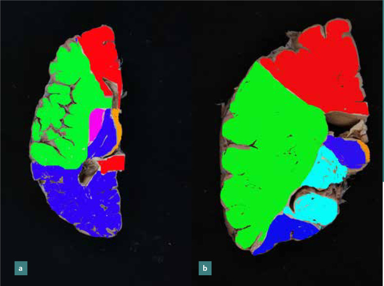
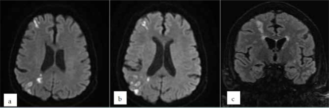
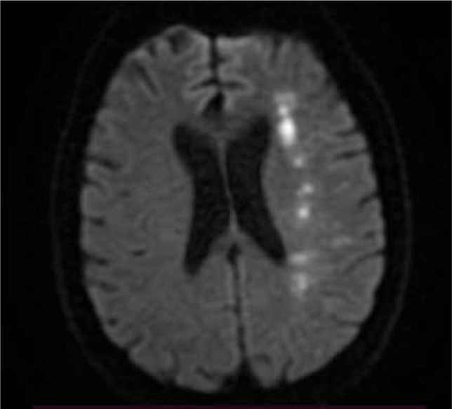
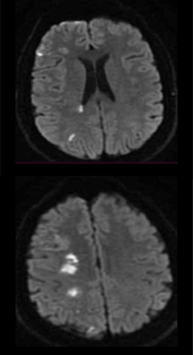
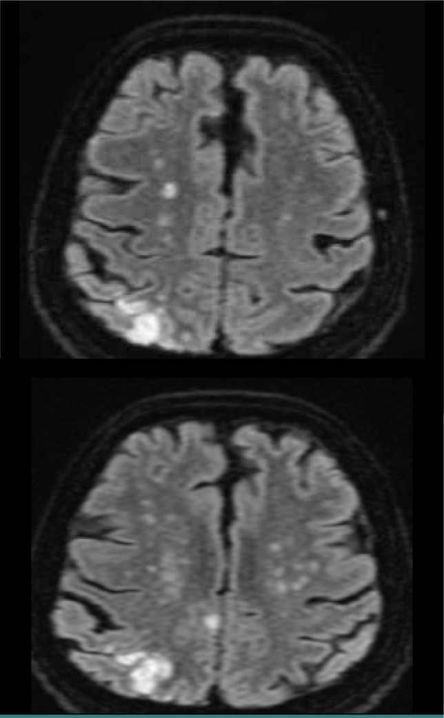
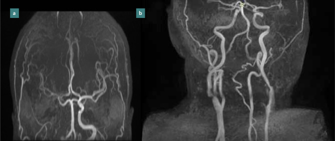
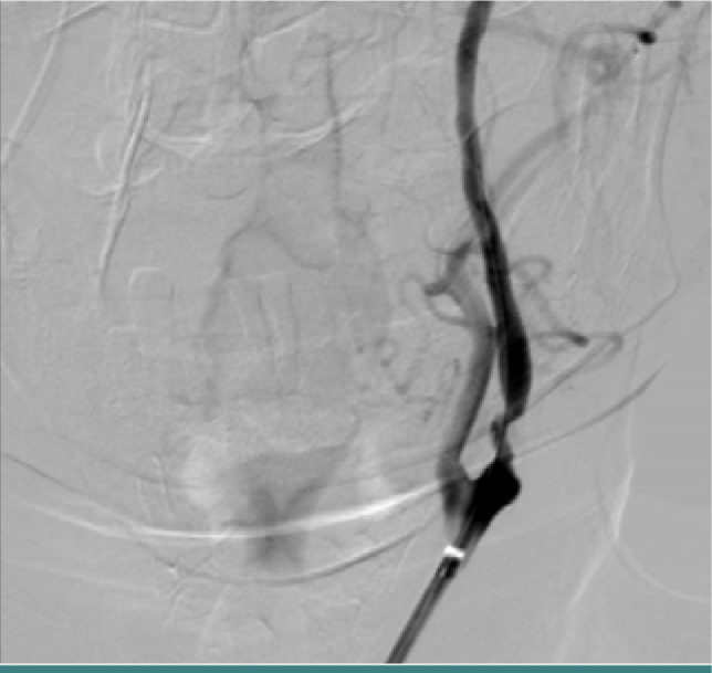
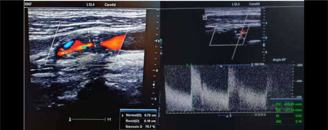
Watershed strokes have been described previously as ischemic strokes located in vulnerable border zones between brain tissue supplied by the anterior, posterior, and middle cerebral arteries in the distal junction between two non-anastomotic arterial territories. Ischemic strokes in border zones are well-recognized entities and well-described in terms of imaging features, but the pathophysiological mechanism of brain injury production is not fully defined. Border zone ischemia is caused by cerebral hypoperfusion through decreased cerebral blood flow and arterial embolism in unstable atheroma plaque. It is often difficult to say which mechanisms are fully responsible for producing cerebral ischemic lesions. This review aimed to highlight the imaging aspect of watershed strokes and to correlate the clinical characteristics of this type of stroke with the diagnostic algorithm for optimal therapeutic management. Neurologists should promptly recognize this type of stroke and investigate its etiology in the shortest possible time.

Keywords: ACA - anterior cerebral artery; BMT - best medical treatment; CAS - carotid artery stentings; CBF - cerebral blood flow; CBV - cerebral blood volume; CBZ - cortical border-zone; CEA - carotid endarterectomy; CT - computer tomography; DWI - diffusion-weighted imaging; ECST - European Carotid Surgery Trial; FLAIR - fluid attenuation inversion recovery; IBZ - internal border-zone; ICA - internal carotid artery; LVO - large vessel oclussion; MCA - middle cerebral artery; MRI - magnetic resonance imaging; MS - multiple sclerosis; NIHSS - National Institute of Health Stroke Scale; PCA - posterior cerebral artery; PET - positron emission tomography; SPECT - single photon emission tomography; TIA - transient ischemic attack; TOF - time of flight; WI - Watershed infarcts; Watershed infarcts; carotid stenosis; hypoperfusion; ischemic stroke.


Conflict of interest statement

The authors declare no conflict of interest.

Figures

Figure 1
Figure 1
Arterial blood distribution: a - axial section at the level of the genu of the corpus callosum; b - coronal section at the level of the head of the caudate nucleus (anterior cerebral artery in red, the middle cerebral artery in green, the posterior cerebral artery in blue, the anterior choroidal artery in purple in the image a and light blue in image b, posterior communicating artery in orange). Regions between arterial territories (blue arrows) are prone to ischemia.
Figure 2
Figure 2
Brain MRI of a patient with external cortical border zone strokes in the watershed territories between right MCA/ACA and MCA/PCA on a symptomatic ICA stenosis (a, b - DWI axial sections, c - FLAIR coronal section)
Figure 3
Figure 3
Brain MRI, axial DWI – Patient with confluent deep watershed ischemic stroke
Figure 4
Figure 4
Brain MRI of a patient with acute ischemic stroke in the deep and superficial watershed territories of right ICA (DWI, axial sections).
Figure 5
Figure 5
Brain MRI of a patient with internal (deep) border zone strokes (FLAIR axial sections). We can see confluent lesions in a band-like distribution parallel with the lateral ventricle (characteristic "string of pearls" sign). The lesions are more than 3 mm long, suggesting deep watershed stroke lesions.
Figure 6
Figure 6
MRI 3D TOF with signal loss of the ICA in the petrous segment (a); 3D TOF with severe right internal carotid artery stenosis at the bifurcation (b).
Figure 7
Figure 7
Digital subtraction angiography of a patient with severe left ICA stenosis in the proximal portion of the artery
Figure 8
Figure 8
Doppler ultrasound of a patient with high-grade left ICA stenosis (>70% reduced diameter). The peak systolic and end-diastolic velocities are increased. The patient had an episode of amaurosis fugax and then developed hemiparesis due to a lesion in the border zone between MCA/ACA
Figure 9
Figure 9
Comparative between AHA [38] (red) and ESO [44,45] (blue) guideline recommendations of management measurements in carotid stenosis, which can be responsible for watershed strokes.

References

    1. Mangla R, Kolar B, Almast J, Ekholm SE. Border zone infarcts: pathophysiologic and imaging characteristics. Radiographics. 2011;31(5):1201–14. doi: 10.1148/rg.315105014. - DOI - PubMed
    1. Bogousslavsky J, Regli F. Unilateral watershed cerebral infarcts. Neurology. 1986 Mar;36(3):373–7. doi: 10.1212/wnl.36.3.373. - DOI - PubMed
    1. Momjian-Mayor I, Baron JC. The pathophysiology of watershed infarction in internal carotid artery disease: review of cerebral perfusion studies. Stroke. 2005;36(3):567–77. doi: 10.1161/01.STR.0000155727.82242.e1. - DOI - PubMed
    1. Denier C, Masnou P, Mapoure Y, Souillard-Scemama R, et al. Watershed infarctions are more prone than other cortical infarcts to cause early-onset seizures. Arch Neurol. 2010 Oct;67(10):1219–23. doi: 10.1001/archneurol.2010.263. - DOI - PubMed
    1. Ringelstein EB, Zeumer H, Angelou D. The pathogenesis of strokes from internal carotid artery occlusion. Diagnostic and therapeutical implications. Stroke. 1983;14(6):867–75. doi: 10.1161/01.str.14.6.867. - DOI - PubMed