

A simple free flap strategy using end-to-side anastomosis to the main vessels in injured extremity
- PMID: 37675275
- PMCID: PMC10477053
- DOI: 10.1016/j.jpra.2023.08.003
A simple free flap strategy using end-to-side anastomosis to the main vessels in injured extremity
Abstract
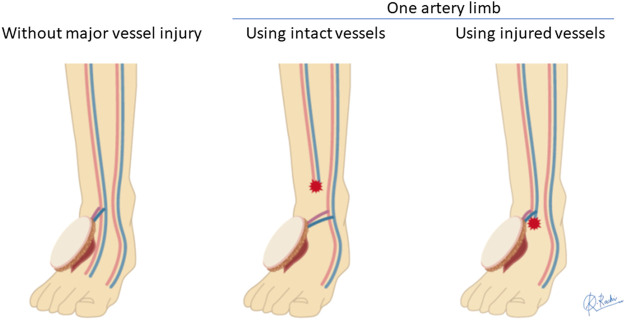
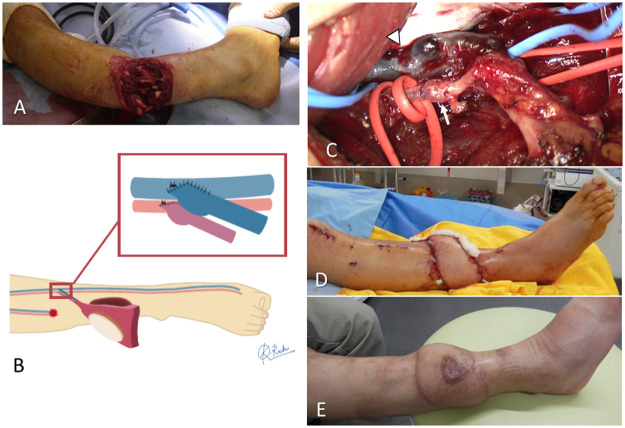
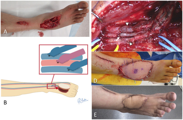
Background: During free flap surgery, the surgeon sometimes encounters problems with anastomosis such as intractable arterial spasms or vessel size discrepancy in venous anastomoses. End-to-side (ETS) anastomosis has the advantages of limited chance of vessel spasm and easy handling by adjusting for vessel size discrepancy. We introduced the arterial and venous end-to-side anastomosis (AV-ETS) strategy, which is based on the ETS anastomosis to the main artery and accompanying veins, to avoid intraoperative anastomotic problems when creating a free flap. The aim of this study was to compare flap outcomes and intraoperative anastomotic problems before and after introduction of the AV-ETS strategy in extremity free flap surgery.
Materials and methods: We retrospectively examined 72 consecutive extremity free flaps. Before introducing the AV-ETS strategy, we used the conventional strategy in which the recipient artery was selected according to the number of the remaining main artery and the anastomosis technique was flexibly changed, although the end-to-end (ETE) technique was used in most cases.
Results: The conventional group had 18 flaps and the AV-ETS group had 54 flaps. The rate of flap survival did not differ between these groups, and there were no cases of flap failure after the introduction of the AV-ETS strategy. The AV-ETS group had significantly fewer flaps that required a change in preoperative planning for the recipient artery or anastomotic site of the artery.
Conclusions: The AV-ETS strategy may facilitate reliable preoperative planning and the performance of stable free flap surgery without requiring a flexible response during surgery.
Keywords: End-to-side anastomosis; Extremities soft tissue defect; Free flap; Main artery; Recipient vessel; Strategy.
© 2023 The Authors.
Conflict of interest statement
None.
Figures
Similar articles
-
Microvascular Anastomoses Using End-to-End versus End-to-Side Technique in Lower Extremity Free Tissue Transfer.J Reconstr Microsurg. 2016 Feb;32(2):114-20. doi: 10.1055/s-0035-1563397. Epub 2015 Aug 31. J Reconstr Microsurg. 2016. PMID: 26322491
-
The suitability of end-to-side microvascular anastomosis in free flap transfer for limb reconstruction.Ann Plast Surg. 2012 Feb;68(2):171-4. doi: 10.1097/SAP.0b013e3182275cf8. Ann Plast Surg. 2012. PMID: 21785334
-
Efficacy of the microscopic parachute end-to-side technique for creating large-to-small venous anastomoses in free flaps in the extremities.JPRAS Open. 2022 Oct 8;34:189-198. doi: 10.1016/j.jpra.2022.10.003. eCollection 2022 Dec. JPRAS Open. 2022. PMID: 36393895 Free PMC article.
-
End to end versus end to side microvascular anastomosis for traumatic lower limb free flap reconstructions: A systematic review and meta-analysis.J Hand Microsurg. 2024 Mar 29;16(4):100065. doi: 10.1016/j.jham.2024.100065. eCollection 2024 Oct. J Hand Microsurg. 2024. PMID: 39234389 Free PMC article. Review.
-
Free flaps for head and neck cancer reconstruction: Does the use of both large cervical vessels as recipient vessels and the employment of end-to-side technique enhance flap survival?Can J Plast Surg. 2003 Fall;11(3):141-2. doi: 10.1177/229255030301100307. Can J Plast Surg. 2003. PMID: 24115856 Free PMC article. Review.
Cited by
-
The sucker-like end-to-side arterial anastomosis for free flap in extremities reconstruction: a retrospective study of 78 cases.J Orthop Surg Res. 2024 Feb 4;19(1):119. doi: 10.1186/s13018-024-04597-z. J Orthop Surg Res. 2024. PMID: 38311748 Free PMC article.
-
Free Flap Surgery for Elbow Soft Tissue Reconstruction Using the Brachial Artery as Recipient Vessel: Evaluation of MPETS Cases and Comparative Literature Review.Medicina (Kaunas). 2025 Feb 8;61(2):295. doi: 10.3390/medicina61020295. Medicina (Kaunas). 2025. PMID: 40005414 Free PMC article.
References
-
- Pollak AN, McCarthy ML, Burgess AR. Short-term wound complications after application of flaps for coverage of traumatic soft-tissue defects about the tibia. The Lower Extremity Assessment Project (LEAP) Study Group. J Bone Joint Surg Am. 2000;82:1681–1691. - PubMed
-
- Khouri RK. Avoiding free flap failure. Clin Plast Surg. 1992;19:773–781. - PubMed
-
- Lorenzo AR, Lin CH, Lin CH, et al. Selection of the recipient vein in microvascular flap reconstruction of the lower extremity: analysis of 362 free-tissue transfers. J Plast Reconstr Aesthet Surg. 2011;64:649–655. - PubMed
LinkOut - more resources
Full Text Sources
