. 2023 Sep 29;133(8):674-686.
doi: 10.1161/CIRCRESAHA.123.322737. Epub 2023 Sep 7.

ADAMTS-7 Modulates Atherosclerotic Plaque Formation by Degradation of TIMP-1

Affiliations

ADAMTS-7 Modulates Atherosclerotic Plaque Formation by Degradation of TIMP-1

M Amin Sharifi et al. Circ Res. .

Abstract

Background: The ADAMTS7 locus was genome-wide significantly associated with coronary artery disease. Lack of the ECM (extracellular matrix) protease ADAMTS-7 (A disintegrin and metalloproteinase-7) was shown to reduce atherosclerotic plaque formation. Here, we sought to identify molecular mechanisms and downstream targets of ADAMTS-7 mediating the risk of atherosclerosis.

Methods: Targets of ADAMTS-7 were identified by high-resolution mass spectrometry of atherosclerotic plaques from Apoe-/- and Apoe-/-Adamts7-/- mice. ECM proteins were identified using solubility profiling. Putative targets were validated using immunofluorescence, in vitro degradation assays, coimmunoprecipitation, and Förster resonance energy transfer-based protein-protein interaction assays. ADAMTS7 expression was measured in fibrous caps of human carotid artery plaques.

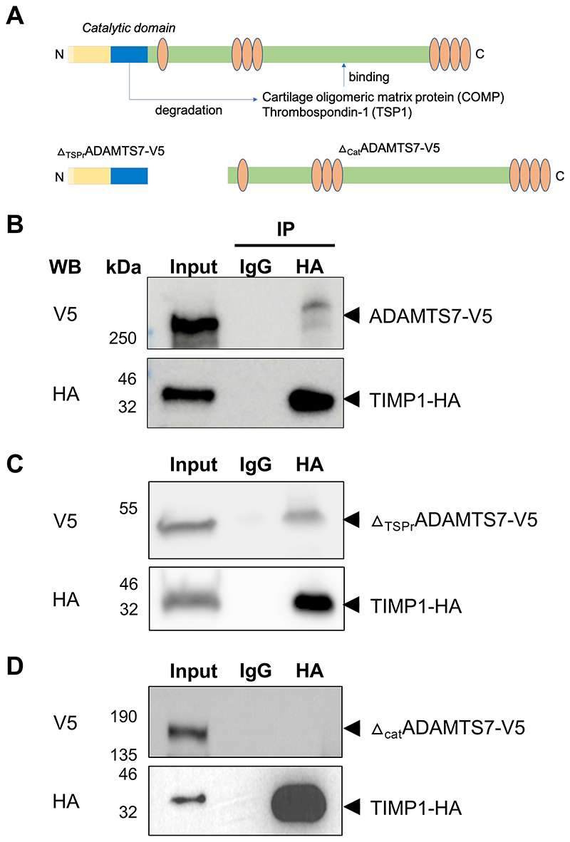
Results: In humans, ADAMTS7 expression was higher in caps of unstable as compared to stable carotid plaques. Compared to Apoe-/- mice, atherosclerotic aortas of Apoe-/- mice lacking Adamts-7 (Apoe-/-Adamts7-/-) contained higher protein levels of Timp-1 (tissue inhibitor of metalloprotease-1). In coimmunoprecipitation experiments, the catalytic domain of ADAMTS-7 bound to TIMP-1, which was degraded in the presence of ADAMTS-7 in vitro. ADAMTS-7 reduced the inhibitory capacity of TIMP-1 at its canonical target MMP-9 (matrix metalloprotease-9). As a downstream mechanism, we investigated collagen content in plaques of Apoe-/- and Apoe-/-Adamts7-/- mice after a Western diet. Picrosirius red staining of the aortic root revealed less collagen as a readout of higher MMP-9 activity in Apoe-/- as compared to Apoe-/- Adamts7-/- mice. To facilitate high-throughput screening for ADAMTS-7 inhibitors with the aim of decreasing TIMP-1 degradation, we designed a Förster resonance energy transfer-based assay targeting the ADAMTS-7 catalytic site.

Conclusions: ADAMTS-7, which is induced in unstable atherosclerotic plaques, decreases TIMP-1 stability reducing its inhibitory effect on MMP-9, which is known to promote collagen degradation and is likewise associated with coronary artery disease. Disrupting the interaction of ADAMTS-7 and TIMP-1 might be a strategy to increase collagen content and plaque stability for the reduction of atherosclerosis-related events.

Keywords: aorta; atherosclerosis; disintegrins; mice; solubility.


Conflict of interest statement

Disclosures Dr Schunkert has received personal fees from MSD SHARP & DOHME, AMGEN, Bayer Vital GmbH, Boehringer Ingelheim, Daiichi-Sankyo, Novartis, Servier, Brahms, Bristol-Myers-Squibb, Medtronic, Sanofi Aventis, Synlab, Pfizer, and Vifor T as well as grants and personal fees from Astra-Zeneca outside the submitted work. Drs Schunkert and Kessler are named inventors on a patent application for prevention of restenosis after angioplasty and stent implantation outside the submitted work. Dr Kessler received lecture fees from Bayer, Bristol-Myers Squibb, Abbott, and Astra-Zeneca, which are unrelated to this work. Dr Bernhagen is an inventor on patents related to anti-MIF and antichemokine strategies in inflammation and CVD (cardiovascular diseases) outside the submitted work. The other authors report no conflicts.

Figures

Graphical Abstract
Graphical Abstract
Figure 1
Figure 1. Proteome profiling of atherosclerotic aortas in Apoe-/- (n=3) and Apoe-/-Adamts7-/- (n=4) mice.
A. Volcano plot displaying proteins that were more abundant in Apoe-/-Adamts7- /- mice (right) as compared to Apoe-/- mice (left). Red and blue dots represent proteins which were significantly different between the genotypes after correction for multiple testing. B. The known ADAMTS-7 targets Comp and thrombospondin 1 (Thbs1) were detected at numerically lower levels in aortic tissue of Apoe-/- as compared to Apoe-/-Adamts7-/- mice. C. As putative novel targets of ADAMTS-7, we observed higher levels for the endogenous tissue inhibitors of MMPs (Timp) Timp-2 and, in particular, Timp-1 (Timp2 was detected in only one of three aortas of Apoe-/- mice). Unpaired t-tests with permutation-based FDR correction for multiple hypothesis testing.
Figure 2
Figure 2. Binding of TIMP-1 to ADAMTS-7.
A. ADAMTS-7 constructs. We cloned full-length ADAMTS-7 containing a C-terminal V5-tag (ADAMTS7-V5) and two constructs lacking the C-terminal disintegrin domain and thrombospondin repeats (ΔTSPrADAMTS7-V5) or the N-terminal catalytic domain (ΔcatADAMTS7-V5), respectively. The constructs were ectopically expressed in HEK293 cells. B. Co-IP of ADAMTS-7 and TIMP-1-HA. After precipitation of TIMP-1-HA, ADAMTS7-V5 was detectable. C, D. Binding of TIMP-1-HA to different parts of ADAMTS-7. After precipitation of TIMP-1-HA, ΔTSPrADAMTS7-V5 was detectable (C) while in contrast ΔcatADAMTS7-V5 was not detectable (D) indicating binding of TIMP-1 to the N-terminal part containing the catalytic domain.
Figure 3
Figure 3. Degradation of TIMP-1 in the presence of ADAMTS-7.
A, B. In vitro degradation assay. TIMP-1-HA was either overexpressed with full-length ADAMTS7-V5 (TS7-V5), the C-terminal part lacking the N-terminal catalytic domain (ΔcatTS7-V5) or an empty vector (mock). ADAMTS-7 constructs and TIMP-1 were detected by immunoblotting in cell lysates (CL) and the supernatant (SN). Vinculin served as housekeeping control. After co-expression with fulll-length ADAMTS7-V5, less TIMP-1-HA was detectable compared to mock or ΔcatTS7-V5 co-expression. Four independent experiments. C, D. Fluorescence intensity of TIMP-1-GFP after co-expression with an empty vector (mock), TS7-V5 or ΔcatTS7-V5 indicates reduced TIMP-1 levels in the presence of full-length ADAMTS-7. Five independent experiments. Scale bar: 100 μm. E, F. GFP-positive cells after overexpression of TIMP-1-GFP with either empty vector (mock), TS7-V5 or ΔcatTS7-V5. Presence of full-length ADAMTS-7 leads to reduced TIMP-1-GFP fluorescence. Six independent experiments. One-way ANOVA with Sidak’s multiple comparisons test. Data are mean and s.e.m.
Figure 4
Figure 4. Influence of ADAMTS-7 on inhibition of MMP-9 by TIMP-1.
A. Endogenous MMP-2-/MMP-9-activity of hCASMC in the presence (TS7-V5) or absence (mock transfection) of full-length ADAMTS-7. Presence of full-length ADAMTS-7 reduced the inhibitory potential of TIMP-1. Nine (TS7-V5) and ten (mock) independent experiments. Student’s t-test. B-D. Gel zymography with endogenous (pro-) MMP-2 and MMP-9 in the presence of either mock or TS7-V5 (B). Quantification of MMP-9 and MMP-2 bands reveals higher MMP-9 (C) and MMP-2 activity (D) in the presence of full-length ADAMTS-7, respectively. Eight independent experiments. Mann-Whitney (C) and Student’s t-test (D). E. Reduced inhibition of recombinant MMP-9 (rMMP-9) by TIMP-1 in the presence (TS7-V5) as compared to the absence (mock) of full-length ADAMTS-7. Five independent experiments. Student’s t-test. F, G. Secondary to precipitating FLAG-tagged MMP-9 (MMP-9-FLAG) more TIMP-1-HA could be recovered in the presence of full-length ADAMTS-7 (TS7-V5) as compared to ΔcatTS7-V5. Representative Western Blot. Three independent experiments. Student’s t-test. H, I. Picosirius red staining of collagen fibers in aortic root sections of Apoe-/- (n=14) and Apoe-/- Adamts7-/- mice (n=9) which were fed a Western diet. Mice lacking Adamts-7 displayed increased collagen content as compared to Apoe-/- mice. Student’s t-test. Scale bar, 500 μM. Data are mean and s.e.m.
Figure 5
Figure 5. ADAMTS7 expression in human atherosclerotic carotid plaques.
ADAMTS7 mRNA was detected at higher levels in caps of unstable (n=10) as compared to caps of stable plaques (n=10). Mann-Whitney test.
Figure 6
Figure 6. ADAMTS-7-TIMP-1 screening assay.
A. Left: After binding of ADAMTS-7 and TIMP-1 and excitation of cryptate linked to ADAMTS-7, FRET takes place between cryptate and d2. The ratio between emission of d2 and cryptate is used as the readout for interaction between ADAMTS-7 and TIMP-1. Right: 665/620 nm ratios for ADAMTS-7 and TIMP-1 and the negative controls, i.e., TIMP-1 or ADAMTS-7 alone. Four (TS7-FLAG-Crypt.+mock) and six (TS7-FLAG-Crypt.+TIMP-1-HA-d2, mock+TIMP1-HA-d2) independent experiments. One-way ANOVA with Sidak’s multiple comparisons test. B. Competition assay. Left: In the presence of untagged TIMP-1, emission of cryptate at 620 nm wavelength increases and FRET-induced emission of d2 at 665 nm wavelength decreases. Right: 665/620 nm ratios for ADAMTS-7 and TIMP-1 in the presence (400 or 800 ng) or absence (0 ng) of untagged TIMP-1. Seven independent experiments. One-way ANOVA with post-test for linear trend.

Similar articles

Cited by

References

    1. Virani SS, Alonso A, Aparicio HJ, Benjamin EJ, Bittencourt MS, Callaway CW, Carson AP, Chamberlain AM, Cheng S, Delling FN, et al. Heart Disease and Stroke Statistics—2021 Update: A Report From the American Heart Association. Circulation. 2021;143:e254–e743. - PubMed
    1. Yusuf S, Hawken S, Ounpuu S, Dans T, Avezum A, Lanas F, McQueen M, Budaj A, Pais P, Varigos J, et al. Effect of potentially modifiable risk factors associated with myocardial infarction in 52 countries (the INTERHEART study): case-control study. Lancet. 2004;364:937–952. - PubMed
    1. Kessler T, Schunkert H. Coronary Artery Disease Genetics Enlightened by Genome-Wide Association Studies. Jacc Basic Transl Sci. 2021;6:610–623. - PMC - PubMed
    1. Coronary Artery Disease (C4D) Genetics Consortium. A genome-wide association study in Europeans and South Asians identifies five new loci for coronary artery disease. Nat Genet. 2011;43:339–344. - PubMed
    1. Schunkert H, König IR, Kathiresan S, Reilly MP, Assimes TL, Holm H, Preuss M, Stewart AFR, Barbalic M, Gieger C, et al. Large-scale association analysis identifies 13 new susceptibility loci for coronary artery disease. Nat Genet. 2011;43:333–338. - PMC - PubMed

Publication types

MeSH terms