

Iron overload induces cerebral endothelial senescence in aged mice and in primary culture in a sex-dependent manner
- PMID: 37675802
- PMCID: PMC10652299
- DOI: 10.1111/acel.13977
Iron overload induces cerebral endothelial senescence in aged mice and in primary culture in a sex-dependent manner
Abstract
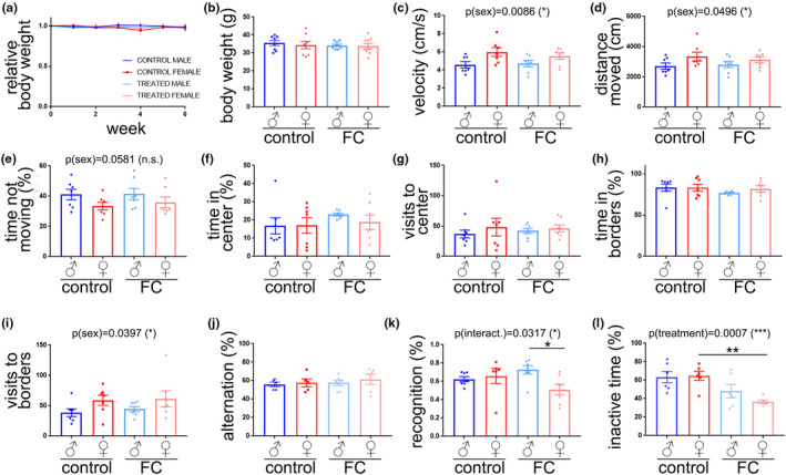
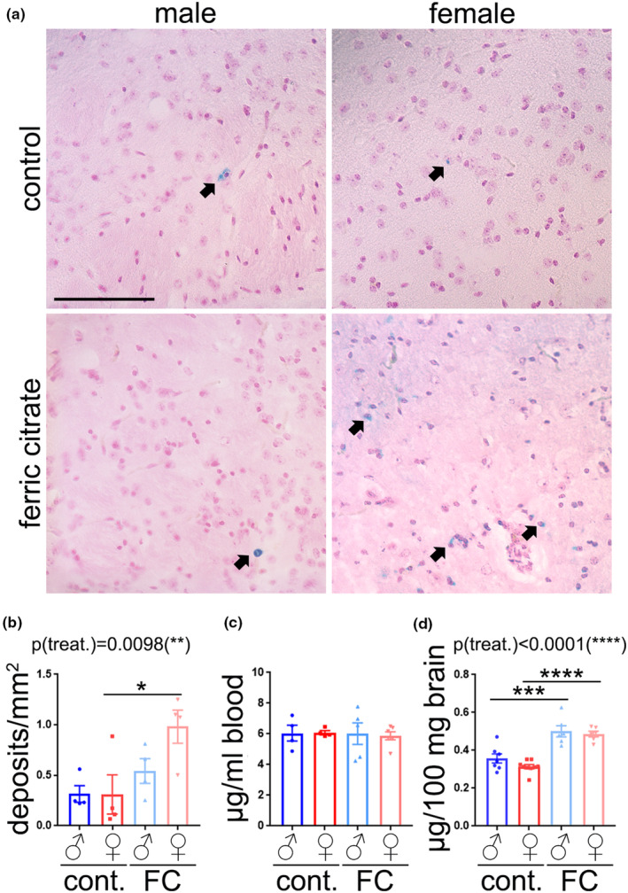
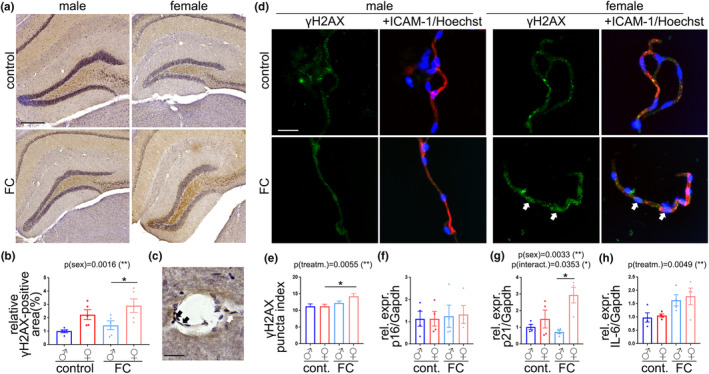
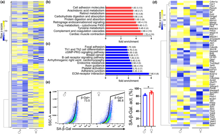
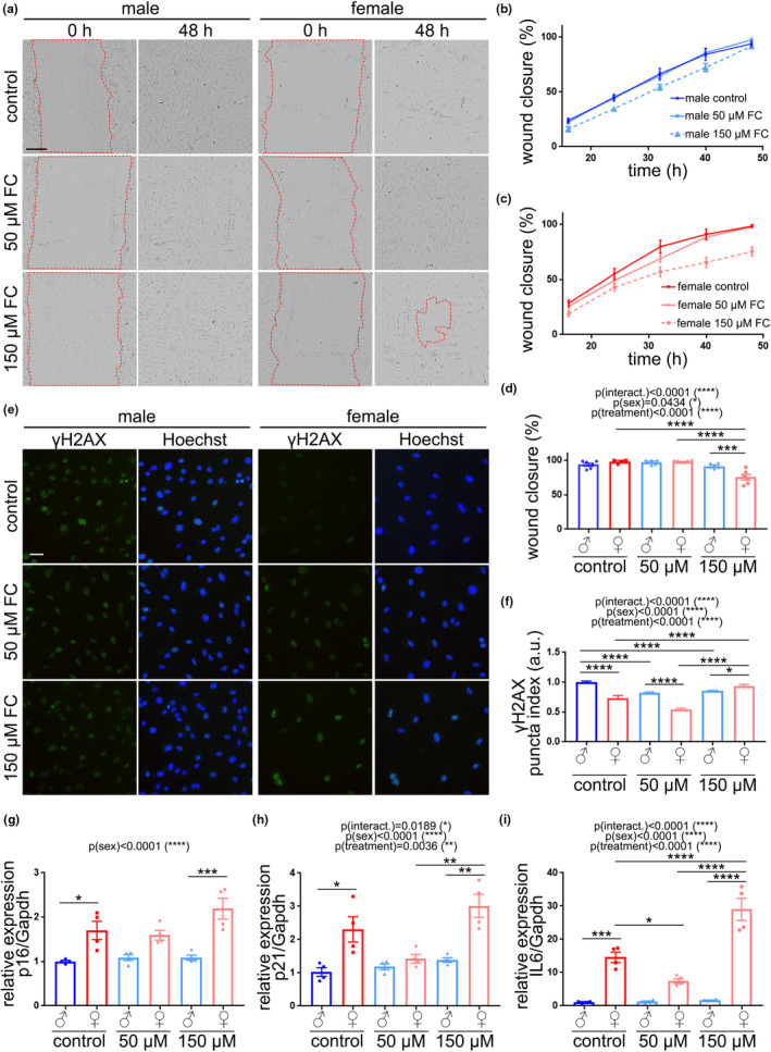
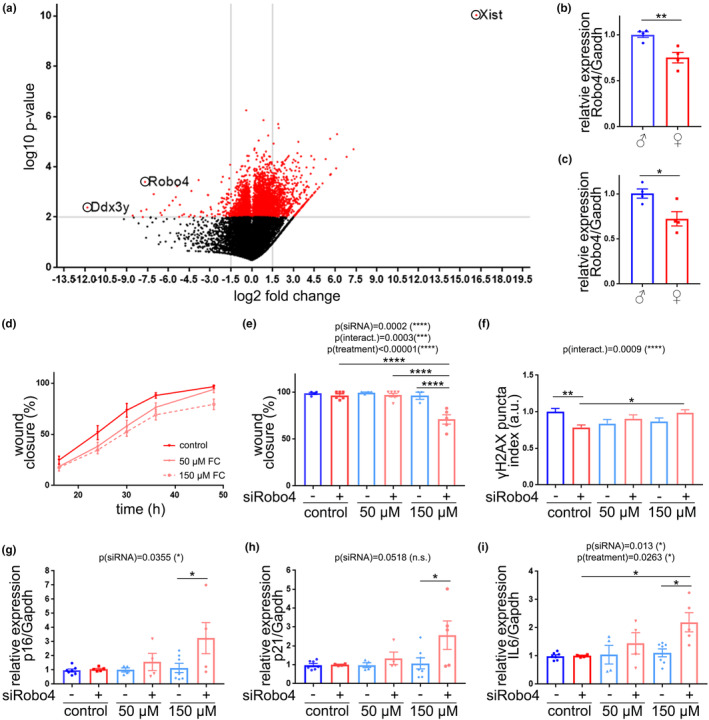
Iron imbalance in the brain negatively affects brain function. With aging, iron levels increase in the brain and contribute to brain damage and neurological disorders. Changes in the cerebral vasculature with aging may enhance iron entry into the brain parenchyma, leading to iron overload and its deleterious consequences. Endothelial senescence has emerged as an important contributor to age-related changes in the cerebral vasculature. Evidence indicates that iron overload may induce senescence in cultured cell lines. Importantly, cells derived from female human and mice generally show enhanced senescence-associated phenotype, compared with males. Thus, we hypothesize that cerebral endothelial cells (CEC) derived from aged female mice are more susceptible to iron-induced senescence, compared with CEC from aged males. We found that aged female mice, but not males, showed cognitive deficits when chronically treated with ferric citrate (FC), and their brains and the brain vasculature showed senescence-associated phenotype. We also found that primary culture of CEC derived from aged female mice, but not male-derived CEC, exhibited senescence-associated phenotype when treated with FC. We identified that the transmembrane receptor Robo4 was downregulated in the brain vasculature and in cultured primary CEC derived from aged female mice, compared with those from male mice. We discovered that Robo4 downregulation contributed to enhanced vulnerability to FC-induced senescence. Thus, our study identifies Robo4 downregulation as a driver of senescence induced by iron overload in primary culture of CEC and a potential risk factor of brain vasculature impairment and brain dysfunction.
Keywords: cellular senescence; cerebral endothelial cells; iron overload; molecular biology of aging; sex characteristics.
© 2023 The Authors. Aging Cell published by Anatomical Society and John Wiley & Sons Ltd.
Conflict of interest statement
All authors declare no competing financial interests.
Figures
References
-
- Algarin, C. , Karunakaran, K. D. , Reyes, S. , Morales, C. , Lozoff, B. , Peirano, P. , & Biswal, B. (2017). Differences on brain connectivity in adulthood are present in subjects with iron deficiency anemia in infancy. Frontiers in Aging Neuroscience, 9, 54. 10.3389/fnagi.2017.00054 - DOI - PMC - PubMed
-
- Block, G. A. , Fishbane, S. , Rodriguez, M. , Smits, G. , Shemesh, S. , Pergola, P. E. , Wolf, M. , & Chertow, G. M. (2015). A 12‐week, double‐blind, placebo‐controlled trial of ferric citrate for the treatment of iron deficiency anemia and reduction of serum phosphate in patients with CKD stages 3‐5. American Journal of Kidney Diseases, 65(5), 728–736. 10.1053/j.ajkd.2014.10.014 - DOI - PubMed
Publication types
MeSH terms
Substances
Grants and funding
LinkOut - more resources
Full Text Sources
