. 2023 Sep 7:12:e83189.
doi: 10.7554/eLife.83189.

Translation of dipeptide repeat proteins in C9ORF72 ALS/FTD through unique and redundant AUG initiation codons

Affiliations

Translation of dipeptide repeat proteins in C9ORF72 ALS/FTD through unique and redundant AUG initiation codons

Yoshifumi Sonobe et al. Elife. .

Abstract

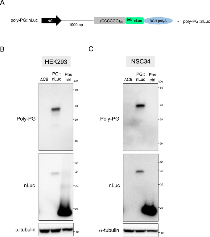
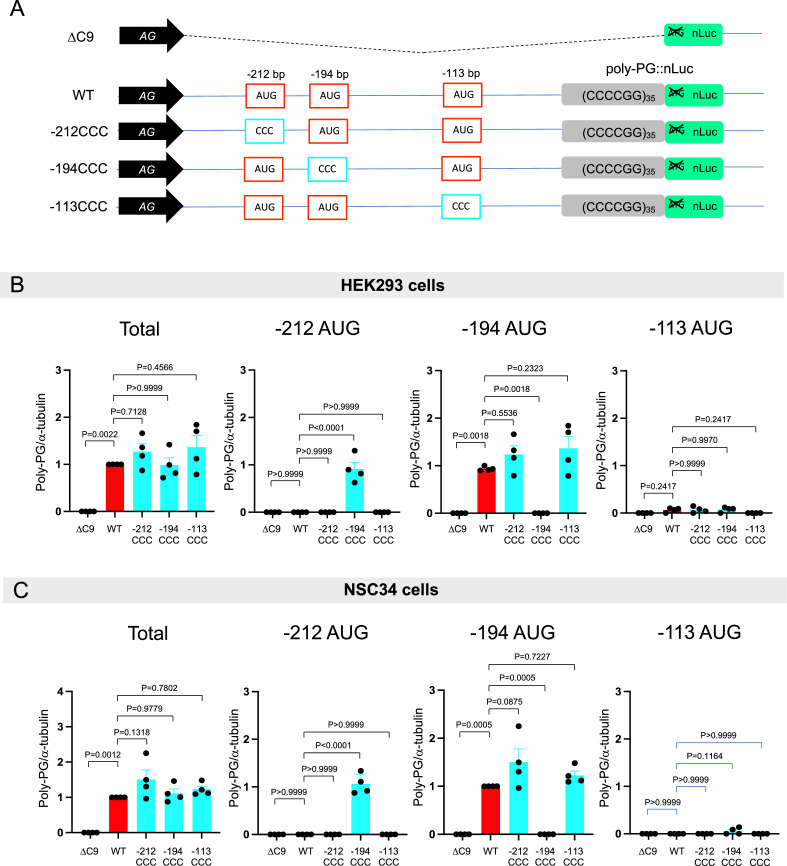
A hexanucleotide repeat expansion in C9ORF72 is the most common genetic cause of amyotrophic lateral sclerosis (ALS) and frontotemporal dementia (FTD). A hallmark of ALS/FTD pathology is the presence of dipeptide repeat (DPR) proteins, produced from both sense GGGGCC (poly-GA, poly-GP, poly-GR) and antisense CCCCGG (poly-PR, poly-PG, poly-PA) transcripts. Translation of sense DPRs, such as poly-GA and poly-GR, depends on non-canonical (non-AUG) initiation codons. Here, we provide evidence for canonical AUG-dependent translation of two antisense DPRs, poly-PR and poly-PG. A single AUG is required for synthesis of poly-PR, one of the most toxic DPRs. Unexpectedly, we found redundancy between three AUG codons necessary for poly-PG translation. Further, the eukaryotic translation initiation factor 2D (EIF2D), which was previously implicated in sense DPR synthesis, is not required for AUG-dependent poly-PR or poly-PG translation, suggesting that distinct translation initiation factors control DPR synthesis from sense and antisense transcripts. Our findings on DPR synthesis from the C9ORF72 locus may be broadly applicable to many other nucleotide repeat expansion disorders.

Keywords: AUG initiation codons; C9ORF72; amyotrophic lateral sclerosis; dipeptide protein repeats; frontotemporal dementia; genetics; genomics; human; iPSC-derived neurons; mouse; neuroscience.


Conflict of interest statement

YS, SL, GK, FG, RR No competing interests declared, YG, DK affiliated with Biogen. The authors have no financial interests to declare, PK Reviewing editor, eLife

Figures

Figure 1.
Figure 1.. Poly-PR and poly-PG are translated from antisense CCCCGG repeats.
(A) Schematic diagram of the constructs with 35 CCCCGG repeats preceded by 1000-bp-long intronic sequence from human C9ORF72, and then followed by nanoluciferase (nLuc). (B) HEK293 and (C) NSC34 cells were cotransfected with fLuc along with either ΔC9 or AS-C9 plasmids. The levels of luciferase activity were assessed by dual luciferase assays (mean ± s.e.m.). The experiments were repeated four times. One-way ANOVA with Tukey’s multiple comparison test was performed. (D–E) HEK293 and NSC34 cells were transfected with either ΔC9 or AS-C9 plasmids. Cell lysates were processed for western blotting, and immunostained with antibodies to (D) poly-PR, (E) poly-PG, and α-tubulin. (F–G) NSC34 cells transfected with either ΔC9, (F) poly-PR::nLuc, or (G) poly-PG::nLuc were stained with a nuclear marker (4′,6-diamidino-2-phenylindole [DAPI]: blue) and with antibodies against poly-PR (F: green) or poly-PG (G: green). Scale bars indicate 20 μm. (H–I) NSC34 cells were transfected with either ΔC9, (H) poly-PR::nLuc, or (I) poly-PG::nLuc plasmids. WST-8 assay was performed to assess the cell viability. The experiments were repeated five times. Unpaired t test was performed.
Figure 1—figure supplement 1.
Figure 1—figure supplement 1.. Nanoluciferase (nLuc) is fused to dipeptide repeats (DPRs) translated from antisense C9 plasmids containing CCCCGG repeats.
(A) Schematic diagram of the construct. (B) HEK293 and (C) NSC34 cells were transfected with either ΔC9, PG::nLuc, or positive control (Pos Ctrl) plasmids. The nanoluciferase (nLuc) expression plasmid pNL1.1.[Nluc/CMV] from Promega was used as positive control. Cell lysates were processed for western blotting, and immunostained with antibodies to poly-PG, nLuc, and α- tubulin.
Figure 1—figure supplement 2.
Figure 1—figure supplement 2.. Expression levels of poly-PR and poly-PG in the RIPA-insoluble fraction.
(A) Schematic diagram of the constructs. (B, D, F, H) HEK293 and (C, E, G, I) NSC34 cells were transfected with either ΔC9 (blue) or AS-C9 (red) plasmids. Cell lysates were fractionated into RIPA-soluble (S) and -insoluble (I) fraction. Western blotting was performed and immunostained with antibodies to poly-PR (B–C), poly-PG (F–G), α-tubulin, and H3K4me2. H3K4me2 is a reliable marker for the RIPA-insoluble fraction (see Materials and methods). (D–E, H–I) The signal intensity of the bands was quantified. The expression levels of poly-PR (D–E) or poly-PG (H–I) in AS-C9 of RIPA-soluble fraction were set to 1.0. Experiments were repeated four times. Two-way ANOVA with Tukey’s multiple comparison test was performed.
Figure 1—figure supplement 3.
Figure 1—figure supplement 3.. Poly-PA is not detected by western blotting upon transfection of antisense C9 plasmids containing CCCCGG repeats.
(A) Schematic diagram of the constructs. (B) HEK293 and (C) NSC34 cells were transfected with either ΔC9, AS-C9, or positive control plasmids. For positive control, we generated a plasmid that has an AUG start codon located at –67 bp upstream from CCCCGG repeats. This AUG is in poly-PA frame. Cell lysates were processed for western blotting, and immunostained with antibodies to poly-PA and α-tubulin.
Figure 2.
Figure 2.. An AUG at –273 bp position is the start codon for poly-PR translation.
(A) Schematic diagram showing constructs with mutations in the putative start codons for poly-PR. HEK293 (B–C) and NSC34 (D–E) cells were transfected with indicated plasmids. Cell lysates were processed for western blotting, and immunostained with antibodies to poly-PR and α -tubulin. (B, D) Representative blots are shown. (C, E) The signal intensity of the bands were quantified (mean ± s.e.m.). The experiments were repeated four times. One-way ANOVA with Tukey’s multiple comparison test was performed. (F) HEK293 and (G) NSC34 cells were cotransfected with the plasmids along with fLuc. The levels of luciferase activity were assessed by dual luciferase assays (mean ± s.e.m.). The experiments were repeated four times. One-way ANOVA with Tukey’s multiple comparison test was performed. (H) NSC34 cells transfected with either ΔC9, poly-PR::nLuc, or –273 AUG ->UAG plasmids were stained with 4′,6-diamidino-2-phenylindole [DAPI] (blue) and immunostained with a poly-PR antibody (green). Scale bars show 20 μm. (I) NSC34 cells were transfected with either ΔC9, wild type (WT), or –273 AUG ->UAG plasmids. WST-8 assay was performed to assess the cell viability. The experiments were repeated five times. One-way ANOVA with Tukey’s multiple comparison test was performed. In ΔC9 and WT, the same datasets as Figure 1H were used (mean ± s.e.m.). The experiments were repeated five times. One-way ANOVA with Tukey’s multiple comparison test was performed.
Figure 3.
Figure 3.. Mutation of AUG codons to CCC fails to suppress poly-PG translation.
(A) Schematic diagram showing mutants with changes in the putative start codons for poly-PG. (B) HEK293 and NSC34 cells were transfected with indicated plasmids. Cell lysates were processed for western blotting, and immunostained with antibodies to poly-PG and α-tubulin. (C) HEK293 and (D) NSC34 cells were cotransfected with fLuc plasmid along with other indicated plasmids. The level of luciferase activity was assessed by dual luciferase assay (mean ± s.e.m.). The experiments were repeated four times. One-way ANOVA with Tukey’s multiple comparison test was performed.
Figure 3—figure supplement 1.
Figure 3—figure supplement 1.. Quantification of data from Figure 3B.
(A) Schematic diagram of the constructs. (B–C) The signal intensity of total poly-PG and poly-PG translated from each of the AUGs (−212,–194, or –113) in (B) HEK293 and (C) NSC34 was quantified. One-way ANOVA with Tukey’s multiple comparison test was performed. The experiments were repeated four times. Data are presented as mean ± s.e.m.
Figure 4.
Figure 4.. An AUG at –194 bp position is the primary start codon for poly-PG translation.
(A) Schematic diagram of the constructs. (B) HEK293 and NSC34 cells were transfected with indicated plasmids. Cell lysates were processed for western blotting, and immunostained with antibodies to poly-PG and α-tubulin. (C) HEK293 and (D) NSC34 cells were cotransfected with fLuc plasmid along with indicated plasmids. The level of luciferase activity was assessed by dual luciferase assays (mean ± s.e.m.). The experiments were repeated four times. One-way ANOVA with Tukey’s multiple comparison test was performed.
Figure 4—figure supplement 1.
Figure 4—figure supplement 1.. Quantification of data from Figure 4B.
(A) Schematic diagram of the constructs. (B–C) The signal intensity of poly-PG and poly-PG translated from each of the AUGs (−212, –194, or –113) in (B) HEK293 and (C) NSC34 cells was quantified. One-way ANOVA with Tukey’s multiple comparison test was performed. The experiments were repeated four times. Data are presented as mean ± s.e.m.
Figure 5.
Figure 5.. Redundancy of start codon usage in poly-PG translation.
(A) Schematic diagram of the constructs. (B) HEK293 and NSC34 cells were transfected with indicated plasmids. Cell lysates were processed for western blotting, and immunostained with antibodies to poly-PG and α-tubulin. (C) HEK293 and (D) NSC34 cells were cotransfected with fLuc plasmid along with indicated plasmids. The level of luciferase activity was assessed by dual luciferase assays (mean ± s.e.m.). The experiments were repeated four times. One-way ANOVA with Tukey’s multiple comparison test was performed. (E) NSC34 cells transfected with indicated plasmids were stained with 4′,6-diamidino-2-phenylindole [DAPI] (blue) and immunostained with a poly-PG antibody (green). Scale bars show 20 μm. (F) NSC34 cells were transfected with indicated plasmids. WST-8 assay was performed to assess the cell viability (mean ± s.e.m.). The experiments were repeated five times. One-way ANOVA with Tukey’s multiple comparison test was performed. In ΔC9 and wild type (WT), the same datasets as Figure 1I were used.
Figure 5—figure supplement 1.
Figure 5—figure supplement 1.. Quantification of data from Figure 5B and C.
(A) Schematic diagram of the constructs. (B–C) The signal intensity of total poly-PG and poly-PG translated from each of the AUGs (−212, –194, or –113) in (B) HEK293 and (C) NSC34 cells was quantified. One-way ANOVA with Tukey’s multiple comparison test was performed. The experiments were repeated four times. Data are presented as mean ± s.e.m.
Figure 6.
Figure 6.. Downregulation of EIF2D does not reduce expression levels of poly-PG and poly-PR.
(A) A gRNA targeted the second exon of human EIF2D (see Materials and methods). (B) After CRISPR/Cas9-mediated gene editing, the EIF2D knockout (EIF2DKO) HEK293 cells carried different mutations on each allele. (C) Cell lysates from wild type (WT) and EIF2DKO HEK293 cells were processed for western blotting, and immunostained with antibodies to eIF2D and α-tubulin. (D–F) WT and EIF2DKO HEK293 cells were cotransfected with fLuc plasmid along with either (D–E) AS-C9 plasmids or (F) C9 plasmids containing 75 GGGGCC repeats. The level of luciferase activity was assessed by dual luciferase assays. (G–I) WT HEK293 cells were transfected with fLuc and either (G–H) AS-C9 plasmids or (I) C9 monocistronic plasmids containing 75 GGGGCC repeats along with anti-EIF2D short hairpin RNA (shRNA). The level of luciferase activity was assessed by dual luciferase assays (mean ± s.e.m.). The experiments were repeated three times. Unpaired t test was performed. The poly-GA reduction upon EIF2D shRNA is consistent with our previous observations (Sonobe et al., 2021), albeit more modest - likely due to a technical reason (a bicistronic construct containing 75 GGGGCC repeats was used in Sonobe et al., 2021).
Figure 7.
Figure 7.. Dipeptide repeat (DPR) levels in human iPSC-derived neurons upon eIF2D knockdown.
(A) The EIF2D, EIF2A, and actin mRNA levels were assessed by real-time quantitative PCR on either isogenic control (26Z90) or C9ORF72 human motor neurons (patient line 26#6) upon small interfering RNA (siRNA) transfection (scramble or EIF2D siRNA-1). The eIF2D and eIF2A mRNA levels were normalized to actin. The experiments were repeated twice. p<0.05 by one-way ANOVA with Tukey’s post hoc test. (B) Poly-GA, poly-GR, and poly-GP levels in motor neurons differentiated independently (twice) from isogenic control and one C9ORF72 iPSC line. DPR levels were measured using an Meso Scale Discovery (MSD) immunoassay in a blinded manner. Data presented as mean ± SD. p-Values were calculated using two-way ANOVA with Dunnett’s multiple comparison test using Prism (9.1) software. (C–D) The EIF2D and actin mRNA levels were assessed by real-time quantitative PCR on C9ORF72 human motor neurons (two patient lines) upon siRNAs transfection (scramble, EIF2D siRNA-1 or EIF2D siRNA-2). The eIF2D mRNA levels were normalized to actin. The experiments were repeated three times. *p<0.05, ***p<0.001, ns, not significant by two-tailed unpaired t tests were used for two groups and a one-way ANOVA followed by Dunnett’s post hoc analysis was used for more than two groups. (E) Poly-GA, poly-GR, and poly-GP levels in motor neurons differentiated independently (n=3 times) from isogenic or healthy control lines and total two C9ORF72 patient iPSC lines (lines 27#11 and 40#3). DPR levels were measured using an MSD immunoassay in a blinded manner. For poly(GA) assay, total protein normalized poly(GA) concentrations were converted to percentage and presented as mean ± SE. For poly(GR), poly(GP) assay, total protein normalized electrochemiluminescence (ECL) values were converted to percentage and presented as mean ± SE. p-Values were calculated using one-way ANOVA with Dunnnett’s T3 multiple comparisons test .
Figure 7—figure supplement 1.
Figure 7—figure supplement 1.. The small interfering RNA (siRNA) against eIF2D knocks down eIF2D protein levels.
siRNA (s4496) against eIF2D knocks down eIF2D protein levels. 3×10⁵ HEK293 cells were plated into six-well cell culture plate. 60 pmol siRNAs were transfected into the cells using 6.25 μl Lipofectamine RNAiMAX. 72 hr later, protein was extracted using RIPA buffer and western blotting was performed.

Update of

  • doi: 10.1101/2022.08.06.503063

Similar articles

Cited by

References

    1. Almeida S, Gascon E, Tran H, Chou HJ, Gendron TF, Degroot S, Tapper AR, Sellier C, Charlet-Berguerand N, Karydas A, Seeley WW, Boxer AL, Petrucelli L, Miller BL, Gao F-B. Modeling key pathological features of frontotemporal dementia with C9ORF72 repeat expansion in iPSC-derived human neurons. Acta Neuropathologica. 2013;126:385–399. doi: 10.1007/s00401-013-1149-y. - DOI - PMC - PubMed
    1. Almeida S, Krishnan G, Rushe M, Gu Y, Kankel MW, Gao F-B. Production of poly(GA) in C9ORF72 patient motor neurons derived from induced pluripotent stem cells. Acta Neuropathologica. 2019;138:1099–1101. doi: 10.1007/s00401-019-02083-z. - DOI - PMC - PubMed
    1. Ayhan F, Perez BA, Shorrock HK, Zu T, Banez-Coronel M, Reid T, Furuya H, Clark HB, Troncoso JC, Ross CA, Subramony SH, Ashizawa T, Wang ET, Yachnis AT, Ranum LP. SCA8 RAN polySer protein preferentially accumulates in white matter regions and is regulated by eIF3F. The EMBO Journal. 2018;37:e99023. doi: 10.15252/embj.201899023. - DOI - PMC - PubMed
    1. Banerjee P, Mehta AR, Nirujogi RS, Cooper J, James OG, Nanda J, Longden J, Burr K, McDade K, Salzinger A, Paza E, Newton J, Story D, Pal S, Smith C, Alessi DR, Selvaraj BT, Priller J, Chandran S. Cell-autonomous immune dysfunction driven by disrupted autophagy in C9orf72-ALS iPSC-derived microglia contributes to neurodegeneration. Science Advances. 2023;9:eabq0651. doi: 10.1126/sciadv.abq0651. - DOI - PMC - PubMed
    1. Boivin M, Pfister V, Gaucherot A, Ruffenach F, Negroni L, Sellier C, Charlet-Berguerand N. Reduced autophagy upon C9ORF72 loss synergizes with dipeptide repeat protein toxicity in G4C2 repeat expansion disorders. The EMBO Journal. 2020;39:e100574. doi: 10.15252/embj.2018100574. - DOI - PMC - PubMed

Publication types

MeSH terms