Review
. 2023 Oct;23(10):593-605.
doi: 10.1007/s11910-023-01297-9. Epub 2023 Sep 7.

Brain Sagging Dementia

Affiliations
Review

Brain Sagging Dementia

Aslan Lashkarivand et al. Curr Neurol Neurosci Rep. 2023 Oct.

Abstract

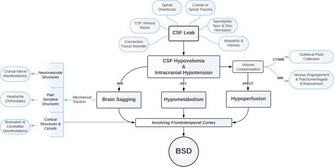
Purpose of review: Brain sagging dementia (BSD) is a rare but devastating form of early-onset dementia characterized by intracranial hypotension and behavioral changes resembling behavioral variant frontotemporal dementia. This review aims to provide a comprehensive overview of BSD, highlighting its pathomechanism, diagnostic tools, and available treatment options.

Recent findings: BSD exhibits a complex clinical manifestation with insidious onset and gradual progression of behavioral disinhibition, apathy, inertia, and speech alterations. Additionally, patients may exhibit brainstem and cerebellar signs such as hypersomnolence and gait disturbance. Although headaches are common, they may not always demonstrate typical orthostatic features. Recent radiological advances have improved the detection of CSF leaks, enabling targeted treatment and favorable outcomes. Understanding the pathomechanism and available diagnostic tools for BSD is crucial for a systematic approach to timely diagnosis and treatment of this reversible form of early-onset dementia, as patients often endure a complex and lengthy clinical course.

Keywords: Behavioral variant frontotemporal dementia; Brain sagging dementia (BSD); Early-onset dementia; Intracranial hypotension.


Conflict of interest statement

The authors declare no competing interests.

Figures

Fig. 1
Fig. 1
Schematic diagram depicting etiology and pathophysiological mechanisms of clinical and radiological changes in brain sagging dementia (BSD). MRI, magnetic resonance imaging; PET, positron emission tomography; SPECT, single-photon emission computed tomography; BSD, brain sagging dementia
Fig. 2
Fig. 2
Neuroimaging and intracranial pressure (ICP) observations in intracranial hypotension. Brain T1-weighted magnetic resonance images in A sagittal, B coronal, and C axial planes demonstrating imaging signs of spontaneous intracranial hypotension (SIH), including enlargement of the pituitary (P) gland, brainstem sagging (BS) toward the clival bone, downward tonsillar (T) ectopy, smaller pontomesencephalic angle (shown by one asterisk), decreased angle between vein of Galen and straight sinus (shown by two asterisks), enhancement of the dura mater (DM), dural venous engorgement, here indicated by rounding of the cross-section of the dural venous sinuses (S). There may as well be reduced space for the mesencephalon (M). Sometimes dural venous engorgement may be associated with subdural effusion/hematoma, which was not the case for the present individual. D The overnight ICP in an individual with signs of intracranial hypotension was measured in the right frontal lobe using a Codman ICP sensor, illustrated by the trend plots of mean ICP (MeanP, light green) and mean ICP wave amplitude (Mean Wave AMP, MWA darker green). The position of the patient is indicated, illustrating that in the supine position mean ICP was positive (5 to 10 mmHg), while mean ICP declined to about − 3 mmHg while sitting up, and further declining to below − 5 mmHg when standing up. In the upright position, the mean ICP was about − 10 mmHg. Negative mean ICP scores like these can be seen in shunted patients with overdrainage [87]. The MWA was on average 3.6 mmHg, independent of position, but severe negative mean ICP scores may be accompanied with altered MWA [86]. Image: Sensometrics RD Software (dPCom AS, Oslo, Norway). E In this individual, a spinal myelography revealed a CSF-venous fistula (CSFVF) at the level of Th1/Th12; type 3 leakage where the CSF leakage was not associated with extradural CSF collections [38]. It was occluded by transvenous embolization of the fistula via the azygos vein

References

    1. Lashkarivand A, Eide PK. Brain sagging dementia – diagnosis, treatment, and outcome: a review. Neurology. 2022;25:25. - PubMed
    1. Lashkarivand A, Eide PK. The first report on brain sagging dementia caused by a cranial leak: a case report. Frontiers Neurol [electronic resource] 2022;13:1006060. - PMC - PubMed
    1. Schievink WI, Maya MM, Barnard ZR, Moser FG, Jean-Pierre S, Waxman AD, et al. Behavioral variant frontotemporal dementia as a serious complication of spontaneous intracranial hypotension. Operative Neurosurg. 2018;15(5):505–515. doi: 10.1093/ons/opy029. - DOI - PubMed
    1. Schievink WI, Maya M, Barnard Z, Tache RB, Prasad RS, Wadhwa VS, et al. The reversible impairment of behavioral variant frontotemporal brain sagging syndrome: challenges and opportunities. Alzheimers Dement (N Y) 2022;8(1):e12367. doi: 10.1002/trc2.12367. - DOI - PMC - PubMed
    1. Vetrugno R, Mascalchi M, Chierichetti F, Gallassi R, Alessandria M, Guerrini L, et al. Hypoactive-hypoalert behavior (“psychic akinesia”) in intracranial hypotension syndrome. Neurology. 2008;71(18):1452–1454. doi: 10.1212/01.wnl.0000327869.39403.82. - DOI - PubMed