Review
. 2022 Oct 1;2(1):42.
doi: 10.1007/s44154-022-00064-z.

Electrophysiology and fluorescence to investigate cation channels and transporters in isolated plant vacuoles

Affiliations
Review

Electrophysiology and fluorescence to investigate cation channels and transporters in isolated plant vacuoles

Antonella Gradogna et al. Stress Biol. .

Abstract

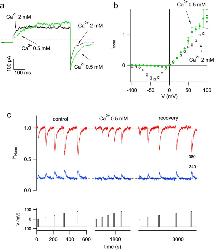
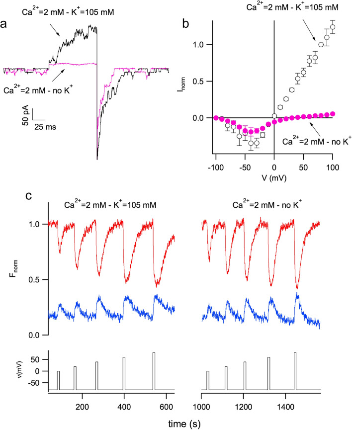
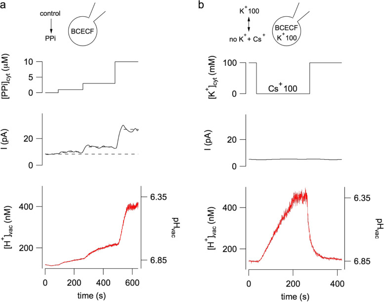
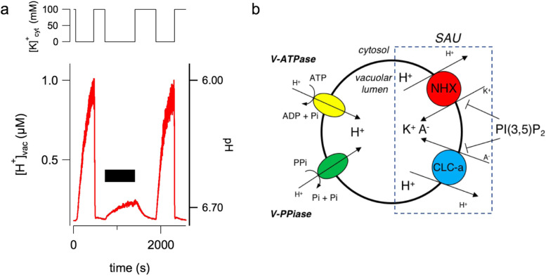
The plant vacuole plays a fundamental role in cell homeostasis. The successful application of patch-clamp technique on isolated vacuoles allows the determination of the functional characteristics of tonoplast ion channels and transporters. The parallel use of a sensor-based fluorescence approach capable of detecting changes in calcium and proton concentrations opens up new possibilities for investigation. In excised patch, the presence of fura-2 in the vacuolar solution reveals the direct permeation of calcium in plant TPC channels. In whole-vacuole, the activity of non-electrogenic NHX potassium proton antiporters can be measured by using the proton sensitive dye BCECF loaded in the vacuolar lumen by the patch pipette. Both vacuolar NHXs and CLCa (chloride/nitrate antiporter) are inhibited by the phosphoinositide PI(3,5)P2, suggesting a coordinated role of these proteins in salt accumulation. Increased knowledge in the molecular mechanisms of vacuolar ion channels and transporters has the potential to improve our understanding on how plants cope with a rapidly changing environment.

Keywords: BCECF; Calcium; Fura-2; NHX antiporters; Patch-clamp; Potassium; Proton; TPC channels.


Conflict of interest statement

The authors declare that they have no competing interests.

Figures

Fig. 1
Fig. 1
The plant vacuole can occupy most of the intracellular volume and is easy to isolate. a Confocal images showing an Arabidopsis thaliana mesophyll protoplast transiently expressing a tonoplast-localized AtTPC1-EGFP fusion protein (left, green signal) and stained with the plasma membrane marker FM4–64 (middle, red signal). The right panel displays merged signals. Scale bar 7 μm (see Supplemental material of Picco et al. for experimental details). b Protoplasts from Arabidopsis mesophyll cells were obtained by enzymatic treatment with cellulase and pectolyase (Scholz-Starke et al. 2006). Upon application of the vacuole release solution VRS (see text) they burst and release the vacuoles. Scale bar 10 μm
Fig. 2
Fig. 2
Cartoon of the patch-clamp technique applied on plant vacuoles. The patch clamp technique is applied in the whole-vacuole (cytosolic side-out) configuration. Positive currents correspond to the movement of cations from the cytosolic to the luminal side of the vacuole (or to the opposite movement of anions)
Fig. 3
Fig. 3
Plant TPC channels are modulated by cytosolic calcium. a Current recordings of carrot TPC channels in the presence of 2 mM (black trace) and 0.5 mM (green trace) cytosolic calcium. Main voltage pulse of + 80 mV; holding voltage was − 80 mV. b Stationary currents in 2 mM (black open circles) and 0.5 mM (green solid circles) cytosolic calcium were normalized to the value at + 80 mV in 2 mM Ca2+ and displayed versus voltage. Voltage pulses were ranging from − 100 mV to + 100 mV in 10 mV increments. c Patch pipette was filled with 100 μM of the calcium-sensitive dye fura-2. Fluorescence signals (upper panel) were induced by excitation light at 380 nm (red trace) and 340 nm (blue trace), respectively. The lower panel displayed 10 s voltage pulses of 0, + 20, + 40, + 60, + 80 mV, which were applied starting in 2 mM Ca2+ (control, left panel), at low calcium (Ca2+ 0.5 mM, middle panel), and again in 2 mM Ca2+ (recovery, left panel). Modification of Figs. 1 and 2, from Carpaneto and Gradogna (2018), Biophysical Chemistry, 236:1–7, reprinted by permission from Elsevier (license number 5333730921940)
Fig. 4
Fig. 4
Removal of cytosolic potassium strongly reduced TPC currents but did not change calcium permeation. a Currents of carrot TPC channels recorded in control condition (Ca2+ = 2 mM – K+ = 105 mM, black trace) and in the absence of cytosolic K+ (Ca2+ = 2 mM – no K+, violet trace). Main voltage pulse of + 80 mV lasting 100 ms. Holding and tail voltage of − 80 mV. b Stationary currents (normalized to the value at + 80 mV in control) in the presence (open black circles) and absence of cytosolic potassium (solid violet circles) were plotted versus applied voltage. c In the upper panel, fura-2 signals did not change significantly in control condition (Ca2+ = 2 mM – K+ = 105 mM, left panel) and upon removal of cytosolic potassium (Ca2+ = 2 mM – no K+, right panel). The lower panel displays the applied voltage (10 s voltage pulses from 0 mV to + 80 mV, in 20 mV steps, from a holding potential of − 80 mV). Modification of Figs. 4 and 5, from Carpaneto and Gradogna (2018), Biophysical Chemistry, 236:1–7, reprinted by permission from Elsevier (license number 5333730921940)
Fig. 5
Fig. 5
The loading phase of the vacuole with the proton sensitive fluorophore BCECF. a The left panel shows a bright-field image of a micropipette placed on an isolated vacuole. Scale bar, 10 μm. The whole-vacuole configuration allows the loading of the fluorophore inside the vacuolar lumen. In the right panel the fluorescence image of the same vacuole was obtained with a 490 nm excitation light and detected using a 515-nm bandpass emission filter. The red circle is the region of interest (ROI) where fluorescence is evaluated. b After establishment of the whole-vacuole configuration it is possible to follow the time course of BCECF fluorescence emission signals, F490 (excitation at 490 nm, upper panel), F440 (excitation at 440 nm, middle panel) and fluorescence ratio (F490/F440 and pH, lower panel). Seal and break-in are obtained in VRS, i.e. the solution able to blast the protoplast, which is changed to control bath solution after about 250 s. Modification of Fig. S2 and S4, from Gradogna et al. (2021), New Phytologist, 229:3026–3036, reprinted by permission from John Wiley and Sons (license number 5333730373195)
Fig. 6
Fig. 6
Vacuolar acidification revealed by the proton-sensitive fluorescent dye BCECF. a Inorganic pyrophosphate (PPi) is added to the bath solution (top panel) at concentrations of 1, 3 and 10 μM (dotted lines indicate that the switching of the bath solution is irrespective to the real change due to the perfusion system, see Gradogna et al. for a discussion about the effects of the perfusion). Middle panel shows the correspondent time course of tonoplast membrane current due to vacuolar proton-pumping pyrophosphatase activation. Bis(2-carboxyethyl)-5(6)-carboxyfluorescein (BCECF) loaded inside the vacuole through the patch pipette allows the determination of luminal proton concentration changes (bottom panel). b Substitution of cytosolic bath solution potassium with an equimolar amount of caesium ions is schematically displayed in the top panel. The middle panel shows that there is no change in background current (holding voltage of 0 mV). However a significant acidification of the luminal solution is apparent (bottom panel). Modification of Fig. 1, from Gradogna et al. (2021), New Phytologist, 229:3026–3036, reprinted by permission from John Wiley and Sons (license number 5333730373195)
Fig. 7
Fig. 7
The phosphoinositide PI(3,5)P2 inhibits NHX activity. a PI(3,5)P2 added at a concentration of 200 nM in the bath solution lacking potassium, which is substituted by the large, membrane- impermeable cation BTP+ (no K+ + BTP+), induces a strong and reversible inhibition of the vacuolar acidification mediated by NHX activity. b Scheme of the tonoplast key players responsible of vacuolar salt uptake. A Salt Accumulation Unit (SAU) is formed by NHXs together with CLC-a. V-ATPase, vacuolar H+-ATPase; V-PPiase, vacuolar H+- pyrophosphatase. A, H+ and K+ indicates respectively anions, protons and potassium ions; the dimension of the letters for the ions is proportional to their concentration. Modification of Fig. 6, from Gradogna et al. (2021), New Phytologist, 229:3026–3036, reprinted by permission from John Wiley and Sons (license number 5333730373195)

Similar articles

Cited by

References

    1. Bak G, Lee E-J, Lee Y, Kato M, Segami S, Sze H, Maeshima M, Hwang J-U, Lee Y (2013) Rapid structural changes and acidification of guard cell vacuoles during stomatal closure require phosphatidylinositol 3,5-bisphosphate. Plant Cell 25:2202–2216. 10.1105/tpc.113.110411 - PMC - PubMed
    1. Balla T (2013) Phosphoinositides: tiny lipids with giant impact on cell regulation. Physiol Rev 93:1019–1137. 10.1152/physrev.00028.2012 - PMC - PubMed
    1. Benkerrou D, Minicozzi V, Gradogna A, Milenkovic S, Bodrenko IV, Festa M, Lagostena L, Cornara L, D’Amore A, Ceccarelli M et al (2019) A perspective on the modulation of plant and animal two pore channels (TPCs) by the flavonoid naringenin. Biophys Chem 254:106246. 10.1016/j.bpc.2019.106246 - PubMed
    1. Bertl A, Blumwald E, Coronado R, Eisenberg R, Findlay G, Gradmann D, Hille B, Köhler K, Kolb HA, MacRobbie E (1992) Electrical measurements on endomembranes. Science 258:873–874. 10.1126/science.1439795 - PubMed
    1. Boccaccio A, Scholz-Starke J, Hamamoto S, Larisch N, Festa M, Gutla PVK, Costa A, Dietrich P, Uozumi N, Carpaneto A (2014) The phosphoinositide PI (3, 5) P2 mediates activation of mammalian but not plant TPC proteins: functional expression of endolysosomal channels in yeast and plant cells. Cell Mol Life Sci 71:4275–4283. 10.1007/s00018-014-1623-2 - PMC - PubMed