. 2023 Mar 23;3(1):3.
doi: 10.1007/s44154-023-00084-3.

Coat protein of rice stripe virus enhances autophagy activity through interaction with cytosolic glyceraldehyde-3-phosphate dehydrogenases, a negative regulator of plant autophagy

Affiliations

Coat protein of rice stripe virus enhances autophagy activity through interaction with cytosolic glyceraldehyde-3-phosphate dehydrogenases, a negative regulator of plant autophagy

Wanying Zhao et al. Stress Biol. .

Abstract

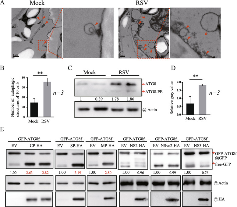
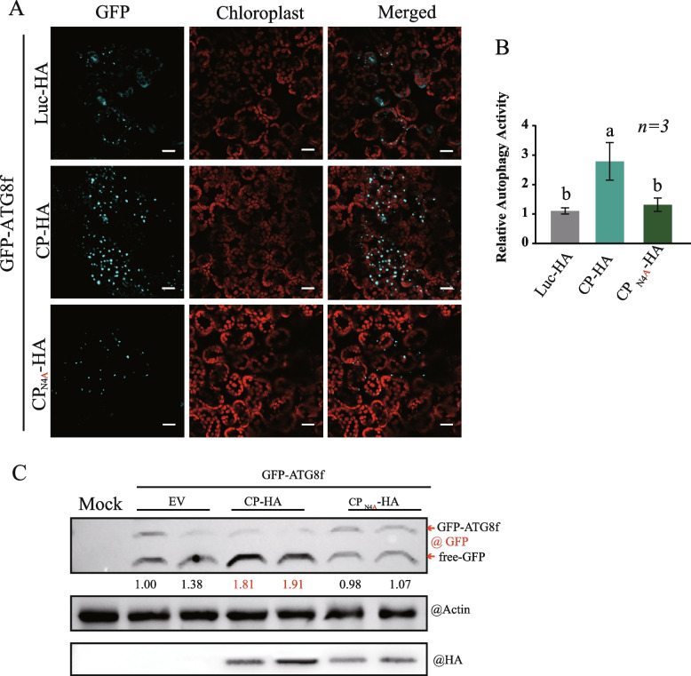
Viral infection commonly induces autophagy, leading to antiviral responses or conversely, promoting viral infection or replication. In this study, using the experimental plant Nicotiana benthamiana, we demonstrated that the rice stripe virus (RSV) coat protein (CP) enhanced autophagic activity through interaction with cytosolic glyceraldehyde-3-phosphate dehydrogenase 2 (GAPC2), a negative regulator of plant autophagy that binds to an autophagy key factor, autophagy-related protein 3 (ATG3). Competitive pull-down and co-immunoprecipitation (Co-IP)assays showed that RSV CP activated autophagy by disrupting the interaction between GAPC2 and ATG3. An RSV CP mutant that was unable to bind GAPC2 failed to disrupt the interaction between GAPC2 and ATG3 and therefore lost its ability to induce autophagy. RSV CP enhanced the autophagic degradation of a viral movement protein (MP) encoded by a heterologous virus, citrus leaf blotch virus (CLBV). However, the autophagic degradation of RSV-encoded MP and RNA-silencing suppressor (NS3) proteins was inhibited in the presence of CP, suggesting that RSV CP can protect MP and NS3 against autophagic degradation. Moreover, in the presence of MP, RSV CP could induce the autophagic degradation of a remorin protein (NbREM1), which negatively regulates RSV infection through the inhibition of viral cell-to-cell movement. Overall, our results suggest that RSV CP induces a selective autophagy to suppress the antiviral factors while protecting RSV-encoded viral proteins against autophagic degradation through an as-yet-unknown mechanism. This study showed that RSV CP plays dual roles in the autophagy-related interaction between plants and viruses.

Keywords: ATG3; Autophagy; Coat protein; GAPC; RSV.


Conflict of interest statement

The authors declare no compete of interests.

Figures

Fig. 1
Fig. 1
RSV and RSV coding proteins induce autophagy. A Representative transmission electron microscope (TEM) images of N. benthamiana leaf cells with (right picture) or without (left picture) RSV infection. The ultrastructure of autophagic bodies was observed in the cells (indicated by arrows). B Quantification of the number of autophagosomes in the cells of leaves described in (A). Each bar represents the total number of autophagic bodies from 10 cells obtained from three independent experiments. “**” indicates P < 0.01 (Student’s t-test). C Western blotting analyses of the accumulation of ATG8 and its lipidated form (ATG8-PE) in RSV-infected N. benthamiana plants. Total protein samples were extracted from the upper leaves of plants and subjected to immunoblotting analysis with anti-ATG8 and anti-actin antibodies. D Quantification of the relative autophagic activity in (C). Gray value statistics were normalized to the internal control actin and control plant values, which were set to 1.0. Values represent the standard deviation obtained from three independent experiments. “*” and “**” indicate P < 0.05 and P < 0.01, respectively (Student’s t-test). E Western blotting analysis of GFP accumulation in N. benthamiana leaves co-expressing RSV proteins CP-HA, SP-HA, MP-HA, NS2-HA, NSvc2-HA, and NS3-HA with GFP-ATG8f. The accumulation of GFP-ATG8f and free GFP was detected with an anti-GFP antibody, and RSV coding proteins were detected via their fused tag using an anti-HA antibody. Sample loading was normalized to the endogenous actin protein using an anti-actin antibody
Fig. 2
Fig. 2
RSV CP interacts with GAPCs. A BiFC assays to examine the interaction between RSV CP and GAPCs. RSV CP and GAPCs (NbGAPC1, NbGAPC2, NbGAPC3, OsGAPC1, OsGAPC2, and OsGAPC3) were fused to the N-terminal or C-terminal portions of the split yellow fluorescent protein (nYFP or cYFP, respectively) and transiently co-expressed in N. benthamiana. The reconstituted YFP fluorescence in epidermal cells was observed by confocal laser scanning microscopy. Scale bars, 20 μm. B and C In vivo co-immunoprecipitation assay to examine the interaction of CP with NbGAPC2 or OsGAPC2. RSV CP fused to an HA tag or GFP, NbGAPC2 fused to GFP, and OsGAPC2 fused to Flag were transiently expressed in N. benthamiana leaves, and immunoprecipitations were performed with an anti-GFP antibody. Protein samples before and after immunoprecipitation were analyzed by western blot with anti-HA, anti-Flag, and anti-GFP antibodies. D and E Yeast two-hybrid analysis of the interaction between CP and NbGAPC2 or OsGAPC2. The CP and NbGAPC2 or OsGAPC2 were inserted into pGADT7(AD) and pGBKT7(BD) plasmids. The combination plasmids were co-introduced into the yeast AH109 strain and cultured on a selective medium lacking SD-Leu-Trp and SD-Leu-Trp-His-Ade for 3–5 days
Fig. 3
Fig. 3
The fourth amino acid (Asn) of CP is essential for binding to NbGAPC2. A Schematic representation of the CP mutants analyzed in this study. The amino acid positions of deletions and point mutations in the CP sequence are shown above the diagram. N/A indicates that Asn (N) was replaced by Ala (A). B BiFC assay to examine the interaction of NbGAPC2 and CP or CP mutants. NbGAPC2 and CP or CP mutants (CPΔ58, CPΔ59-322, CPΔ4, CPΔ3, and CPN4A) were fused to the N-terminal or C-terminal portions of the split yellow fluorescent protein nYFP or cYFPnd transiently co-expressed in N. benthamiana. The reconstituted YFP fluorescence in epidermal cells were observed by confocal laser scanning microscopy. Scale bars, 20 μm. C In vivo co-immunoprecipitation assay to examine the interaction of NbGAPC2 with CP or CPN4A. CP and CPN4A fused to an HA tag and NbGAPC2 fused to GFP were transiently expressed in N. benthamiana leaves, and immunoprecipitations were performed with an anti-GFP antibody. Protein samples before and after immunoprecipitation were analyzed by western blot with anti-HA and anti-GFP antibodies
Fig. 4
Fig. 4
Activation of autophagy by transiently expressed RSV CP. A The autophagy activity of N. benthamiana leaves expressing CP or CPN4A was assessed by the autophagy marker GFP-NbATG8f. Luc-HA, CP-HA, or CPN4A-HA were co-expressed with GFP-ATG8f in N. benthamiana using Agrobacterium infiltration. The GFP fluorescence in epidermal cells was observed by confocal laser scanning microscopy. GFP-NbATG8f fusion proteins are in cyan, and chloroplasts are in red. Scale bars, 20 μm. B The quantification of the numbers of autophagic structures in the cells of leaves described in (A). More than 100 mesophyll cells for each treatment were used for the quantification. The relative autophagic activity in Luc-HA-infected plants was normalized to control plant values, which were set to 1.00. Values represent the means from three independent experiments. The significant differences are marked with different lowercase letters over the columns (P < 0.05, one-way ANOVA). C The protein samples obtained from (A) were subjected to western blot analysis using anti-GFP, anti-HA, and anti-actin antibodies. The accumulation of GFP was normalized to actin. The ratio of free GFP via total GFP (GFP-ATG8f plus free-GFP) was compared to the control sample (EV), which was set to 1.00
Fig. 5
Fig. 5
RSV CP disrupts the interaction between NbGAPC2 and NbATG3. A The interaction of NbGAPC2 and NbATG3 was evaluated by BiFC assay. The recombinant plasmids of NbGAPC2-nYFP and NbATG3-cYFP were co-expressed with CP-HA or CPN4A-HA in N. benthamiana leaves. Luc-HA was used as the control. The reconstituted YFP fluorescence in epidermal cells were observed by confocal laser scanning microscopy. Scale bars, 20 μm. B The intensity of reconstituted YFP fluorescence (BiFC) described in (A) was normalized to the control sample (Luc-HA). The error bar represents the mean intensity of YFP fluorescence quantified by ImageJ from 30 pictures obtained from three independent experiments. Different letters above the bars indicate significant differences (P < 0.05, one-way ANOVA). C The protein accumulation of experiment described in (A) examined by western blot using GFP and CP antibodies. D Competitive pull-down assay. NbGAPC2-His, MBP-NbATG3, MBP, MBP-CP were expressed and purified from E. coli BL21(DE3). An equivalent amount (20 μg) of NbGAPC2-His and MBP-NbATG3 was incubated with MBP (40, 30, 10, 0 ng), MBP-CP (0, 10 30 , 40 ng) at 4 ℃ for 1 hour. A pull-down assay was performed with His-specific affinity resin (Ni-NTA agarose) and analyzed by western blotting using His, and MBP antibodies. The accumulation of MBP-NbATG3 protein was quantified by Image J and set to 1.00 for the control sample (MBP). The number indicated the relative MBP-NbATG3 accumulation, which was normalized to NbGAPC2-His and compared to the control sample. E Competitive pull-down assay. NbGAPC2-His, GST-NbATG3, MBP, MBP-CP, and MBP-CPN4A were expressed and purified from E. coli BL21(DE3). An equivalent amount (20 μg) of NbGAPC2-His and GST-NbATG3 was incubated with MBP, MBP-CP, or MBP-CPN4A protein at 4 ℃ for 1 hour. A pull-down assay was performed with His-specific affinity resin (Ni-NTA agarose) and analyzed by immunoprecipitation with anti-GST, anti-His, and anti-MBP antibodies. The accumulation of GST-NbATG3 protein was quantified by ImageJ and set to 1.00 for the control sample (MBP). The number indicated the relative GST-NbATG3 accumulation, which was normalized to NbGAPC2-His and compared to the control sample. F Competitive Co-IP assay. NbGAPC2-GFP and HA-NbATG3 were co-expressed in N. benthamiana leaves and immunoprecipitated with anti-GFP beads incubated with purified MBP, MBP-CP, MBP-CPN4A proteins at 4 ℃. Input and IP proteins were analyzed by western blot with anti-HA, anti-GFP, and anti-MBP antibodies. The quantification of HA-NbATG3 protein accumulation is presented. The results are representative of three independent experiments
Fig. 6
Fig. 6
The expression of the NbATG3 is upregulated by RSV or RSV CP. A and B RT-qPCR showed the upregulation of NbATG3 in RSV infection (A) or transiently expressed CP and CPN4A-HA(B) in N. benthamiana. The error bars represent the mean from three biological replicates. “*” indicates P < 0.05 (Student’s t-test) (A). Different letters above the bars indicate significant differences (P < 0.05, one-way ANOVA) (B). C NbATG3-3XFlag was co-expressed with CP-HA and CPN4A-HA, and its expression was examined by western blot with an anti-Flag antibody. The accumulation of NbATG3-3XFlag protein was quantified by ImageJ and normalized to actin. The number indicates the relative NbATG3-3XFlag accumulation compared to the control sample (EV), which was set to 1.00. C The results are representative of three independent experiments
Fig. 7
Fig. 7
Effects of RSV CP on protein autophagic degradation. A and B CP-HA was expressed in N. benthamiana leaves and treated with 100 μM E64d, an autophagy inhibitor (A), or 100 μM MG132, a proteasome inhibitor (B). The protein accumulation was examined by western blot with an anti-HA antibody. The quantification of protein was performed by ImageJ and normalized to actin. C-E CP-HA was co-expressed with the viral proteins CLBV-MP (C), RSV-MP (D), or RSV-NS3 (E) with GFP tags in N. benthamiana and treated with 100 μM E64d. F and G CP-HA was co-expressed with the host protein NbREM1-GFP in the absence (F) or presence (G) of RSV-MP tagged with mCherry in N. benthamiana and treated with 100 μM E64d. The total protein was extracted and detected by western blot with anti-GFP, anti-mCherry, and anti-HA antibodies. Coomassie blue-stained proteins are shown as the loading control. The quantification of protein was performed by ImageJ and normalized to actin or the loading control. The number indicated the relative protein accumulation compared to the control sample (Luc-HA or EV), which was set to 1.00. The results are representative of three independent experiments

Similar articles

Cited by

References

    1. Adachi A, Koizumi M, Ohsumi Y. Autophagy induction under carbon starvation conditions is negatively regulated by carbon catabolite repression. J Biol Chem. 2017;292(48):19905–19918. doi: 10.1074/jbc.M117.817510. - DOI - PMC - PubMed
    1. Avin-Wittenberg T. Autophagy and its role in plant abiotic stress management. Plant Cell Environ. 2019;42(3):1045–1053. doi: 10.1111/pce.13404. - DOI - PubMed
    1. Barbier P, Takahashi M, Nakamura I, Toriyama S, Ishihama A. Solubilization and promoter analysis of RNA polymerase from rice stripe virus. J Virol. 1992;66(10):6171. doi: 10.1128/jvi.66.10.6171-6174.1992. - DOI - PMC - PubMed
    1. Cerff R (1978) Glyceraldehyde-3-phosphate dehydrogenase (NADP) from Sinapis alba: Steady state kinetics. Phytochemistry 17(12):2061–2067. 10.1016/S0031-9422(00)89281-X
    1. Chen H, Dong J, Wang T (2021) Autophagy in Plant Abiotic Stress Management. Int J Mol Sci. 22 (8). 10.3390/ijms22084075 - PMC - PubMed