. 2023 Sep 7;8(19):e173374.
doi: 10.1172/jci.insight.173374.

Epithelial Yap/Taz are required for functional alveolar regeneration following acute lung injury

Affiliations

Epithelial Yap/Taz are required for functional alveolar regeneration following acute lung injury

Gianluca T DiGiovanni et al. JCI Insight. .

Abstract

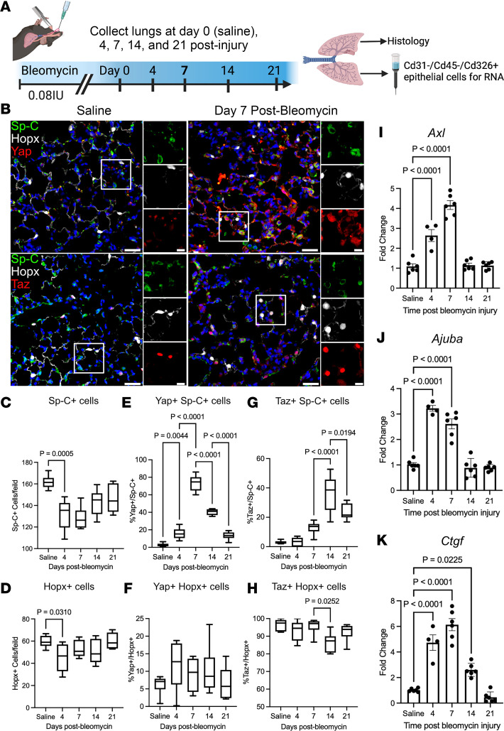
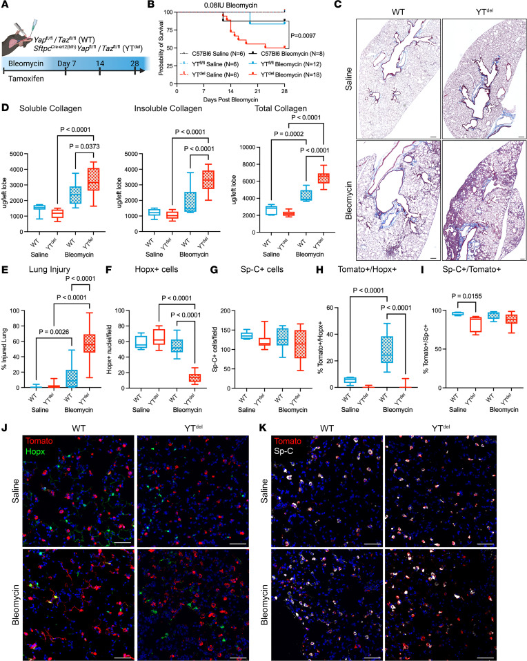
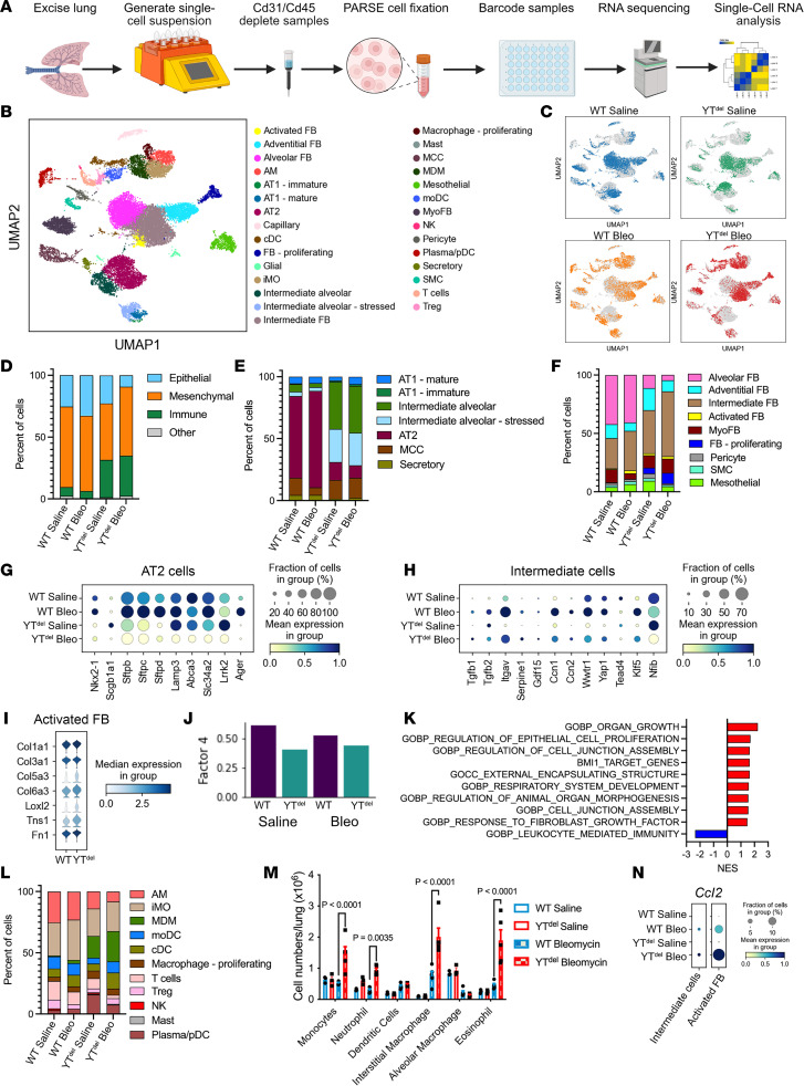
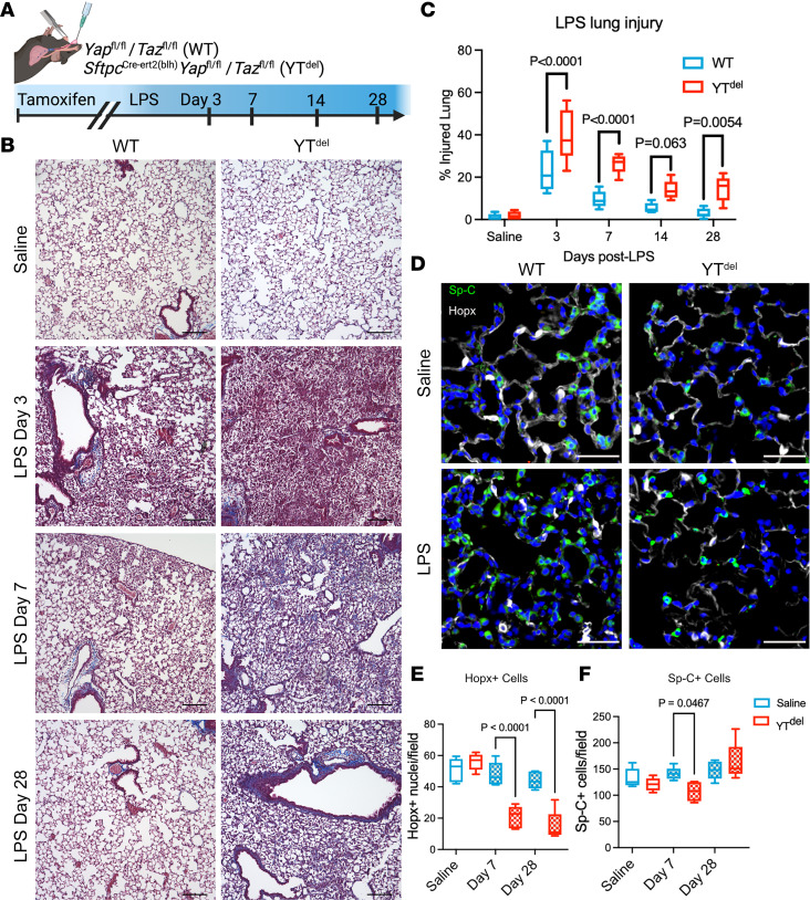
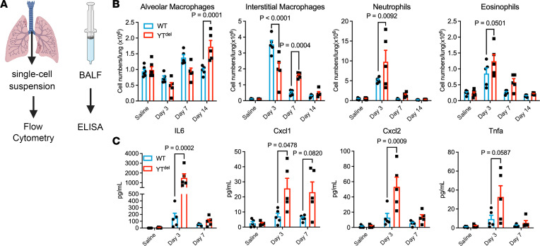
A hallmark of idiopathic pulmonary fibrosis (IPF) and other interstitial lung diseases is dysregulated repair of the alveolar epithelium. The Hippo pathway effector transcription factors YAP and TAZ are implicated as essential for type 1 and type 2 alveolar epithelial cell (AT1 and AT2) differentiation in the developing lung, yet aberrant activation of YAP/TAZ is a prominent feature of the dysregulated alveolar epithelium in IPF. In these studies, we sought to define the functional role of YAP/TAZ activity during alveolar regeneration. We demonstrated that Yap and Taz were normally activated in AT2 cells shortly after injury, and deletion of Yap/Taz in AT2 cells led to pathologic alveolar remodeling, failure of AT2-to-AT1 cell differentiation, increased collagen deposition, exaggerated neutrophilic inflammation, and increased mortality following injury induced by a single dose of bleomycin. Loss of Yap/Taz activity prior to an LPS injury prevented AT1 cell regeneration, led to intraalveolar collagen deposition, and resulted in persistent innate inflammation. These findings establish that AT2 cell Yap/Taz activity is essential for functional alveolar epithelial repair and prevention of fibrotic remodeling.

Keywords: Fibrosis; Mouse stem cells; Pulmonology.


Figures

Figure 1
Figure 1. YAP/TAZ are dynamically regulated during acute bleomycin lung injury.
(A) Schematic of injury model and time points when lungs were assessed. (B) Immunofluorescence analysis of Yap or Taz (red) in Sp-C+ (green) AT2 and Hopx+ (white) AT1 cells. (C and D) Quantification of Sp-C+ AT2 and Hopx+ AT1 cells at respective injury repair time points. (E and F) Quantification of Yap+ nuclei in Sp-C+ AT2 and Hopx+ AT1 cells. (G and H) Quantification of Taz+Sp-C+ AT2 and Hopx+ AT1 cells. Scale bars: 50 µm, 10 µm (insets). In box-and-whisker plots, whiskers are minimum and maximum, and data represent n = 6 mice. (I, J, and K) qPCR analysis of Yap/Taz target genes Axl (I), Ajuba (J), and Ctgf (K) during bleomycin injury repair. n = 6 mice for saline, 7, 14, and 21 days after injury and n = 4 mice at day 4. Data are shown as mean ± SEM. Statistical analysis was performed using 1-way ANOVA and Tukey’s post hoc comparison.
Figure 2
Figure 2. Deletion of YAP/TAZ leads to failed alveolar repair following single-dose bleomycin induced lung injury.
(A) Schematic of injury model in which mice were treated with tamoxifen the same day as bleomycin-induced lung injury. (B) Survival curve of WT and YTdel bleomycin- or saline-treated mice out to 28 days after injury. Statistics determined using Mantel-Cox test. (C) Masson’s trichrome staining of tissue sections from WT and YTdel mice at day 28 after bleomycin or PBS. (D) Soluble, insoluble, and total collagen quantification from WT and YTdel at day 28 after saline or bleomycin. (E) Quantification of total injured lung area in respective groups at day 28 after saline/bleomycin. n = 7 WT saline, n = 9 YTdel saline, n = 9 WT bleomycin, and n = 12 YTdel bleomycin mice. (F and G) Quantification of total Hopx+ cells or Sp-C+ cells per 20× field of view in each group. n = 10 WT saline-, n = 9 YTdel saline-, n = 12 WT bleomycin-, and n = 18 YTdel bleomycin-treated mice. (H and I) Quantification of Sp-Ctomato lineage–labeled Hopx+ or Sp-C+ AT2 cells 28 days after bleomycin. (J) Immunofluorescence analysis of Sp-Ctomato lineage labeled cells (red) and Hopx+ (green) AT1 cells at 28 days after injury. (K) Immunofluorescence analysis of Sp-Ctomato lineage–labeled cells (red) and Sp-C+ (white) AT2 cells at 28 days after injury. n = 5 WT saline, n = 5 YTdel saline, n = 10 WT bleomycin, and n = 15 YTdel bleomycin mice. Scale bars: 200 µm (C), 50 µm (J and K). Statistical analysis in DI was performed using 1-way ANOVA and Tukey’s post hoc comparison.
Figure 3
Figure 3. YAP/TAZ deletion prior to bleomycin injury leads to altered epithelial cell populations, activated fibroblast, and disrupted immune response.
(A) Schematic of experimental strategy to generate single-cell RNA-Seq analysis from mouse lungs. (B and C) UMAP embedding demonstrating clustering of the 29 cell types identified in the mouse lungs (B) and depicting cells recovered from respective genotype and treatment groups (C). (DF) Relative proportions of each cell types/states within total cell populations (D), epithelial cell populations (E), and fibroblast cell populations between each treatment group (F). (G and H) Dot plot showing expression levels of AT2 cell lineage markers (G) and intermediate epithelial cell marker expression (H) in each treatment group. (I) Violin plot showing relative expression of activated fibroblast markers in WT or YTdel bleomycin-treated mice. (J) Output from LIANA demonstrating loss of Factor 4 ligand-receptor signaling in YTdel mice. (K) Gene ontology–associated programs identified as misregulated by LIANA. (L) Relative proportions of immune cell populations from each treatment group. (M) Flow cytometry analysis of immune cell subpopulations present in mice 7 days after bleomycin injury. n = 3 WT saline- and YTdel saline-, n = 5 WT bleomycin- and YTdel bleomycin-treated mice. (N) Dot plot showing expression of Ccl2 in intermediate alveolar epithelial cells and activated (including both intermediate and activated from F) fibroblast population.
Figure 4
Figure 4. Loss of YAP/TAZ prior to LPS-induced lung injury leads to failed alveolar regeneration and increased collagen deposition.
(A) Schematic of LPS lung injury model in which mice were treated with tamoxifen 2 weeks prior to LPS lung injury. (B) Masson’s trichrome staining of WT and YTdel mice treated with saline or 3, 7, or 28 days after LPS injury. n = 6 mice per group. (C) Quantification of injured lung area in WT and YTdel mice 3 to 28 days after LPS. (D) Immunofluorescence analysis of Hopx+ (white) AT1 cells and Sp-C+ (green) AT2 cells 7 days after LPS injury. (E and F) Quantification of Hopx+ and Sp-C+ cells in WT or YTdel lungs treated with saline or LPS at 7 or 28 days after injury; n = 5 mice per group. Statistics were performed using 1-way ANOVA and Tukey’s post hoc comparison. Scale bars: 50 µm (D), 200 µm (B).
Figure 5
Figure 5. AT2 cell–specific deletion of YAP/TAZ leads to altered immune response after LPS injury.
(A) Schematic showing experimental strategy to assess immune response in mice injured with LPS. (B) Quantification of immune cell types by flow cytometry in saline- or LPS-treated mouse lungs at 3, 7, or 14 days after injury. n = 6 saline-treated and n = 5 LPS-treated mice per group. (C) ELISA of immune cytokines isolated from bronchoalveolar lavage fluid from mice 3 or 7 days after LPS injury or saline controls. n = 5 mice per group. One-way ANOVA and Tukey’s post hoc comparison were used to assess statistical significance. Dots represent individual values per mouse.

References

    1. Barkauskas CE, et al. Type 2 alveolar cells are stem cells in adult lung. J Clin Invest. 2013;123(7):3025–3036. doi: 10.1172/JCI68782. - DOI - PMC - PubMed
    1. Hogan BL, et al. Repair and regeneration of the respiratory system: complexity, plasticity, and mechanisms of lung stem cell function. Cell Stem Cell. 2014;15(2):123–138. doi: 10.1016/j.stem.2014.07.012. - DOI - PMC - PubMed
    1. LaCanna R, et al. Yap/Taz regulate alveolar regeneration and resolution of lung inflammation. J Clin Invest. 2019;129(5):2107–2122. doi: 10.1172/JCI125014. - DOI - PMC - PubMed
    1. Gokey JJ, et al. YAP regulates alveolar epithelial cell differentiation and AGER via NFIB/KLF5/NKX2-1. iScience. 2021;24(9):102967. doi: 10.1016/j.isci.2021.102967. - DOI - PMC - PubMed
    1. Nantie LB, et al. Lats1/2 inactivation reveals Hippo function in alveolar type I cell differentiation during lung transition to air breathing. Development. 2018;145(21):dev163105. doi: 10.1242/dev.163105. - DOI - PMC - PubMed

Publication types

MeSH terms