. 2023 Oct;24(10):1735-1747.
doi: 10.1038/s41590-023-01604-z. Epub 2023 Sep 7.

CXCR6 orchestrates brain CD8+ T cell residency and limits mouse Alzheimer's disease pathology

Affiliations

CXCR6 orchestrates brain CD8+ T cell residency and limits mouse Alzheimer's disease pathology

Wei Su et al. Nat Immunol. 2023 Oct.

Abstract

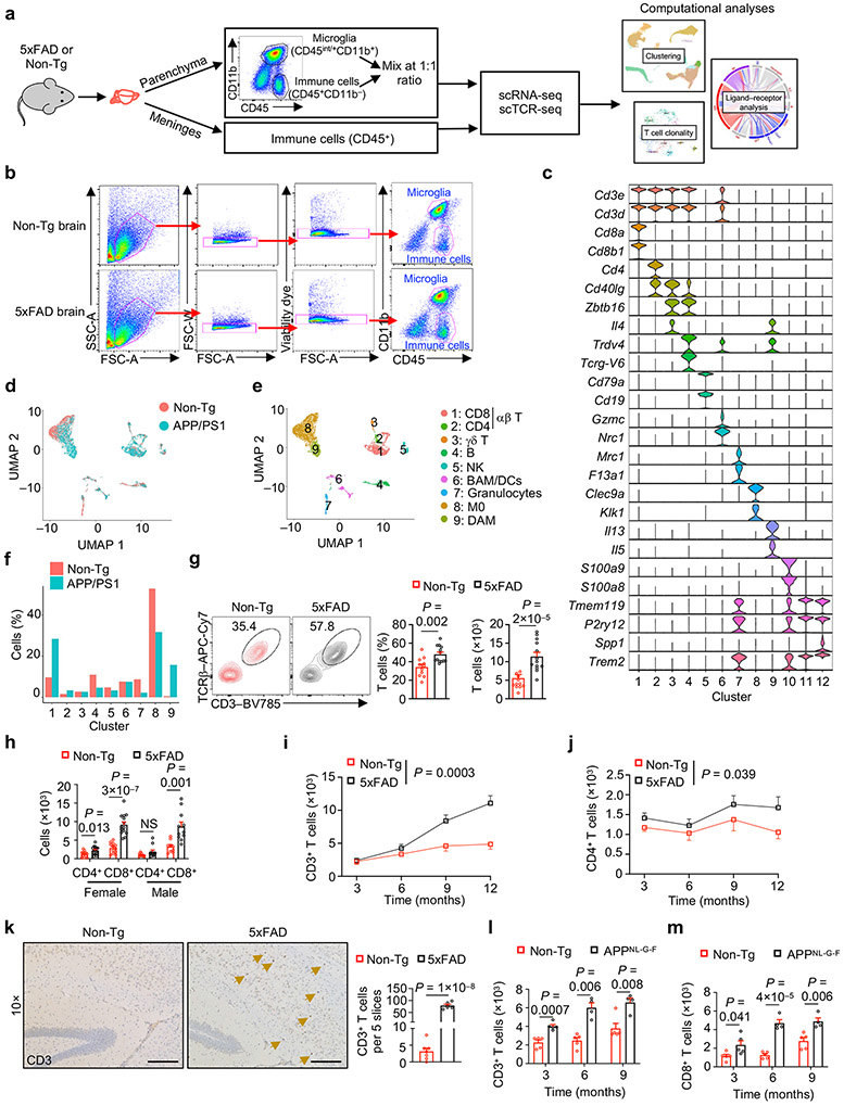
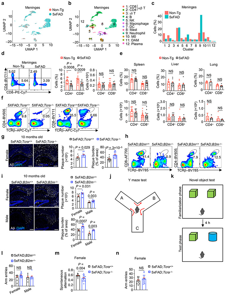
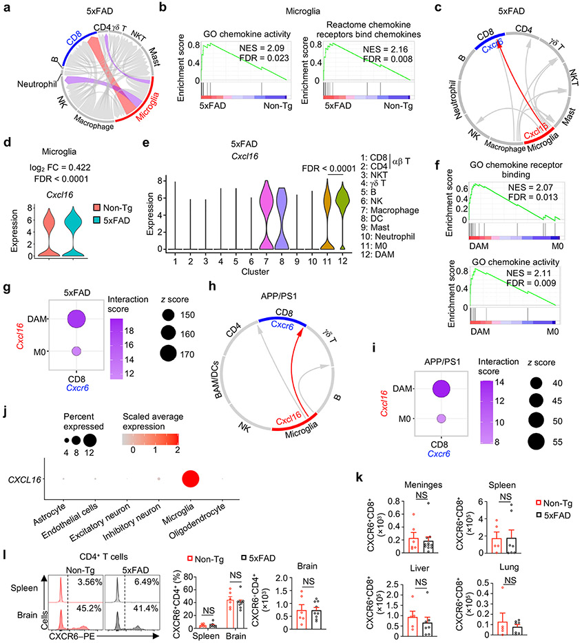
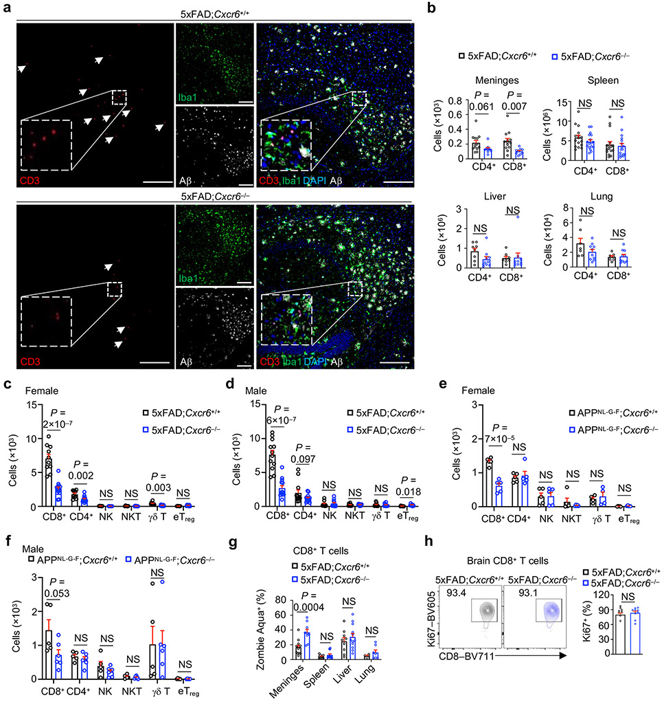
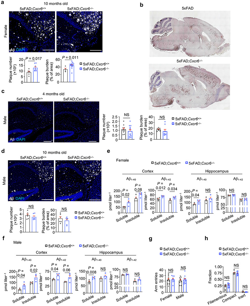
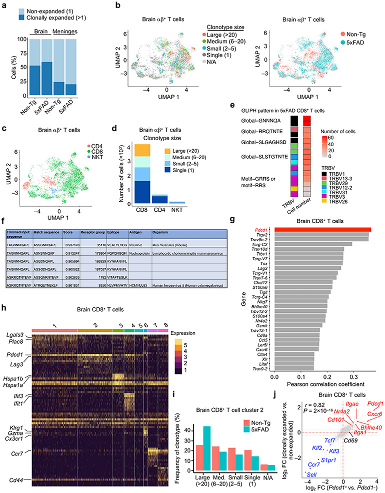
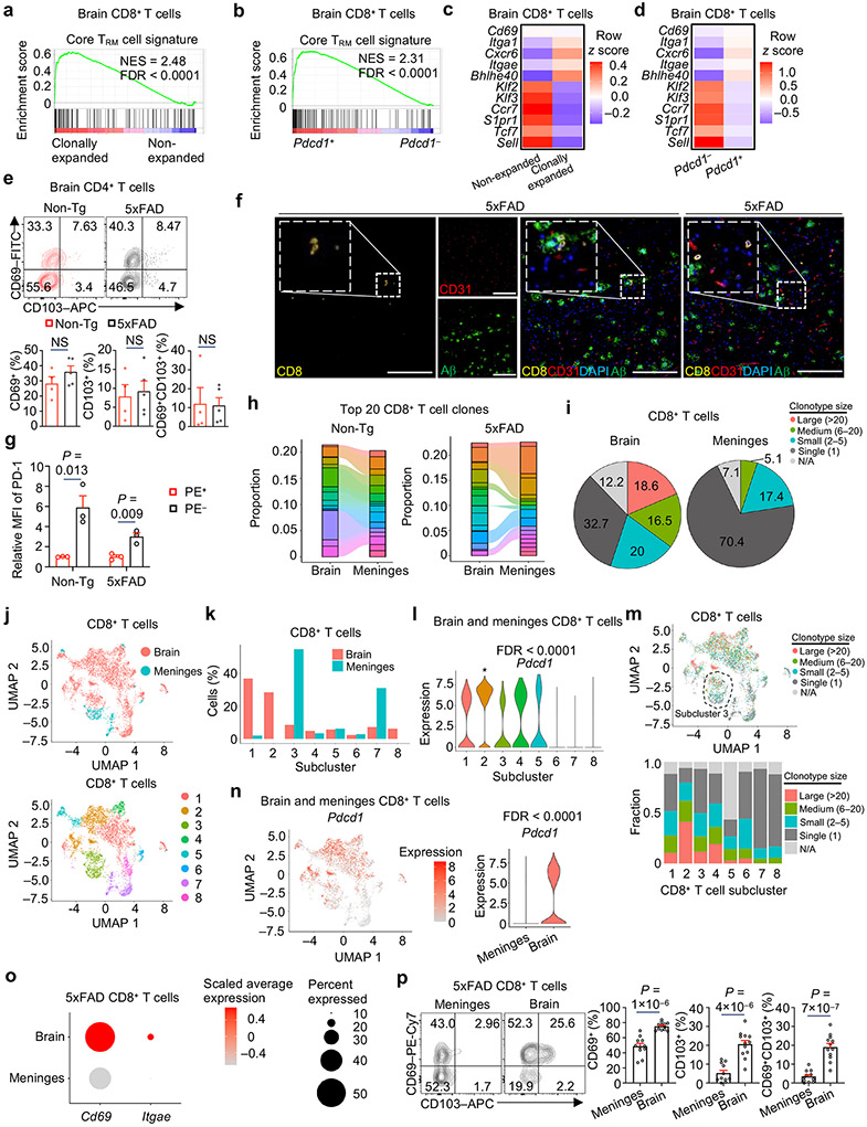
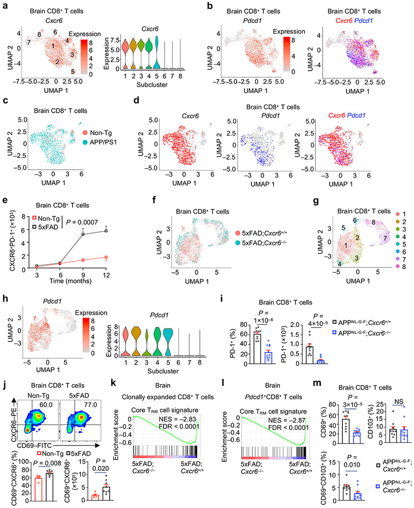
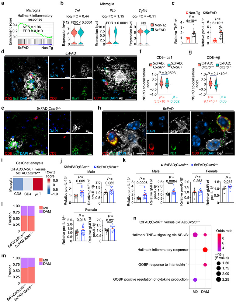
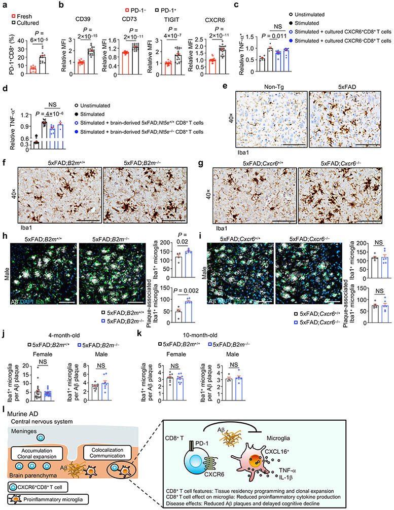
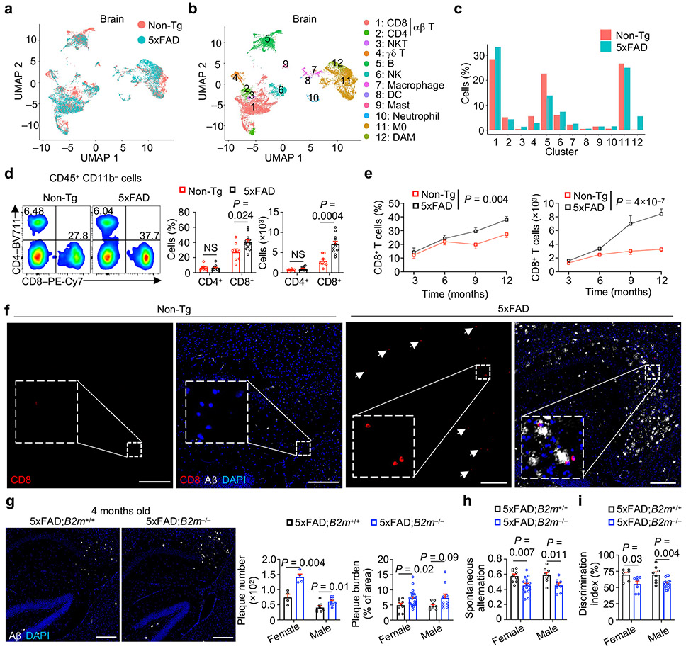
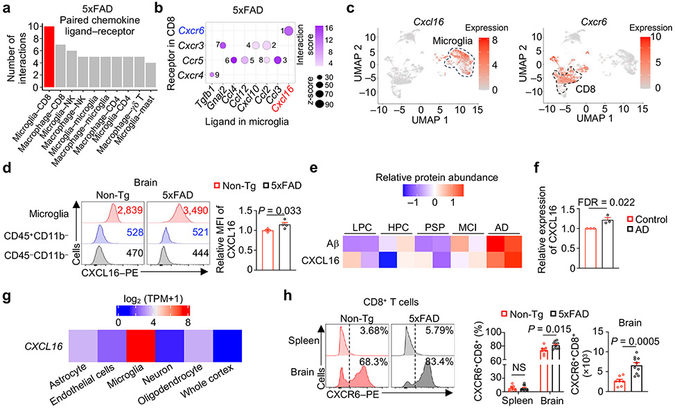
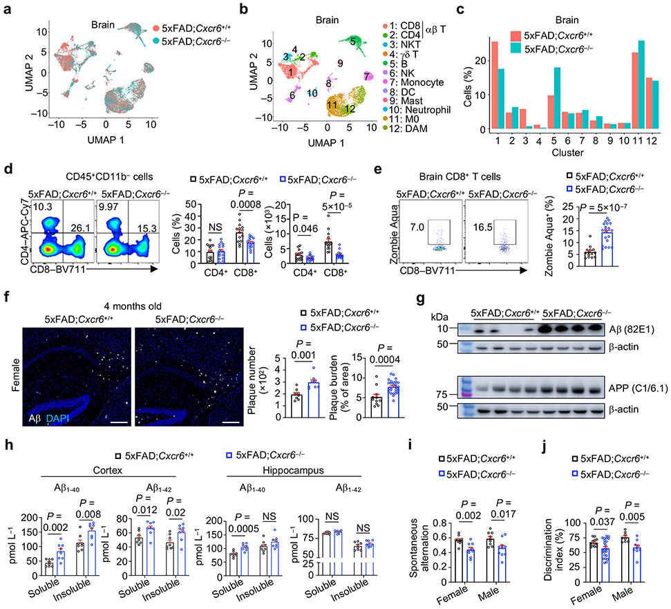
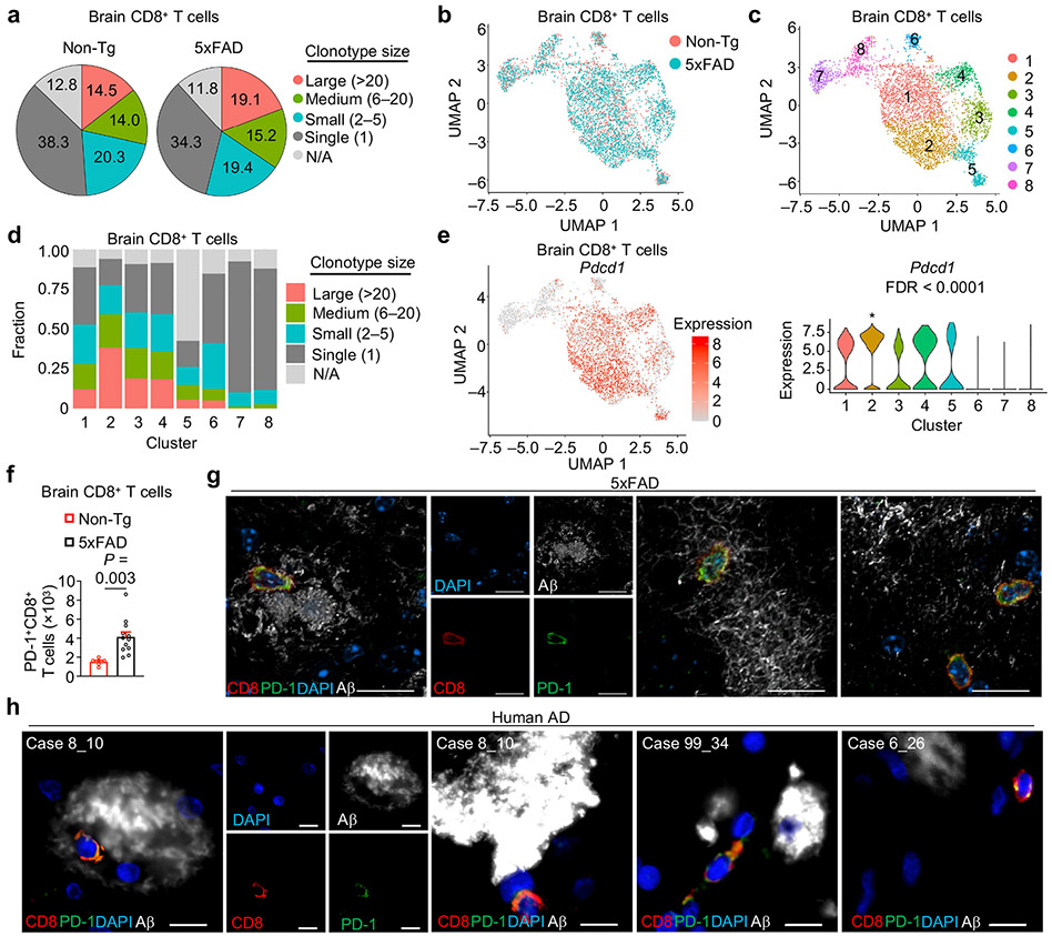
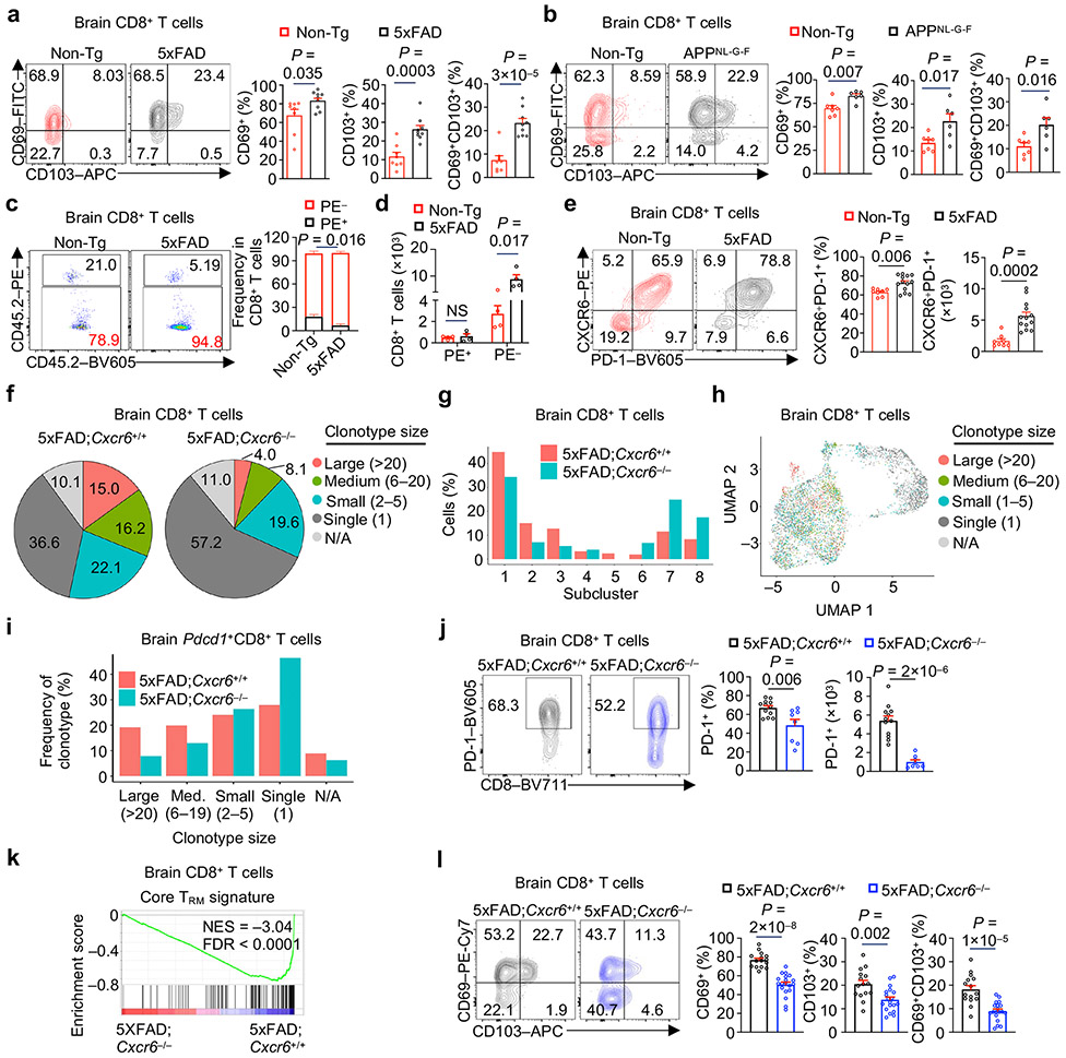
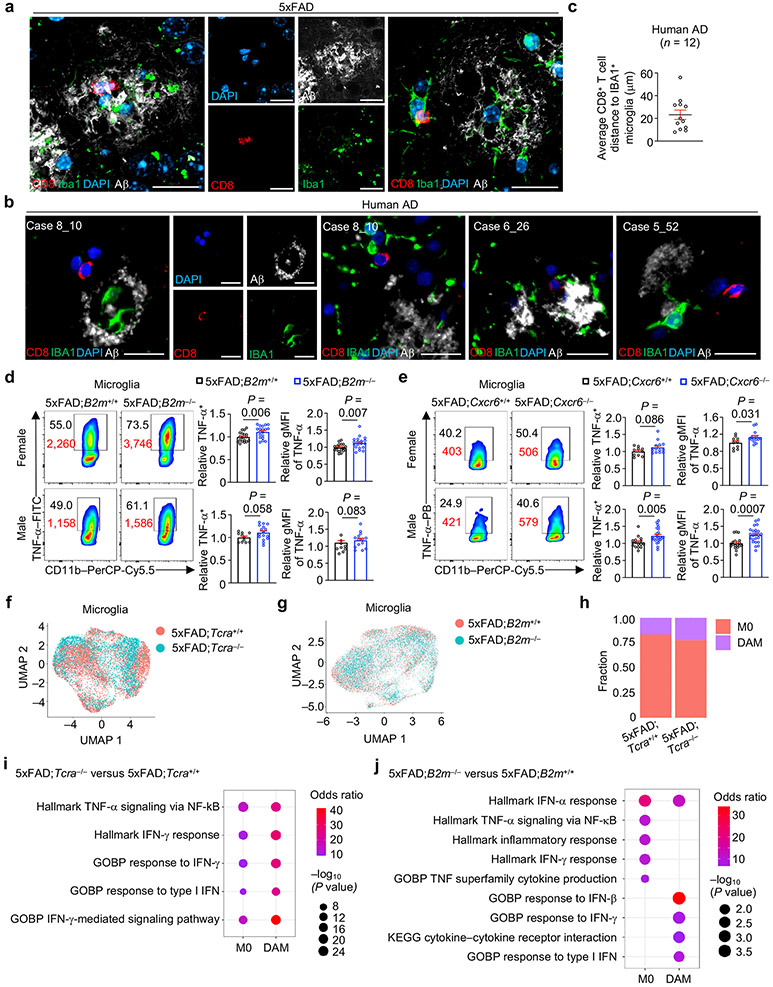
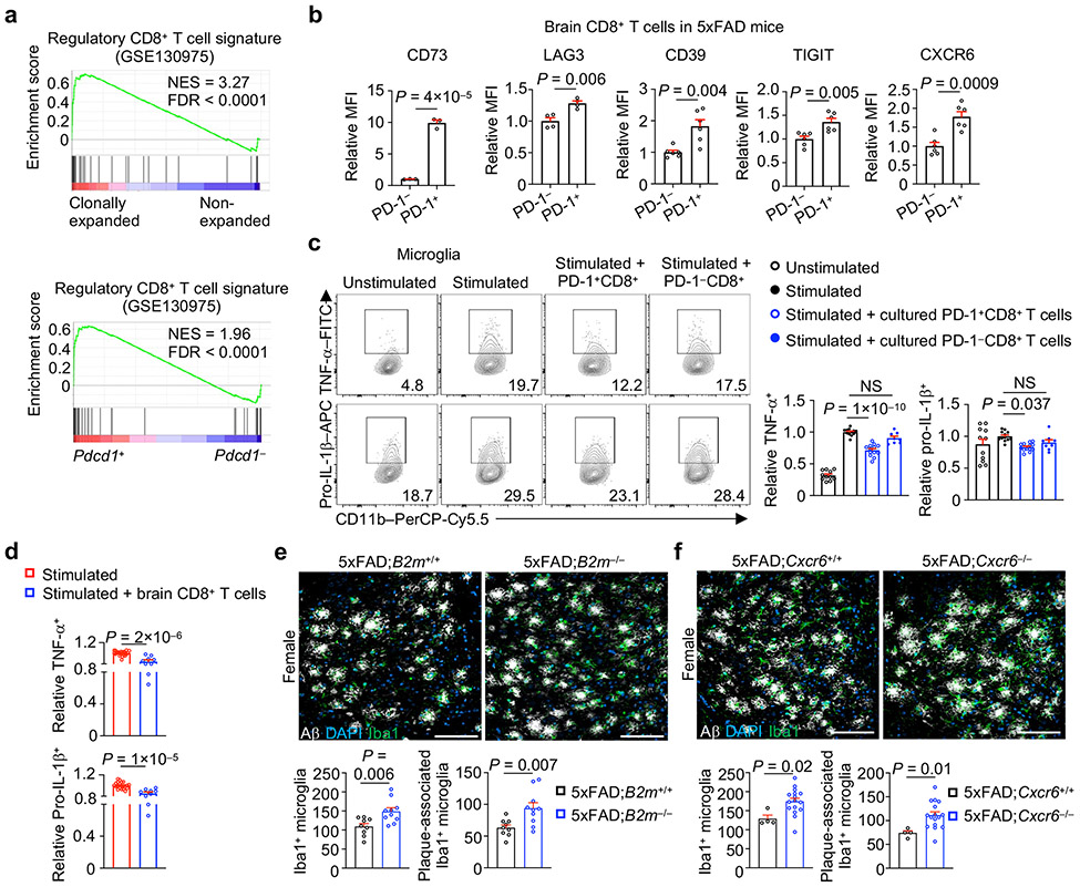
Neurodegenerative diseases, including Alzheimer's disease (AD), are characterized by innate immune-mediated inflammation, but functional and mechanistic effects of the adaptive immune system remain unclear. Here we identify brain-resident CD8+ T cells that coexpress CXCR6 and PD-1 and are in proximity to plaque-associated microglia in human and mouse AD brains. We also establish that CD8+ T cells restrict AD pathologies, including β-amyloid deposition and cognitive decline. Ligand-receptor interaction analysis identifies CXCL16-CXCR6 intercellular communication between microglia and CD8+ T cells. Further, Cxcr6 deficiency impairs accumulation, tissue residency programming and clonal expansion of brain PD-1+CD8+ T cells. Ablation of Cxcr6 or CD8+ T cells ultimately increases proinflammatory cytokine production from microglia, with CXCR6 orchestrating brain CD8+ T cell-microglia colocalization. Collectively, our study reveals protective roles for brain CD8+ T cells and CXCR6 in mouse AD pathogenesis and highlights that microenvironment-specific, intercellular communication orchestrates tissue homeostasis and protection from neuroinflammation.


Figures

Extended Data Fig. 1
Extended Data Fig. 1. T cells accumulate in mouse models of AD.
a, Schematic workflow of scRNA-seq and scTCR-seq experiments, showing tissue collection sites, cell sorting strategies, and computational analyses of Non-Tg and 5xFAD mice. b, Gating strategy for sorting microglia and non-microglia immune cells used for scRNA-seq and scTCR-seq analysis. Sorting gates for Non-Tg and 5xFAD samples were set according to the following parameters (in order): 1) cell size (to exclude debris); 2) singlet; 3) viable cells; 4) CD45int/+CD11b+ (for microglia) and CD45+CD11b (for non-microglia immune cells). c, Violin plots showing expression of cell type-defining genes in clusters from Fig. 1b. d, e, CD45+ immune cell clusters colored by genotype (d) or cell types (e) from a public dataset of indicated mice. f, Frequencies of cells described in d and e. g, Brain T cells (CD3+TCRβ+) from 10-month-old Non-Tg (n = 11) and 5xFAD (n = 13) mice. h, CD4+ and CD8+ T cells in the brains of female and male 10-month-old Non-Tg (n = 12 for female and 9 for male) and 5xFAD (n = 14 for female and 11 for male) mice. i, j, CD3+ [i, 3 months: Non-Tg (n = 4) and 5xFAD (n = 4); 6 months: Non-Tg (n = 8) and 5xFAD (n = 9); 9 months: Non-Tg (n = 8) and 5xFAD (n = 13); 12 months: Non-Tg (n = 11) and 5xFAD (n = 13)] or CD4+ [j, 3 months: Non-Tg (n = 6) and 5xFAD (n = 6); 6 months: Non-Tg (n = 13) and 5xFAD (n = 16); 9 months: Non-Tg (n = 8) and 5xFAD (n = 9); 12 months: Non-Tg (n = 14) and 5xFAD (n = 17)] T cells in the brains of Non-Tg and 5xFAD mice at indicated time points. k, Immunohistochemical analysis of CD3+ T cells in the brains of 10-month-old Non-Tg (n = 7) and 5xFAD (n = 6) mice. Scale bar, 200 μm. Arrows indicate positive staining for CD3+ T cells. l, m, CD3+ (l) or CD8+ (m) T cells in the brains of Non-Tg and APPNL-G-F mice [3 months: Non-Tg (n = 5) and APPNL-G-F (n = 5); 6 months: Non-Tg (n = 5) and APPNL-G-F (n = 4); 9 months: Non-Tg (n = 5) and APPNL-G-F (n = 4)]. Data were analyzed by two-tailed unpaired Student’s t-test (g, h, km) or two-way ANOVA (i, j). Data are shown as mean ± s.e.m. in gm; NS, not significant. Data were pooled from at least three (gj) or two (km) independent experiments.
Extended Data Fig. 2
Extended Data Fig. 2. Single-cell immune profiling of meninges and functional characterization of T cells by multiple genetic models
a, b, scRNA-seq profiling of CD45+ immune cells from the meninges of male Non-Tg or 5xFAD mice (n = 2 biological replicates for each genotype) at 8 months of age. UMAP showing CD45+ immune cell clusters colored by genotype (a) or cell type (b). c, Frequencies (normalized to total cells from each genotype) of annotated clusters described in b. d, CD4+ and CD8+ T cells in the meninges of 10-month-old Non-Tg (n = 13) and 5xFAD (n = 13) mice. e, CD4+ and CD8+ T cells in the indicated tissues of 10-month-old Non-Tg and 5xFAD mice [spleen: Non-Tg (n = 11), 5xFAD (n = 16); liver: Non-Tg (n = 7), 5xFAD (n = 8); lung: Non-Tg (n = 5), 5xFAD (n = 6)]. f, Representative flow cytometry plots demonstrating a lack of CD8+TCRβ+ or CD4+TCRβ+ T cells in the brains of 5xFAD;Tcra−/− mice. g, Immunofluorescence analysis of the number and burden of Aβ plaques in 10-month-old female 5xFAD;Tcra+/+ (n = 8 sections for plaque number; n = 15 sections for plaque burden) and 5xFAD;Tcra−/− (n = 9 sections for plaque number; n = 15 sections for plaque burden) mice. Scale bar, 250 μm. h, Representative flow cytometry plots demonstrating a lack of CD8+ T cells (CD8+TCRβ+) but not CD4+ T cells (CD4+TCRβ+) in the brains of 5xFAD;B2m−/− mice. i, Immunofluorescence analysis of number and burden of Aβ plaques in 10-month-old male and female 5xFAD;B2m+/+ (n = 9 sections for female and 8 for male for plaque number; n = 10 sections for female and 4 for male for plaque burden) and 5xFAD;B2m−/− (n = 10 sections for female and 12 for male for plaque number; n = 10 sections for female and 6 for male for plaque burden) mice. Scale bar, 250 μm. j, k, Illustrations of Y maze (j) and novel objection recognition (k) behavioral tests. l, Total arm entries from spontaneous Y maze alternation testing in 4-month-old 5xFAD;B2m+/+ (n = 9 for female and 8 for male) and 5xFAD;B2m−/− (n = 15 for female and 8 for male) mice. m, n, Spontaneous alternation (m) and total arm entries (n) from spontaneous Y maze alternation testing in 4-month-old female 5xFAD;Tcra+/+ (n = 15) and 5xFAD;Tcra−/− (n = 15) mice. Data were analyzed by two-tailed unpaired Student’s t-test (d, e; g, i for plaque burden; ln) or two-tailed unpaired t-test with Welch’s correction for plaque number (g, i). Data are shown as mean ± s.e.m. in d, e, g, i, and ln; NS, not significant. Data were pooled from at least three (d, e, m, n) or two (g, i, l), or are representative of at least two (f, h) independent experiments.
Extended Data Fig. 3
Extended Data Fig. 3. CXCL16–CXCR6 inter-cellular communication axis between microglia and T cells
a, Circle plot of predicted cell type interactions based on ligand–receptor pairs in brain immune cells in 5xFAD mice. Line thickness indicates number of ligand–receptor interactions; red color indicates CD8+ T cells and microglia as the cell-type pair with the highest number of interactions; purple color indicates comparatively weaker interactions present between CD4+ T or NK cells and microglia. b, GSEA enrichment plots showing upregulation of chemokine-associated signatures in microglia from 5xFAD mice compared to Non-Tg mice. c, Circle plot of predicted cell–cell interactions based on CXCL16 and CXCR6 pairing in brain immune cells of 5xFAD mice (red arrow indicates strongest interaction). d, Violin plot of Cxcl16 expression in microglia from Non-Tg (n = 1,782 cells) and 5xFAD (n = 2,870 cells) mice. e, Violin plot of Cxcl16 expression in immune cell clusters (n = 4,105 cells for M0 and 547 cells for DAM) in 5xFAD mice. f, GSEA enrichment plots showing upregulation of chemokine-associated signatures in DAM compared to M0. g, Bubble plot showing stronger predicted CXCL16–CXCR6 interaction between DAM and CD8+ T cells than M0 and CD8+ T cells in 5xFAD mice. h, Circle plot of predicted cell–cell interactions based on CXCL16 and CXCR6 pairing in brain immune cells of APP/PS1 mice (red arrow indicates the strongest interaction); BAM/DCs: border-associated macrophages/dendritic cells. i, Bubble plot showing stronger predicted CXCL16–CXCR6 interaction between DAM and CD8+ T cells than M0 and CD8+ T cells in APP/PS1 mice. j, Bubble plot of CXCL16 expression in brain cell types from a public human single-nuclear RNA-seq dataset. k, CXCR6+CD8+ T cells in the meninges [Non-Tg (n = 13) and 5xFAD (n = 13)], spleen [Non-Tg (n = 5) and 5xFAD (n = 7)], liver [Non-Tg (n = 5) and 5xFAD (n = 7)], and lung [Non-Tg (n = 5) and 5xFAD (n = 7)] of 10-month-old Non-Tg and 5xFAD mice. l, CXCR6+CD4+ T cells from the spleens and brains of 10-month-old Non-Tg (n = 6) and 5xFAD (n = 9) mice. Data were analyzed by two-tailed normalized weighted Kolmogorov–Smirnov test (NES for b and f), two-tailed Benjamini–Hochberg adjusted P value (FDR for b and f), two-tailed Bonferroni correction (d), two-tailed Wilcoxon rank-sum test (e), or two-tailed unpaired Student’s t-test (k, l). Data are shown as mean ± s.e.m. in k and l; NS, not significant. Data were pooled from at least three (k, l) independent experiments.
Extended Data Fig. 4
Extended Data Fig. 4. CXCR6 promotes brain CD8+ T cell accumulation
a, Widefield immunofluorescence images of CD3+ T cells, Aβ, and Iba1+ microglia in brains of indicated 10-month-old mice. Scale bar, 200 μm. White arrows indicate positive staining for CD3+ T cells. Boxed areas show CD3 staining insets. b, CD4+ and CD8+ T cells in the meninges [upper left, 5xFAD;Cxcr6+/+ (n = 13) and 5xFAD;Cxcr6−/− (n =9)], spleen [upper right, 5xFAD;Cxcr6+/+ (n = 16) and 5xFAD;Cxcr6−/− (n = 17)], liver [lower left, 5xFAD;Cxcr6+/+ (n = 8) and 5xFAD;Cxcr6−/− (n = 10)], or lung [lower right, 5xFAD;Cxcr6+/+ (n = 7) and 5xFAD;Cxcr6−/− (n = 10)] of indicated 10-month-old mice. cf, Indicated immune cell populations in the brains of 10-month-old female and male 5xFAD;Cxcr6+/+ (c, d; n = 10, 10, 6, 6, 10, and 6 for CD8+ T cells, CD4+ T cells, NK1.1 cells, NKT cells, γδ T cells, and eTreg cells in female mice, respectively; and 12, 12, 9, 9, 9, and 7 for CD8+ T cells, CD4+ T cells, NK1.1 cells, NKT cells, γδ T cells, and eTreg cells in male mice, respectively) and 5xFAD;Cxcr6−/− mice (c, d; n = 14, 14, 7, 7, 13 and 7 for CD8+ T cells, CD4+ T cells, NK1.1 cells, NKT cells, γδ T cells, and eTreg cells in female mice, respectively; and 13, 13, 11, 11, 11, and 7 for CD8+ T cells, CD4+ T cells, NK1.1 cells, NKT cells, γδ T cells, and eTreg in male mice, respectively), or in 4-month-old APPNL-G-F;Cxcr6+/+ (e, f; n = 5, 5, 5, 5, 4 and 5 for CD8+ T cells, CD4+ T cells, NK1.1 cells, NKT cells, γδ T cells, and eTreg cells in female mice, respectively; and 5, 5, 5, 5, 5, and 5 for CD8+ T cells, CD4+ T cells, NK1.1 cells, NKT cells, γδ T cells, and eTreg cells in male mice, respectively) and APPNL-G-F;Cxcr6−/− (e, f; n = 5, 5, 5, 5, 4 and 5 for CD8+ T cells, CD4+ T cells, NK1.1 cells, NKT cells, γδ T cells, and eTreg cells in female mice, respectively; and 6, 6, 6, 6, 6, and 6 for CD8+ T cells, CD4+ T cells, NK1.1 cells, NKT cells, γδ T cells, and eTreg cells in male mice, respectively) mice. g, Non-viable (Zombie Aqua+) CD8+ T cells in indicated tissues of 10-month-old 5xFAD;Cxcr6+/+ (n = 13, 9, 9, 4 for meninges, spleen, liver, lung, respectively) and 5xFAD;Cxcr6−/− (n = 9, 12, 12, 7 for meninges, spleen, liver, lung, respectively) mice. h, Ki67+ cells among brain CD8+ T cells from 10-month-old 5xFAD;Cxcr6+/+ (n = 8) and 5xFAD;Cxcr6−/− (n = 9) mice. Data were analyzed by two-tailed unpaired Student’s t-test (bh). Data are shown as mean ± s.e.m. in bh; NS, not significant. Data were pooled from at least two (bh) or are representative of two (a) independent experiments.
Extended Data Fig. 5
Extended Data Fig. 5. CXCR6 deletion increases Aβ plaques in 5xFAD mice
a, Representative immunofluorescence images (upper) and quantification of number (lower left) and burden (% of area) (lower right) of Aβ plaques in 10-month-old female 5xFAD;Cxcr6+/+ (n = 6 sections for plaque number; n = 4 sections for plaque burden) and 5xFAD;Cxcr6−/− (n = 8 sections for plaque number; n = 16 sections for plaque burden) mice. Scale bar, 250 μm. b, Representative Aβ immunostaining in composite wide-field brain sections from indicated 10-month-old female mice. c, Immunofluorescence analysis of number and burden of Aβ plaques in 4-month-old male 5xFAD;Cxcr6+/+ (n = 10 sections for plaque number; n = 8 sections for plaque burden) and 5xFAD;Cxcr6−/− (n = 12 sections for plaque number; n = 8 sections for plaque burden) mice. Scale bar, 250 μm. d, Immunofluorescence analysis of number and burden of Aβ plaques in 10-month-old male 5xFAD;Cxcr6+/+ (n = 6 sections for plaque number; n = 6 sections for plaque burden) and 5xFAD;Cxcr6−/− (n = 6 sections for plaque number; n = 6 sections for plaque burden). Scale bar, 250 μm. e, f, Soluble and insoluble fractions of Aβ1-40 and Aβ1-42 protein levels in the hippocampus or cortex of 4-month-old female (e) and male (f) 5xFAD;Cxcr6+/+ (n = 4 for female and 4 for male) and 5xFAD;Cxcr6−/− (n = 4 for female and 4 for male) mice. g, Total arm entries from spontaneous Y maze alternation testing of 5xFAD;Cxcr6+/+ (n = 10 for female and 9 for male) and 5xFAD;Cxcr6−/− (n = 9 for female and 10 for male ) mice. h, Filamentous, compact, and inert Aβ plaque phenotypes in brain sections from 10-month-old female 5xFAD;Cxcr6+/+ (n = 6 sections) and 5xFAD;Cxcr6−/− (n = 8 sections) mice. Data were analyzed by two-tailed unpaired Student’s t-test (a, c, d for plaque burden, eh) or two-tailed unpaired t-test with Welch’s correction for plaque number (a, c, d). Data are shown as mean ± s.e.m. in a and ch; NS, not significant. Data were pooled from at least two (a, c, d, g) or one (b, e, f, h) independent experiment.
Extended Data Fig. 6
Extended Data Fig. 6. Clonal expansion of Pdcd1-expressing brain CD8+ T cells in 5xFAD mice
a, Frequencies of clonally expanded (clonotype size > 1) versus non-expanded (clonotype size = 1) αβ+ T cells in brain and meninges from indicated mice. b, Subclustered brain and meninges αβ+ T cells colored by clonotype sizes and genotypes. c, Identification of CD4+ T cell, CD8+ T cell, and natural killer T (NKT) cell clusters among αβ+ T cells combined from both tissues and genotypes. d, TCR clones detected among CD8+ T cells, CD4+ T cells, and NKT cells, grouped by their clonotype sizes. e, Top 5 GLIPH patterns with TRBV sequence and number of cells in clonally expanded CD8+ T cells in the brains of 5xFAD mice from scTCR-seq analysis. f, TCRMatch analysis for predicted antigen epitopes that are recognized by clonally expanded brain CD8+ T cells from 5xFAD mice. g, Bar plot showing Pearson correlation of gene expression with clonotype sizes, with Pdcd1 identified as the top-ranked gene. h, Heatmap depicting relative expression values of the top 10 differentially expressed genes in each brain CD8+ T cell subcluster shown in Fig. 4c, with selected genes labeled. i, Frequencies of clonotype sizes for brain CD8+ T cells from Fig. 4c cluster 2 (representing the highest expression of Pdcd1) in Non-Tg versus 5xFAD mice. j, Fold change (FC)/FC plot analysis of transcriptomes from clonally expanded versus non-expanded CD8+ T cells compared with Pdcd1+ versus Pdcd1 CD8+ T cells. Red and blue dots indicate shared upregulated and downregulated genes, respectively. Data were analyzed by Pearson correlation coefficient (for r in j) and two-tailed F-test (for P value in j).
Extended Data Fig. 7
Extended Data Fig. 7. Clonal expansion and heterogeneity of CD8+ T cells in the brain and meninges
a, b, GSEA enrichment plots showing increased core TRM signature in clonally expanded versus non-expanded (a) or Pdcd1+ versus Pdcd1 (b) brain CD8+ T cells. c, d, Heatmap of TRM signature genes in clonally expanded versus non-expanded (c) or Pdcd1+ versus Pdcd1 (d) brain CD8+ T cells. e, CD69+ (lower left), CD103+ (lower middle), and CD69+ CD103+ (lower right) among brain CD4+ T cells from 10-month-old Non-Tg (n = 4) and 5xFAD (n = 5) mice. f, Two separate imaging fields depicting brain CD8+ T cells (yellow) in proximity to Aβ (green) and CD31+ intravascular structures (red) in 10-month-old 5xFAD mice. Scale bar, 200 μm. Boxed areas show CD8 staining insets. g, PD-1 expression on PE+ and PE CD8+ T cells from 10-month-old Non-Tg (n = 3) and 5xFAD (n = 3) mice. PE+ and PE populations were identified after 5 min of labeling by intravenous injection of PE-conjugated anti-CD45 antibody; MFI, mean fluorescence intensity. h, Alluvial plots showing the top 20 CD8+ T cell clones (shared TCRα and TCRβ sequences) between brain and meninges from Non-Tg and 5xFAD mice. i, Pie chart summarizing the percentages (normalized to total cells in each compartment) of CD8+ T cell clonotype sizes in brain and meninges. j, Subclustered CD8+ T cells colored by CNS compartments or subclusters. k, Bar plot showing the frequencies (normalized to total cells from each compartment) of cells derived from the brain and meninges in subclusters as shown in j. l, Violin plots of Pdcd1 expression in CD8+ T cell subclusters as shown in j. The asterisk (*) labels significance in indicated subcluster (n = 1,274 cells) compared to all other subclusters (n = 3,900 cells). m, Clonotypes of CD8+ T cells and bar plot showing the distribution of clonotype sizes for CD8+ T cell subclusters shown in j. The encircled region indicates subcluster 3. Clonotype size: Large (>20); Medium (6–20); Small (2–5); and Single (1). n, UMAP and violin plots showing Pdcd1 expression from the brain (n = 4,641 cells) and meninges (n = 533 cells). o, Bubble plot showing relative Cd69 and Itgae expression in CD8+ T cells in the brain and meninges of 5xFAD mice. p, CD69+, CD103+, and CD69+CD103+ cells among CD8+ T cells in the meninges (n = 11) and brains (n = 12) of 10-month-old 5xFAD mice. Data were analyzed by two-tailed normalized weighted Kolmogorov–Smirnov test (NES for a and b), two-tailed Benjamini–Hochberg adjusted P value (FDR for a and b), two-tailed unpaired Student’s t-test (e, g, p), or two-tailed Wilcoxon rank-sum test (l, n). Data are shown as mean ± s.e.m. in e, g and p; NS, not significant. Data were pooled from at least three (p), two (g), or one (e) independent experiments.
Extended Data Fig. 8
Extended Data Fig. 8. CXCR6-dependent clonal expansion and tissue-residency programs in brain CD8+ T cells from 5xFAD mice
a, UMAP and violin plots showing Cxcr6 expression in brain CD8+ T cells. b, UMAP of Pdcd1 expression (also shown in Fig. 4e) or Cxcr6 and Pdcd1 co-expression (purple color) in brain CD8+ T cell subclusters shown in a. c, UMAP of CD8+ T cells from a public dataset of Non-Tg and APP/PS1 mice colored by genotypes. d, Expression of Cxcr6, Pdcd1, or their overlap (purple color) on subclusters shown in c. e, CXCR6+PD-1+CD8+ T cells [3 months: Non-Tg (n = 3) and 5xFAD (n = 3); 6 months: Non-Tg (n = 4) and 5xFAD (n = 4); 9 months: Non-Tg (n = 4) and 5xFAD (n = 4); 12 months: Non-Tg (n = 5) and 5xFAD (n = 10)] in the brains of Non-Tg and 5xFAD mice at indicated time points. f, g, Subclustered brain CD8+ T cells from indicated mice on UMAP colored by genotype (f) or subclusters (g). h, UMAP and violin plot of Pdcd1 expression in brain CD8+ T cell subclusters in g. i, PD-1+CD8+ T cells in the brains of APPNL-G-F;Cxcr6+/+ (n = 10) and APPNL-G-F;Cxcr6−/− (n = 11) mice. j, CD69+CXCR6+CD8+ T cells from 10-month-old Non-Tg (n = 5) and 5xFAD (n = 8) mice. k, l, GSEA enrichment plots showing downregulated core TRM signature in clonally expanded (k) or Pdcd1+ brain CD8+ T cells (l) from 5xFAD;Cxcr6−/− versus 5xFAD;Cxcr6+/+ mice. m, CD69+, CD103+, and CD69+CD103+ among brain CD8+ T cells from 4-month-old APPNL-G-F;Cxcr6+/+ (n = 10) and APPNL-G-F;Cxcr6−/− (n = 11) mice. Data were analyzed by two-way ANOVA (e), two-tailed unpaired Student’s t-test (i, j, m), two-tailed normalized weighted Kolmogorov–Smirnov test (NES for k and l), or two-tailed Benjamini–Hochberg adjusted P value (FDR for k and l). Data are shown as mean ± s.e.m. in e, i, j and m; NS, not significant. Data were pooled from at least three (12 months in e; i,and j), two (6 and 9 months in e and m), or one (3-month-old mice in e) independent experiments.
Extended Data Fig. 9
Extended Data Fig. 9. Brain T cells co-localize with microglia and Aβ plaques in 5xFAD mice and inhibit microglia pro-inflammatory activity
a, GSEA enrichment plot showing upregulation of inflammatory response signatures in microglia from 5xFAD mice versus Non-Tg mice. b, Violin plots showing expression of Tnf, Il1b, and Tgfb1 in microglia from Non-Tg (n = 1,782 cells) and 5xFAD (n = 2,870 cells) mice. c, Relative percentages (normalized to Non-Tg mice) of TNF-α+ and pro-IL-1β+ microglia from 10-month-old Non-Tg (n = 15) and 5xFAD (n = 21) mice. d, Immunofluorescence images of brain CD3+ T cells in proximity to Aβ or Iba1+ microglia in 10-month-old 5xFAD mice. Scale bar, 20 μm. e, Immunofluorescence image of brain CD8+ T cells (red), Aβ (white), and Iba1+ microglia (green) in 10-month-old 5xFAD;Cxcr6−/− mice. Scale bar, 20 μm. f, g, Spatial co-localization index between CD8+ T cells (CD8) and activated microglia (Iba1) (f) or CD8 and Aβ plaques (g) within the brains of 5xFAD;Cxcr6+/+ (n = 105 scored interactions) and 5xFAD;Cxcr6−/− (n = 71 scored interactions) mice. The index values for each target–pair were compared by genotype (P values shown in black color) and against simulated random distribution (P values shown in red or green color) (see Methods). h, Immunofluorescence images of PD-1 (green) and CD8 (red) co-expressing T cells in proximity to Iba1+ microglia (white) in 10-month-old 5xFAD mice. Scale bar, 5 μm. i, Heatmap showing the differential interaction strength, predicted by CellChat, between T cell subsets and microglia in indicated mice. Row z score indicates the differential interaction strengths. j, k, Relative percentages (normalized to sex-matched controls) of pro-IL-1β+ microglia (j, k) and geometric mean fluorescence intensity (gMFI; j, k) from 10-month-old 5xFAD;B2m+/+ (percentage, n = 19 for female and 11 for male; gMFI, n = 19 for female and 9 for male) and 5xFAD;B2m−/− (percentage, n = 19 for female and 16 for male; gMFI, n = 19 for female and 13 for male) mice (j), or 5xFAD;Cxcr6+/+ (n = 10 for female and 16 for male) and 5xFAD;Cxcr6−/− (n = 14 for female and 24 for male) mice (k). l, m Frequencies of M0 and DAM subclusters from scRNA-seq analysis of microglia from indicated mice. n, Functional enrichment analysis of upregulated genes in M0 and DAM from 5xFAD;Cxcr6−/− versus 5xFAD;Cxcr6+/+ mice. Data were analyzed by two-tailed normalized weighted Kolmogorov–Smirnov test (NES for a), two-tailed Benjamini–Hochberg adjusted P value (FDR for a), two-tailed unpaired Student’s t-test (b, c, f, g, j, k), or two-tailed Fisher’s exact test (n). Data are shown as mean ± s.e.m. (c, j, k) or medians with quartiles (f, g); NS, not significant. Data were pooled from at least three (c, j, k) or are representative of three (d, e) or two (fh) independent experiments.
Extended Data Fig. 10
Extended Data Fig. 10. Brain CD8+ T cells mitigate pro-inflammatory features of microglia and limit microgliosis in 5xFAD mice
a, PD-1+CD8+ T cells among freshly isolated splenic CD44hiCD8+ T cells from C57BL/6 mice [Fresh (n = 13)] and splenic CD44hiCD8+ T cells after cytokine culture [Cultured (n = 13)] (see Methods). b, Indicated marker expression on PD-1+ versus PD-1 CD44hiCD8+ T cells from 5xFAD mice (n = 20) after cytokine culture. c, TNF-α+ cells in microglia after in vitro co-culture with indicated in vitro-expanded, spleen-derived CD8+ T cell populations (see Methods for details). Stimulated condition indicates treatment with PMA and ionomycin plus 1 μM Aβ. [Unstimulated (n = 6); Stimulated (n = 5); Stimulated + cultured CXCR6+CD8+ T cells (n = 12); Stimulated + cultured CXCR6CD8+ T cells (n = 12)]. d, TNF-α+ cells in microglia after in vitro co-culture with brain CD8+ T cells from the indicated mice [Unstimulated (n = 28); Stimulated (n = 28); Stimulated + brain-derived 5xFAD;Nt5e+/+ CD8+ T cells (n = 9); Stimulated + brain-derived 5xFAD;Nt5e−/− CD8+ T cells (n = 4)]. eg, Immunohistochemical images of Iba1+ microglia in brains of indicated mice. Scale bar, 200 μm (e) or 100 μm (f, g). h, i, Immunofluorescence analysis of Iba1+ microglia or Aβ-plaque-associated Iba1+ microglia from 10-month-old male 5xFAD;B2m+/+ (n = 4 sections from 3 mice) and 5xFAD;B2m−/− (n = 6 sections from 3 mice) mice (h), or male 5xFAD;Cxcr6+/+ (n = 6 sections from 3 mice) and 5xFAD;Cxcr6−/− (n = 6 sections from 3 mice) mice (i). Scale bar, 100 μm. j, k, Immunofluorescence analysis of Iba1+ microglia cells associated with individual Aβ plaques in 4-month-old (j) or 10-month-old (k) female and male 5xFAD;B2m+/+ (n = 22 sections for 4-month-old female, 10 sections for 4-month-old male, 10 sections for 10-month-old female, 4 sections for 10-month-old male) and 5xFAD;B2m−/− (n = 25 sections for 4-month-old female, 8 sections for 4-month-old male, 10 sections for 10-month-old female, 6 sections for 10-month-old male) mice. l, Model for CD8+ T cell-dependent suppression of pro-inflammatory microglia. CXCR6+CD8+ T cells accumulate in the brains but not meninges of 5xFAD mice, where they co-localize with pro-inflammatory microglia near Aβ plaques. CXCL16-expressing microglia are present in the brains of individuals with AD and mouse models, and CXCR6 is required for CD8+ T cells to undergo enhanced tissue residency programing and clonal expansion (as marked by PD-1 expression) in the brain. These PD-1+CD8+ T cells limit pro-inflammatory microglia that contribute to Aβ plaque accumulation and cognitive impairments. Data were analyzed by two-tailed unpaired Student’s t-test (a, b, hk) or one-way ANOVA (c, d). Data are shown as mean ± s.e.m. in ad and hk; NS, not significant. Data were pooled from at least four (d), three (b), two (a, c), or one (j, k), or are representative of two (ei) independent experiments.
Figure 1.
Figure 1.. CD8+ T cells accumulate in AD and protect mice from AD-associated pathologies.
ac, scRNA-seq analysis of CD45int/+CD11b+ (primarily microglia) and CD45+CD11b non-microglia immune cells from the brains of 8-month-old Non-Tg or 5xFAD mice (n = 2 biological replicates, pooled from 3 mice per group). UMAP showing immune cell clusters colored by genotype (a) or cell types (b) (see Methods). M0: homeostatic microglia; DAM: disease associated microglia; DC: dendritic cells; NK: natural killer cells; NKT: natural killer T cells. c, Cell frequencies (normalized to total cells from each genotype) in each cluster in b. d, Brain CD4+ and CD8+ T cells from 10-month-old Non-Tg (n = 8) and 5xFAD (n = 11) mice. e, Brain CD8+ T cells at 3 (n = 6 per genotype), 6 (n = 13 for Non-Tg and 16 for 5xFAD), 9 (n = 8 for Non-Tg and 10 for 5xFAD), and 12 (n = 15 for Non-Tg and 18 for 5xFAD) months of age. f, Images of CD8+ T cells, Aβ, and DAPI in hippocampus of 12-month-old mice. Scale bars, 250 μm. White arrows indicate positive staining for CD8+ T cells. Boxed areas show the CD8 staining inset. g, Immunohistochemical analysis of Aβ plaques in 5xFAD;B2m−/− and 5xFAD;B2m+/+ mice at 4 months of age (representative images are from females). Number and burden of Aβ plaques in the brains of 5xFAD;B2m+/+ [n = 4 sections for female and 8 for male mice for plaque number; n = 9 sections for female and 8 for male mice for plaque burden] and 5xFAD;B2m−/− [n = 4 sections for female and 10 for male mice for plaque number; n = 24 sections for female and 10 for male mice for plaque burden] mice. Scale bars, 250 μm. A serial section from the same control mouse is shown in Fig. 3f. h, Spontaneous alternation from Y maze testing in 4-month-old 5xFAD;B2m+/+ (n = 9 for female and 8 for male) and 5xFAD;B2m−/− (n = 15 for female and 8 for male) mice. i, Quantified discrimination index from novel objection recognition testing in 4-month-old 5xFAD;B2m+/+ (n = 6 for female and 8 for male) and 5xFAD;B2m−/− (n = 7 for female and 15 for male) mice. Data were analyzed by two-tailed unpaired Student’s t-test (d, g for plaque burden, h, i), two-way analysis of variance (ANOVA; e), or two-tailed unpaired t-test with Welch’s correction for plaque number (g). Data are shown as mean ± s.e.m. in d, e and gi; NS, not significant. Data were pooled from at least three (d, e, g, i), two (h), or are representative of three (f) independent experiments.
Figure 2.
Figure 2.. CXCL16–CXCR6 inter-cellular communication axis and CXCR6-dependent accumulation of CD8+ T cells in AD.
a, Predicted cell–cell interactions based on expression of chemokine ligand–receptor pairs in scRNA-seq from 5xFAD mice. CD8, CD8+ T cells; NK, NK cells (NK); CD4, CD4+ T cells; γδ Τ, γδ T cells; Mast, mast cells. b, Bubble plot depicting ranked chemokine ligand–receptor interactions between microglia and CD8+ T cells, respectively. The numbers indicate rankings of the interactions, in descending order based on z score. c, Cxcl16 and Cxcr6 expression within microglia and CD8+ T cell clusters, respectively (indicated by dashed circles). d, CXCL16 expression in microglia (CD45int/+CD11b+), non-microglia immune cells (CD45+CD11b), and non-immune cells (CD45CD11b) from 10-month-old Non-Tg (n = 4) and 5xFAD (n = 4) mice. MFI: mean fluorescence intensity. e, Heatmap of Aβ and CXCL16 protein expression in individuals with different neurodegenerative conditions. LPC, control individuals with low pathology of plaques; HPC, control individuals with Aβ pathology but no obvious cognitive defects; PSP, individuals with progressive supranuclear palsy who accumulate tau proteins; MCI, individuals with mild cognitive impairment with Aβ plaques; AD, diagnosed AD with cognitive defects, high pathology scores of plaques and tangles. f, Relative abundance of CXCL16 protein in individuals with AD versus healthy individuals. g, CXCL16 expression across cell types or whole-brain cortex isolated from human brains. TPM: transcripts per million. h, CXCR6 expression on CD8+ T cells from spleens and brains of 10-month-old Non-Tg (n = 7) and 5xFAD (n = 10) mice. Data were analyzed by two-tailed unpaired Student’s t-test (d, h) or one-tailed t-test followed by Benjamini–Hochberg false discovery rate correction (f). Data are shown as mean ± s.e.m. in d, f, and h. Data were pooled from at least three (d, h) independent experiments or three independent studies (f).
Figure 3.
Figure 3.. Loss of CXCR6 exacerbates cognitive decline of 5xFAD mice.
a, b, scRNA-seq analysis of microglia and non-microglia immune cells from the brains of 8-month-old 5xFAD;Cxcr6+/+ and 5xFAD;Cxcr6−/− mice (n = 2 biological replicates, pooled from 3 mice each per group). UMAP showing immune cell clusters colored by genotypes (a) or cell types (b). c, Cell frequencies (normalized to total cells from each genotype) in each cluster in b. d, Brain CD4+ and CD8+ T cells from 10-month-old 5xFAD;Cxcr6+/+ mice (n =14) and 5xFAD;Cxcr6−/− (n = 15) mice. e, Non-viable (Zombie-Aqua+) CD8+ T cells from the brains of 10-month-old 5xFAD;Cxcr6+/+ (n = 14) and 5xFAD;Cxcr6−/− (n = 20) mice. f, Immunohistochemical analysis of Aβ plaques in 4-month-old female 5xFAD;Cxcr6+/+ [n = 8 sections for plaque number; n = 11 sections for plaque burden] and 5xFAD;Cxcr6−/− [n = 8 sections for plaque number; n = 28 sections for plaque burden] mice. Number and burden of Aβ plaques were quantified. Scale bars, 250 μm. A serial section from the same control mouse is shown in Fig. 1g. g, Immunoblot analysis of Aβ and APP proteins in homogenized brain tissue from 4-month-old female 5xFAD;Cxcr6+/+ (n = 4) and 5xFAD;Cxcr6−/− (n = 5) mice. h, Soluble and insoluble fractions of Aβ1-40 and Aβ1-42 protein levels in hippocampus or cortex of 4-month-old female and male 5xFAD;Cxcr6+/+ (n = 8) and 5xFAD;Cxcr6−/− (n = 8) mice. i, Quantification of spontaneous alternation from Y maze testing in 4-month-old 5xFAD;Cxcr6+/+ (n = 10 for female and 9 for male) and 5xFAD;Cxcr6−/− (n = 9 for female and 10 for male) mice. j, Quantified discrimination index from novel objection recognition testing in 4-month-old 5xFAD;Cxcr6+/+ (n = 13 for female and 8 for male) and 5xFAD;Cxcr6−/− (n = 23 for female and 9 for male) mice. Data were analyzed by two-tailed unpaired Student’s t-test (d, e and f for plaque burden, h, i and j) or two-tailed unpaired t-test with Welch’s correction for plaque number (f). Data are shown as mean ± s.e.m. in df and hj. Data were pooled from at least three (d, e, i, j), two (f), or one (h) independent experiments.
Figure 4.
Figure 4.. CXCR6 coordinates clonal expansion of brain CD8+ T cells.
a, Pie chart summarizing the percentages of brain CD8+ T cell clonotype sizes in age-matched Non-Tg and 5xFAD mice. N/A: not available. b, c, UMAP plots of brain CD8+ T cell subclusters colored by genotype (b) or subcluster (c). d, Bar plot showing the distribution of clonotype sizes for CD8+ T cell clusters shown in c [Clonotype size: Large (> 20); Medium (6–20); Small (2–5); and Single (1)]. e, Pdcd1 expression in brain CD8+ T cells shown on UMAP and violin plots in discrete subclusters. The asterisk labels significance in indicated subcluster (n = 1,199 cells) compared to all other subclusters (n = 3,442 cells). f, Number of PD-1+CD8+ T cells in the brains of 10-month-old Non-Tg (n = 6) and 5xFAD (n = 12) mice. g, Three separate imaging fields depicting brain CD8+ T cells (red) that co-express PD-1 (green) and are in proximity to β-amyloid (Aβ; white) in 12-month-old 5xFAD mice. Scale bars, 20 μm. h, Four separate imaging fields depicting brain CD8+ T cells (red) that co-express PD-1 (green) and are in proximity to Aβ (white). Case 8_10, Case 99_34, and Case 6_26 denote individual donors. Scale bars, 10 μm. Data were analyzed by two-tailed unpaired Student’s t-test (f) or two-tailed Wilcoxon rank-sum test (e). Data are shown as mean ± s.e.m. in f. Data were pooled from three (f) or are representative of three (g) independent experiments or are depicting images obtained from three separate donors (h).
Figure 5.
Figure 5.. CXCR6 programs tissue residency of brain CD8+ T cells.
a, b, CD69+, CD103+, and CD69+CD103+ cells among brain CD8+ T cells from 10-month-old Non-Tg (n = 8) and 5xFAD (n = 9) mice (a) and 9-month-old Non-Tg (n = 7) and APPNL-G-F (n = 6) mice (b). CD69+ (%) graphs indicate CD69+CD103 plus CD69+CD103+ cells. CD103+ (%) graphs indicate CD69CD103+ plus CD69+CD103+ cells. c, d, Frequencies (c) and numbers (d) of PE+ circulating and PE tissue-resident CD8+ T cells in the brains of 10-month-old Non-Tg (n = 4) and 5xFAD (n = 4) mice. e, Brain CXCR6+PD-1+CD8+ T cells in 10-month-old Non-Tg (n = 8) and 5xFAD (n = 13) mice. f, Pie chart summarizing the percentages (normalized to total cells from each genotype) of brain CD8+ T cell clonotype sizes in 5xFAD;Cxcr6+/+ and 5xFAD;Cxcr6−/− mice. g, Frequencies (normalized to total cells from each genotype) of brain CD8+ T cells in each subcluster shown in Extended Data Fig. 8g. h, Clonotypes of brain CD8+ T cells colored by clonotype sizes (N/A: not available). i, Frequencies (normalized to total cells from each genotype) of clonotype sizes for Pdcd1+CD8+ T cells (subclusters 1–5 in Extended Data Fig. 8h). j, Brain PD-1+CD8+ T cells from 10-month-old 5xFAD;Cxcr6+/+ (n = 12) and 5xFAD;Cxcr6−/− (n = 8) mice. k, GSEA enrichment plot showing downregulation of core tissue-resident memory (TRM) signature in brain CD8+ T cells from 5xFAD;Cxcr6−/− mice versus 5xFAD;Cxcr6+/+ mice. NES, normalized enrichment score; FDR, false discovery rate. l, CD69+, CD103+, and CD69+CD103+ cells among brain CD8+ T cells isolated from 10-month-old 5xFAD;Cxcr6+/+ (n = 15) and 5xFAD;Cxcr6−/− (n = 19) mice. Data were analyzed by two-tailed unpaired Student’s t-test (ae, j, l), two-tailed normalized weighted Kolmogorov–Smirnov test (NES for k), or two-tailed Benjamini–Hochberg adjusted P value test (FDR for k). Data are shown as mean ± s.e.m. in ae, j and l. Data were pooled from at least three (a, b, e, j, l) or two (c, d) independent experiments.
Figure 6.
Figure 6.. CD8+ T cells co-localize with microglia in AD and restrain pro-inflammatory activity of microglia in 5xFAD mice.
a, Three separate imaging fields depicting brain CD8+ T cells (red) in proximity to Aβ (white) and Iba1+ microglia (green) in 10-month-old 5xFAD mice. Scale bars, 20 μm. b, Four separate imaging fields depicting brain CD8+ T cells (red) in proximity to Aβ (white) and IBA1+ microglia (green) in individuals with AD. Case 8_10, Case 6_26, and Case 5_52 denote individual donors. Scale bars, 20 μm. c, Distance between an individual CD8+ T cell and microglia in the brains of individuals with AD (n = 12 individual CD8+ T cells from imaging analysis of four individuals). d, e, Relative percentage (normalized to sex-matched controls) of TNF-α+ microglia (shown in black color text) and geometric mean fluorescence intensity (gMFI; shown in red color text) from 10-month-old 5xFAD;B2m+/+ (percentage, n = 19 for female and 11 for male; gMFI, n = 19 for female and 9 for male) and 5xFAD;B2m−/− (percentage, n = 19 for female and 16 for male; gMFI, n = 19 for female and 13 for male) mice (d), or 5xFAD;Cxcr6+/+ (percentage, n = 10 for female and 16 for male; gMFI, n = 10 for female and 18 for male) and 5xFAD;Cxcr6−/− (n = 14 for female and 24 for male) mice (e). f–h, scRNA-seq analysis of CD45int/+CD11b+ microglia cells from the brains of indicated mice (see Methods). UMAPs shows cells separated by genotype (f, g), and the frequencies of microglia subclusters are presented (h). i, j, Functional enrichment analysis of upregulated genes in M0 and DAM from scRNA-seq analysis of indicated mice with the top enriched pathways shown. IFN, interferon; GOBP, Gene Ontology biological process. Data were analyzed by two-tailed unpaired Student’s t-test (d, e) or two-tailed Fisher’s exact test (i, j). Data are shown as mean ± s.e.m. in ce. Data were pooled from at least three (d, e) or are representative of two (a) independent experiments. Human data are depicting images obtained from three separate donors (b) or quantified from imaging experiments of four separate donors (c).
Figure 7.
Figure 7.. Clonally expanded brain CD8+ T cells show regulatory gene expression and function on microglia in 5xFAD mice.
a, GSEA enrichment plots showing upregulation of a regulatory CD8+ T cell signature in clonally expanded versus non-expanded CD8+ T cells and Pdcd1+ versus Pdcd1 CD8+ T cells. NES, normalized enrichment score; FDR, false discovery rate. b, Relative expression (compared to PD-1CD8+ T cells) of the indicated markers on PD-1+ versus PD-1 CD8+ T cells in the brains of 5xFAD mice (n = 3, 4, 6, 6, 6 for CD73, LAG3, CD39, TIGIT, CXCR6, respectively). MFI, mean fluorescence intensity. c, TNF-α+ and pro-IL-1β+ cells in microglia after in vitro co-culture with indicated in vitro-expanded, spleen-derived CD8+ T cell populations (see Methods for details). Stimulated condition indicates treatment with PMA and ionomycin plus 1 μM Aβ [Unstimulated (n = 11); Stimulated (n = 12); Stimulated + PD-1+CD8+ (n = 13); Stimulated + PD-1CD8+ (n = 8)]. d, TNF-α+ and pro-IL-1β+ in microglia from 5xFAD mice upon their co-culture alone [Stimulated (n = 21)] or co-culture with freshly isolated brain CD8+ T cells from 5xFAD mice [Stimulated + brain CD8+ T cells (n = 9)]. e, f, Iba1+ microglia or Aβ-plaque-associated Iba1+ microglia from 10-month-old female 5xFAD;B2m+/+ (n = 9 sections) and 5xFAD;B2m−/− (n = 10 sections) mice (e), or female 5xFAD;Cxcr6+/+ (n = 4 sections) and 5xFAD;Cxcr6−/− (n = 16 sections) mice (f). One or two sections per mouse were quantified. Scale bars, 100 μm. Data were analyzed by two-tailed normalized weighted Kolmogorov–Smirnov test (NES for a), two-tailed Benjamini–Hochberg adjusted P value (FDR for a), or two-tailed unpaired Student’s t-test (b, df) or one-way ANOVA (c). Data are shown as mean ± s.e.m. in bf. Data were pooled from at least three (c, d) or two (b, e, f) independent experiments.

Comment in

References

    1. Bettcher BM, Tansey MG, Dorothee G & Heneka MT Peripheral and central immune system crosstalk in Alzheimer disease - a research prospectus. Nat Rev Neurol 17, 689–701, doi:10.1038/s41582-021-00549-x (2021). - DOI - PMC - PubMed
    1. Leng F & Edison P Neuroinflammation and microglial activation in Alzheimer disease: where do we go from here? Nat Rev Neurol 17, 157–172, doi:10.1038/s41582-020-00435-y (2021). - DOI - PubMed
    1. Gate D. et al. Clonally expanded CD8 T cells patrol the cerebrospinal fluid in Alzheimer's disease. Nature 577, 399–404, doi:10.1038/s41586-019-1895-7 (2020). - DOI - PMC - PubMed
    1. Phongpreecha T. et al. Single-cell peripheral immunoprofiling of Alzheimer's and Parkinson's diseases. Sci Adv 6, eabd5575, doi:10.1126/sciadv.abd5575 (2020). - DOI - PMC - PubMed
    1. Marsh SE et al. The adaptive immune system restrains Alzheimer's disease pathogenesis by modulating microglial function. Proc Natl Acad Sci U S A 113, E1316–1325, doi:10.1073/pnas.1525466113 (2016). - DOI - PMC - PubMed

Methods-only references

    1. Derecki NC et al. Regulation of learning and memory by meningeal immunity: a key role for IL-4. J Exp Med 207, 1067–1080, doi:10.1084/jem.20091419 (2010). - DOI - PMC - PubMed
    1. Bhat SA, Sood A, Shukla R & Hanif K AT2R Activation Prevents Microglia Pro-inflammatory Activation in a NOX-Dependent Manner: Inhibition of PKC Activation and p47(phox) Phosphorylation by PP2A. Mol Neurobiol 56, 3005–3023, doi:10.1007/s12035-018-1272-9 (2019). - DOI - PubMed
    1. Beach TG et al. Arizona Study of Aging and Neurodegenerative Disorders and Brain and Body Donation Program. Neuropathology 35, 354–389, doi:10.1111/neup.12189 (2015). - DOI - PMC - PubMed
    1. Butler A, Hoffman P, Smibert P, Papalexi E & Satija R Integrating single-cell transcriptomic data across different conditions, technologies, and species. Nat Biotechnol 36, 411–420, doi:10.1038/nbt.4096 (2018). - DOI - PMC - PubMed
    1. Satija R, Farrell JA, Gennert D, Schier AF & Regev A Spatial reconstruction of single-cell gene expression data. Nat Biotechnol 33, 495–502, doi:10.1038/nbt.3192 (2015). - DOI - PMC - PubMed