. 2024 Mar;19(3):553-569.
doi: 10.1007/s11548-023-03012-y. Epub 2023 Sep 7.

A simulation-based phantom model for generating synthetic mitral valve image data-application to MRI acquisition planning

Affiliations

A simulation-based phantom model for generating synthetic mitral valve image data-application to MRI acquisition planning

Chiara Manini et al. Int J Comput Assist Radiol Surg. 2024 Mar.

Abstract

Purpose: Numerical phantom methods are widely used in the development of medical imaging methods. They enable quantitative evaluation and direct comparison with controlled and known ground truth information. Cardiac magnetic resonance has the potential for a comprehensive evaluation of the mitral valve (MV). The goal of this work is the development of a numerical simulation framework that supports the investigation of MRI imaging strategies for the mitral valve.

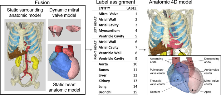
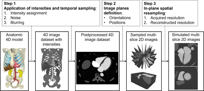
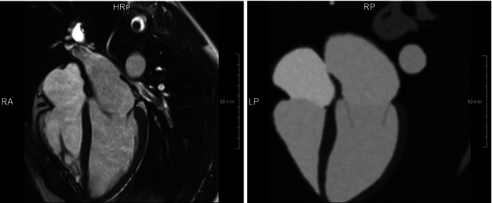
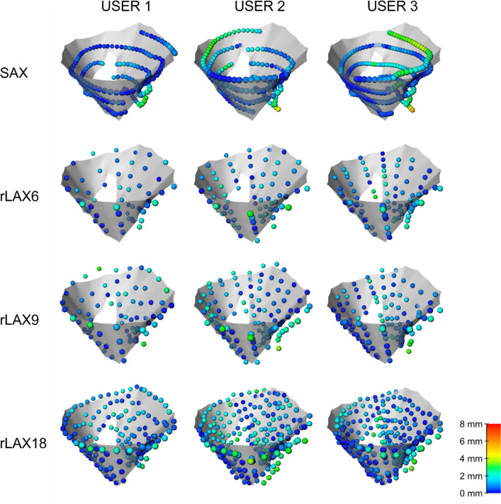
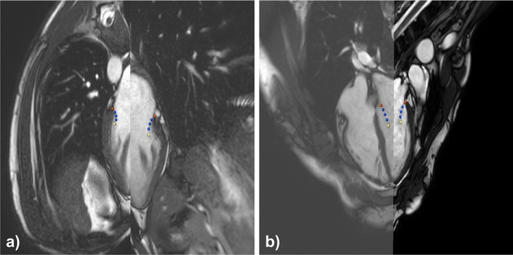
Methods: We present a pipeline for synthetic image generation based on the combination of individual anatomical 3D models with a position-based dynamics simulation of the mitral valve closure. The corresponding images are generated using modality-specific intensity models and spatiotemporal sampling concepts. We test the applicability in the context of MRI imaging strategies for the assessment of the mitral valve. Synthetic images are generated with different strategies regarding image orientation (SAX and rLAX) and spatial sampling density.

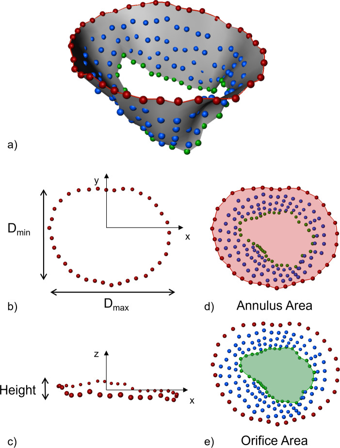
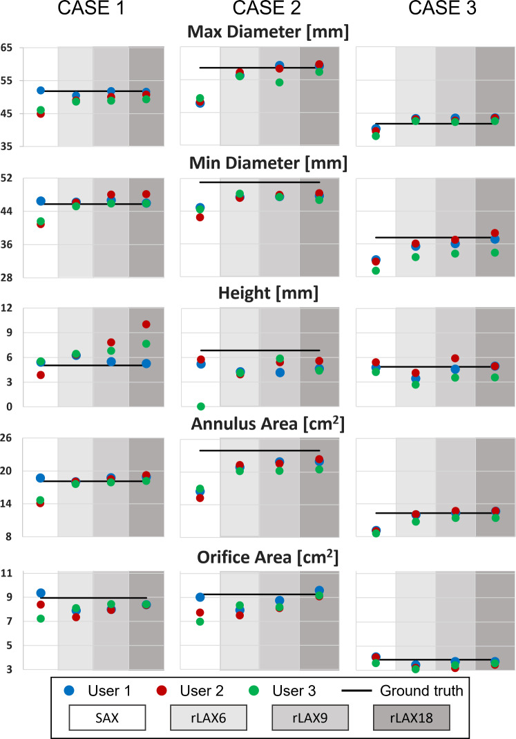
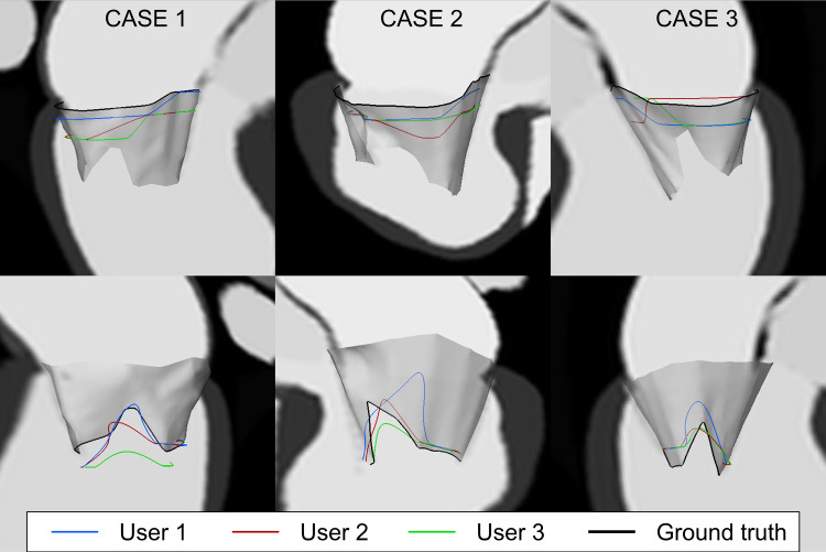
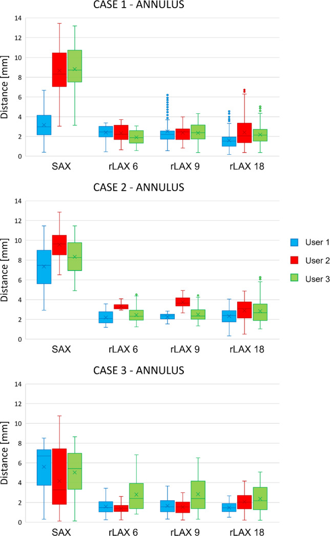
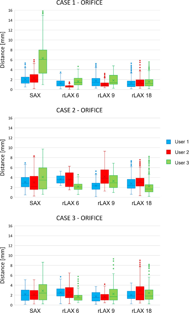
Results: The suitability of the imaging strategy is evaluated by comparing MV segmentations against ground truth annotations. The generated synthetic images were compared to ones acquired with similar parameters, and the result is promising. The quantitative analysis of annotation results suggests that the rLAX sampling strategy is preferable for MV assessment, reaching accuracy values that are comparable to or even outperform literature values.

Conclusion: The proposed approach provides a valuable tool for the evaluation and optimization of cardiac valve image acquisition. Its application to the use case identifies the radial image sampling strategy as the most suitable for MV assessment through MRI.

Keywords: Cardiac phantom; Image simulation; Magnetic resonance imaging; Mitral valve; Modeling; Segmentation.


Conflict of interest statement

The authors declare no conflict of interest.

Figures

Fig. 1
Fig. 1
Model generation. The separate entities (left) are fused, and labels are assigned for all anatomical structures (right). The markers used for the fusion of the heart model with the surrounding structures are shown in the right box
Fig. 2
Fig. 2
Image simulation workflow. Workflow for multi-slice 2D images simulation starting from the anatomic 4D model
Fig. 3
Fig. 3
Segmentation for intensity distribution analysis. Examples of relevant representative regions (first row) for ventricle blood pool (left), and for the myocardium (right). The second row shows the corresponding intensity distributions (yellow) and the two fitted distribution models: Gaussian (blue) and Rician (orange). Mean square distance values (MSD) between the two curves are reported
Fig. 4
Fig. 4
Annotation tools. Annotation software interface for the interactive annotation of the radial long axis (rLAX) images (a) and short axis (SAX) image data (b). Red points indicate the annulus, blue ones the leaflets and the yellow ones the leaflets end (orifice)
Fig. 5
Fig. 5
Clinically established quantitative parameters. Mitral valve model output from annotations (a) and clinically established quantitative parameters: Dmax: maximum diameter (b), Dmin: minimum diameter (b), height (c), annulus 2D area (d) and orifice 2D area (e). The valve axis used to set the plane orientation corresponds to the z-axis of the annulus PCA (c)
Fig. 6
Fig. 6
Real vs synthetic images comparison. MRI acquired on healthy volunteer (left) with similar parameters to our rLAX18 generated synthetic image based on the anatomy derived from CT and intensities from CMR (right)
Fig. 7
Fig. 7
Quantitative parameters for case 1 (left), case 2 (middle) and case 3 (right) for all users (user 1 in blue, user 2 in red and user 3 in green). The black line corresponds to the value computed on the ground truth valve model. SAX: short axis, rLAX6, rLAX9, rLAX18: radial long axis with 6, 9 and 18 planes. The user segmentation values and relative distances from the ground truth are reported in Supplementary Material
Fig. 8
Fig. 8
Annulus and orifice SAX annotations contours. Annulus (top row) and orifice profiles (bottom row) extracted from SAX annotations of all users (blue, red, green) and ground truth profile (black) for case 1 (left), case 2 (middle) and case 3 (right)
Fig. 9
Fig. 9
Annulus (left) and orifice (right) contours from the annotations of all users for case 1. From first row: short axis annotation (SAX), radial long axis with 6, 9 and 18 planes (rLAX6, rLAX9 and rLAX18). The best agreement for the orifice contour is achieved in the annotation of rLAX18
Fig. 10
Fig. 10
Annulus contour distances from the ground truth. Distances values between the ground truth annulus contour and the user annotations on the simulated images with different sampling strategies (SAX, rLAX6, rLAX9 and rLAX18). Mean and standard deviation are reported in Table 3
Fig. 11
Fig. 11
Orifice contour distances from the ground truth. Distances values between the ground truth orifice contour and the user annotations of the image data with different sampling strategies (SAX, rLAX6, rLAX9 and rLAX18). Corresponding mean and standard deviation are reported in Table 4
Fig. 12
Fig. 12
Distances of user annotations from ground truth valve surface for CASE 3. The points are color-coded depending on the distance from the surface, the scale is set according to the minimum and maximum distance values found for all three cases (see Supplementary Material)
Fig. 13
Fig. 13
Valve positions in acquired images. Two different radial LAX images are shown in (a) and (b). Example (a) depicts a visible shift between two rotations. Example (b) shows a proper image acquisition
Fig. 14
Fig. 14
Image annotation results. Reconstructed valves from points segmented on rLAX are shown as gray surfaces. Annotations on SAX are shown as red points. The annotations on the completely misaligned rLAX plane are shown in blue and they have been excluded for the surface generation in (a). The surface borders corresponding to the annulus and the orifice are irregular in the worst case (a) even after excluding the most shifted plane

References

    1. Kainz W, Neufeld E, Bolch WE, Graff CG, Kim CH, Kuster N, Lloyd B, Morrison T, Segars P, Yeom YS, Zankl M, Xu XG, Tsui BMW. Advances in computational human phantoms and their applications in biomedical engineering–a topical review. IEEE Trans Rad Plasma Med Sci. 2019;3(1):1–23. doi: 10.1109/TRPMS.2018.2883437. - DOI - PMC - PubMed
    1. Keenan KE, Ainslie M, Barker AJ, Boss MA, Cecil KM, Charles C, Chenevert TL, Clarke L, Evelhoch JL, Finn P, Gembris D, Gunter JL, Hill DL, Jack CR, Jr, Jackson EF, Liu G, Russek SE, Sharma SD, Steckner M, Stupic KF, Trzasko JD, Yuan C, Zheng J. Quantitative magnetic resonance imaging phantoms: a review and the need for a system phantom. Magn Reson Med. 2018;79(1):48–61. doi: 10.1002/mrm.26982. - DOI - PubMed
    1. Debrun D, Thérain F, Nguyen LD, Léger CP, Visser JJ. Volume measurements in nuclear medicine gated SPECT and 4D echocardiography: validation using a dynamic cardiac phantom. Int J Cardiovasc Imaging. 2005;21:239–247. doi: 10.1007/s10554-004-4014-1. - DOI - PubMed
    1. Zhu Y, Luo XY, Gao H, McComb C, Berry C. A numerical study of a heart phantom model. Int J Comput Math. 2014;91(7):1535–1551. doi: 10.1080/00207160.2013.854337. - DOI
    1. Krakovich A, Zaretsky U, Moalem I, Naimushin A, Rozen E, Scheinowitz M, Goldkorn R. A new cardiac phantom for dynamic SPECT. J Nucl Cardiol. 2021;28(5):2299–2309. doi: 10.1007/s12350-020-02028-0. - DOI - PubMed

MeSH terms

LinkOut - more resources