Review
. 2023 Sep 8;14(1):115.
doi: 10.1186/s40104-023-00918-0.

Integrins and their potential roles in mammalian pregnancy

Affiliations
Review

Integrins and their potential roles in mammalian pregnancy

Gregory A Johnson et al. J Anim Sci Biotechnol. .

Abstract

Integrins are a highly complex family of receptors that, when expressed on the surface of cells, can mediate reciprocal cell-to-cell and cell-to-extracellular matrix (ECM) interactions leading to assembly of integrin adhesion complexes (IACs) that initiate many signaling functions both at the membrane and deeper within the cytoplasm to coordinate processes including cell adhesion, migration, proliferation, survival, differentiation, and metabolism. All metazoan organisms possess integrins, and it is generally agreed that integrins were associated with the evolution of multicellularity, being essential for the association of cells with their neighbors and surroundings, during embryonic development and many aspects of cellular and molecular biology. Integrins have important roles in many aspects of embryonic development, normal physiology, and disease processes with a multitude of functions discovered and elucidated for integrins that directly influence many areas of biology and medicine, including mammalian pregnancy, in particular implantation of the blastocyst to the uterine wall, subsequent placentation and conceptus (embryo/fetus and associated placental membranes) development. This review provides a succinct overview of integrin structure, ligand binding, and signaling followed with a concise overview of embryonic development, implantation, and early placentation in pigs, sheep, humans, and mice as an example for rodents. A brief timeline of the initial localization of integrin subunits to the uterine luminal epithelium (LE) and conceptus trophoblast is then presented, followed by sequential summaries of integrin expression and function during gestation in pigs, sheep, humans, and rodents. As appropriate for this journal, summaries of integrin expression and function during gestation in pigs and sheep are in depth, whereas summaries for humans and rodents are brief. Because similar models to those illustrated in Fig. 1, 2, 3, 4, 5 and 6 are present throughout the scientific literature, the illustrations in this manuscript are drafted as Viking imagery for entertainment purposes.

Keywords: Humans; Implantation; Integrins; Pigs; Pregnancy; Rodents; Sheep.


Conflict of interest statement

The authors declare that they have no competing interests.

Figures
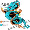
Fig. 1
Fig. 1
The Saga of integrins: Basic integrin structure. Integrins are dominant glycoproteins in adhesion cascades. They comprise a ubiquitous family of cation-dependent, heterodimeric [one α-subunit (chain) non-covalently linked to one β-subunit (chain)], intrinsic transmembrane glycoprotein receptors that mediate cellular differentiation, motility, and adhesion. Integrins are grouped according to the ligands they bind. Those that carry out ligand binding through integrin receptor recognition of small peptide sequences include integrins that bind arginine-glycine-aspartic acid (RGD; depicted here), leucine-aspartic-acid-valine (LDV), and glycine-phenylalanine-hydroxyproline-glycine-glutamic acid-arginine (GFOGER) within collagen. Integrins are also grouped into those that bind laminin, and leukocyte-specific receptors
Fig. 2
Fig. 2
The Saga of integrins: The integrin receptor (integrin) family. The integrin family can form at least 24 distinct pairings of its 18 α-subunits and 8 β-subunits. Integrins that bind to arginine-glycine-aspartic acid (RGD) sequences in ligands include αvβ1, αvβ3, αvβ5, αvβ6, αvβ8, α5β1, α8β1, and αIIbβ3. Those that bind to laminin include α3β1, α6β1, α6β4 and α7β1. Integrins that bind the glycine-phenylalanine-hydroxyproline-glycine-glutamic acid-arginine (GFOGER) sequence in collagen include α1β1, α2β1, α10β1 and α11β1. Those that are leukocyte-specific receptors include α4β1, αLβ2, αMβ2, αDβ2 and αXβ2
Fig. 3
Fig. 3
The Saga of integrins: Integrin iigands. More than one integrin receptor (integrin) can recognize a specific ECM ligand, and more than one ligand can bind a specific integrin. Depicted are Viking ships representing ECM proteins that have been localized to implantation sites in mammals carrying shields representing integrins that potentially bind to these proteins
Fig. 4
Fig. 4
The Saga of integrins: Integrin binding and activation. Shown is the structure of the unliganded (Inactive) and liganded (Active) αvβ3 integrin with the αv-subunit (chain) in orange and the β3-subunit (chain) in blue. In the inactive form the propeller, I/A, hybrid and thigh domains are bent over towards the carboxyl terminal of the legs of the α- and β-subunits which are inserted through the plasma membrane and connected to short cytoplasmic domains. The organization of the domains are difficult to discern in this configuration but are more easily resolved in the active configuration. In the active form the α- and β-subunits are unfolded freeing the propeller and I/A domains for ligand binding. Activation of the integrin is driven by either ligand binding or by effects on the cytoplasmic domains leading to straightening of the α- and β-subunits and separation of the legs. The straightening of the legs separates the cytoplasmic domains and allows binding of cytoplasmic proteins and intracellular signaling. These changes in integrin configuration are reversible and operate in either direction, outside-in or inside-out
Fig. 5
Fig. 5
The Saga of integrins: Integrin adhesion complexes. When the extracellular domains of integrins bind to ECM ligands they cluster within the plasma membrane and the cytoplasmic domains of the integrins become closely associated with the cytoskeleton resulting in the assembly of aggregates of 1) integrins at the surface of the cell, 2) cytoskeletal proteins within the cell, and 3) proteins within the ECM which are large enough to be observed by immunofluorescence microscopy and are known as integrin adhesion complexes (IACs). The proteins recruited to IACs, the integrin adhesome, perform many signaling functions both at the membrane and deeper within the cytoplasm to coordinate processes including cell adhesion, migration, proliferation, survival, differentiation, and metabolism. The integrin adhesome is highly complex and only cryptically alluded to in the figure. The “consensus adhesome” represents links between integrins and the actin cytoskeleton and the adaptor proteins that directly link integrins with actin are α-actinin, filamin, talin, and tensin. Despite the complexity of IACs, they are dynamic, and turnover can occur within a few minutes
Fig. 6
Fig. 6
The Saga of integrins: The implantation cascade. Depicted is a generalized summary of the attachment cascade for implantation in humans in which the conceptus is hypothesized to roll across the uterine surface until it is slowed and tethered to the uterine luminal epithelium (LE) first via interactions with mucins, followed by carbohydrate-lectin binding, and completed when firm adhesion is mediated through integrins binding to ECM bridging ligands, in a manner similar to the extravasation of leukocytes from the vasculature [39, 40]. Similar cascades are postulated for pigs, sheep and mice; however, the conceptuses of pigs and sheep likely do not roll across the uterine surface. Instead, the trophoblast cells proliferate and migrate across the uterine surface as the conceptuses undergo elongation [41, 42]

References

    1. Hynes RO, Ruoslahti E, Springer TA. Reflections on integrins – past, present and future. JAMA. 2022;328:1291–1292. - PubMed
    1. Whittaker CA, Hynes RO. Distribution and evolution of the von Willebrand/Integrin A domain: a widely dispersed domain with roles in cell adhesion and elsewhere. Mol Biol Cell. 2002;13:3369–3387. - PMC - PubMed
    1. Burke RD. Invertebrate integrins: structure, function, and evolution. Int Rev Cytol. 1999;191:257–284. - PubMed
    1. Hughes AL. Evolution of the integrin alpha and beta protein families. J Mol Evol. 2001;53:63–72. - PubMed
    1. Hynes RO, Zhao Q. The evolution of cell adhesion. J Cell Biol. 2000;150:F89–96. - PubMed