. 2023 Aug 9:13:23.
doi: 10.25259/JCIS_46_2023. eCollection 2023.

Magnetic resonance imaging in COVID-19-associated acute invasive fungal rhinosinusitis - Diagnosis and beyond

Affiliations

Magnetic resonance imaging in COVID-19-associated acute invasive fungal rhinosinusitis - Diagnosis and beyond

Gayatri Senapathy et al. J Clin Imaging Sci. .

Abstract

Objectives: The aim of the study was to evaluate the magnetic resonance imaging (MRI) features of acute invasive fungal rhinosinusitis (AIFRS) at presentation and on follow-up imaging when patients receive treatment with systemic antifungal therapy and surgical debridement.

Material and methods: This is a retrospective analysis of imaging data from a cohort of patients diagnosed with AIFRS during the second wave of COVID-19 in single tertiary referral hospital in South India between March 2021 and May 2021 (n = 68). Final diagnosis was made using a composite reference standard which included a combination of MRI findings, clinical presentation, nasal endoscopy and intraoperative findings, and laboratory proof of invasive fungal infection. Analysis included 62 patients with "Definite AIFRS" findings on MRI and another six patients with "Possible AIFRS" findings on MRI and laboratory proof of invasive fungal infection. Follow-up imaging was available in 41 patients.

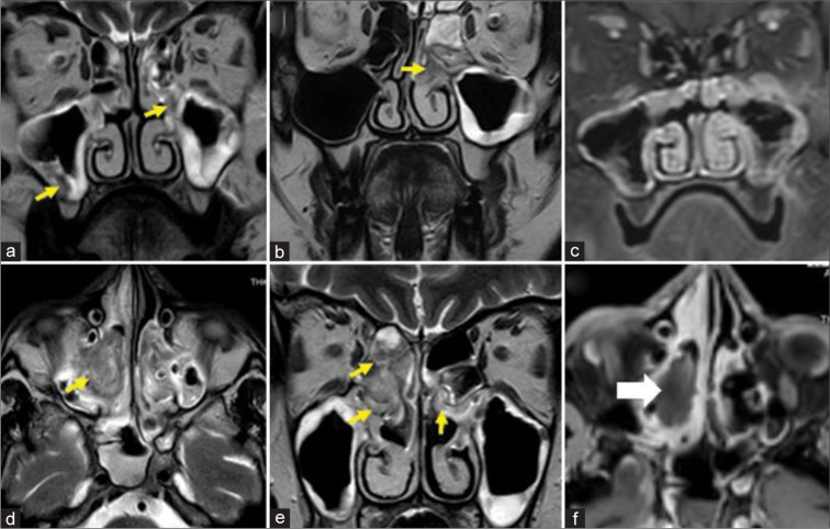
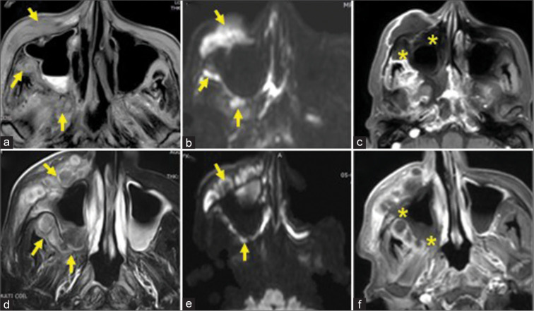
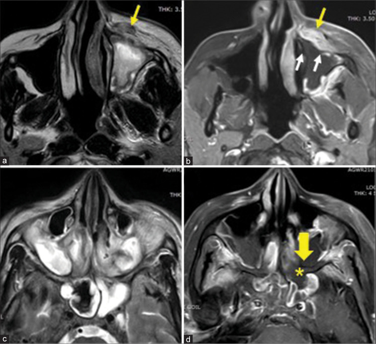
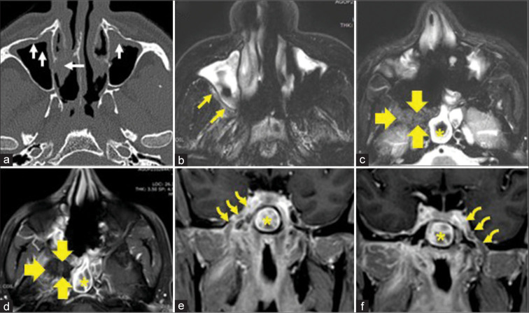
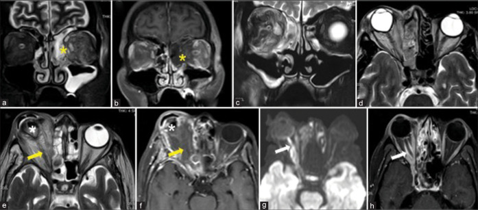
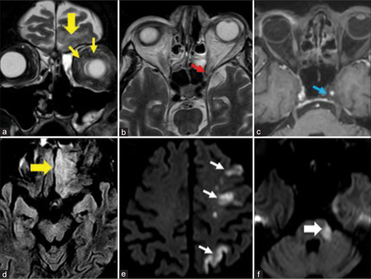
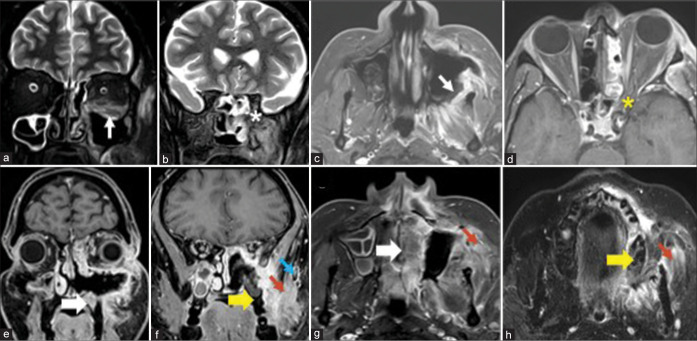
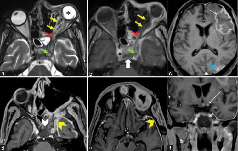
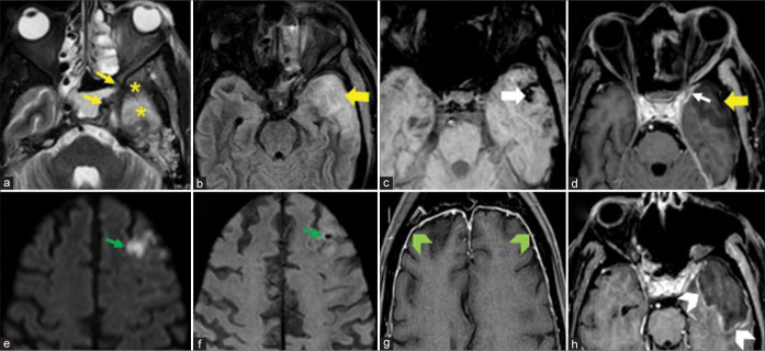
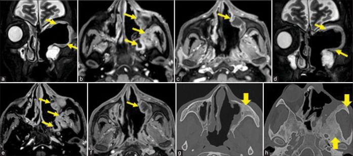
Results: The most frequent MRI finding was T2 hypointensity in the sinonasal mucosa (94%) followed by mucosal necrosis/loss of contrast-enhancement (92.6%). Extrasinosal inflammation with or without necrosis in the pre-antral fat, retroantral fat, pterygopalatine fossa, and masticator space was seen in 91.1% of the cases. Extrasinosal spread was identified on MRI even when the computed tomography (CT) showed intact bone with normal extrasinosal density. Orbital involvement (72%) was in the form of contiguous spread from either the ethmoid or maxillary sinuses; the most frequent presentation being orbital cellulitis and necrosis, with some cases showing extension to the orbital apex (41%) and inflammation of the optic nerve (32%). A total of 22 patients showed involvement of the cavernous sinuses out of which 10 had sinus thrombosis and five patients had cavernous internal carotid artery involvement. Intracranial extension was seen both in the form of contiguous spread to the pachymeninges over the frontal and temporal lobes (25%) and intra-axial involvement in the form of cerebritis, abscesses, and infarcts (8.8%). Areas of blooming on SWI were noted within the areas of cerebritis and infarcts. Perineural spread of inflammation was seen along the mandibular nerves across foramen ovale in five patients and from the cisternal segment of trigeminal nerve to the root exit zone in pons in three patients. During follow-up, patients with disease progression showed involvement of the bones of skull base, osteomyelitis of the palate, alveolar process of maxilla, and zygoma. Persistent hyperenhancement in the post-operative bed after surgical debridement and resection was noted even in patients with stable disease.

Conclusion: Contrast-enhanced MRI must be performed in all patients with suspected AIFRS as non-contrast MRI fails to demonstrate tissue necrosis and CT fails to demonstrate extrasinosal disease across intact bony walls. Orbital apex, pterygopalatine fossa, and the cavernous sinuses form important pathways for disease spread to the skull base and intracranial compartment. While cerebritis, intracranial abscesses, and infarcts can be seen early in the disease due to the angioinvasive nature, perineural spread and skull base infiltration are seen 3-4 weeks after disease onset. Exaggerated soft-tissue enhancement in the post-operative bed after debridement can be a normal finding and must not be interpreted as disease progression.

Keywords: COVID-19; Fungal sinusitis; MRI; Mucormycosis; Rhinosinusitis.


Conflict of interest statement

There are no conflicts of interest.

Figures

Figure 1:
Figure 1:
Early disease in two different patients, confined to the sinonasal cavity with no extrasinosal spread. Case1 (a-c) – A 62-year-old man: Coronal T2-weighted (T2W) magnetic resonance imaging (MRI) (a and b) and coronal post-contrast T1 fat saturation (T1-FS) MRI (c) demonstrate mucosal thickening with areas of T2 hypointense signal in bilateral maxillary and ethmoid sinuses (yellow arrows in a and b) with predominantly preserved enhancement (c). Case 2 (d-f) – A 30-year-old man: Axial T2W (d), coronal T2W (e) and axial post-contrast T1-FS MRI (f) demonstrate mucosal thickening with heterogeneous signal intensity over the bilateral middle turbinates and right ethmoid sinus (yellow arrows in d and e) with a large non-enhancing necrotic area (white block arrow).
Figure 2:
Figure 2:
Extensive extrasinosal disease with necrosis in two different patients with acute invasive fungal rhinosinusitis. Case 1 (a-c) and Case 2 (d-f) with representative axial T2-weighted (a) T2 fat saturation (d), diffusion-weighted imaging (b and e) and post contrast T1 fat saturation magnetic resonance imaging (c and f) demonstrate extensive necrosis in the mucosa and bony walls of right maxillary sinus (yellow * in c and f), extrasinosal necrosis with abscess formation and diffusion restriction in the right pre-antral fat, retroantral fat, and pterygopalatine fossa (yellow arrows).
Figure 3:
Figure 3:
Extrasinosal extension of disease in two different patients. Case 1 (a and b) with representative axial T2-weighted (T2W) magnetic resonance imaging (MRI) (a) and axial post-contrast T1 fat saturation (T1-FS) MRI (b) showing left maxillary sinusitis with necrosis along its anterior and medial walls (white arrows in a and b) and extension of T2 hypointensity with abnormal enhancement of the left pre-antral fat (yellow arrow in a and b). Case 2 (c and d) with representative axial T2W MRI (c) and axial post-contrast T1-FS MRI (d) showing bilateral maxillary and sphenoid sinusitis (c) with devitalization of the posterior wall of left maxillary sinus (yellow block arrow in d) and extension of necrosis into the left retroantral fat (yellow *).
Figure 4:
Figure 4:
A 45-year-old man with diabetes and being treated for COVID-19 pneumonia. Axial computed tomography image (a) shows bilateral maxillary sinus and middle turbinate mucosal thickening (while arrows on a) with intact sinus wall and no apparent extrasinosal abnormality. Axial T2 fat saturation (T2-FS) magnetic resonance imaging (MRI) (b) done on the same day demonstrates unequivocal hyperintensity in the right retroantral fat (yellow arrows in b), indicating spread across intact bony sinus wall. He underwent resection of the right osteomeatal complex and turbinates with sinonasal debridement and was started on systemic antifungal therapy. Follow-up MRI done 3 weeks later (c-f) demonstrates disease progression with abnormal signal intensity on T2-FS axial image (c) and necrosis on post-contrast axial image (d) in the right pterygopalatine fossa and masticator space (yellow block arrows in c and d). There is new development of sphenoid sinusitis (yellow*) and perineural spread in the form of thickening and enhancement along bilateral mandibular nerves across the foramen ovale, into the cavernous sinuses (yellow curved arrows in e and f).
Figure 5:
Figure 5:
Demonstrating the spectrum of orbital involvement in four different acute invasive fungal rhinosinusitis cases. Case 1 (a and b) with coronal T2 fat saturation (T2-FS) and post-contrast T1 fat saturation (T1-FS) magnetic resonance (MR) images showing left maxillary and ethmoid sinusitis with contiguous extension of necrotic tissue from the left ethmoid sinus into the left orbit across the medial wall (yellow *). Case 2 (c and d) with coronal T2-FS and axial T2-weighted (T2W) MR images showing right ethmoid sinusitis, right orbital cellulitis (c), and proptosis with posterior tenting of the globe (d). Case 3 (e and f) with axial T2W and post-contrast T1-FS MR images showing extensive necrosis in the right orbit and optic nerve (yellow arrow in e and f) with globe destruction and deformity (white *). Case 4 (g and h) with axial diffusion-weighted imaging and post-contrast T1-FS MR images showing marked diffusion restriction of the right optic nerve (white arrow in g) with perineural enhancement (white arrow in h).
Figure 6:
Figure 6:
A 74-year-old man on treatment for COVID-19-related acute invasive fungal rhinosinusitis. Representative coronal T2 fat saturation (a), axial T2 (b), axial post-contrast T1 fat saturation (c) images through orbits and para nasal sinuses demonstrate left orbital cellulitis (yellow arrows in a) extending up to the orbital apex (red arrow in b) and left cavernous sinus causing thrombosis of the sinus and narrowing of left cavernous internal carotid artery (ICA) (blue arrow in c). Axial fluid attenuated inversion recovery image through brain (d) and image “a” show intracranial extension of inflammation across the orbital roof with cerebritis in the left frontal lobe (yellow block arrows in a and d). Representative axial diffusion-weighted imaging sequences (e and f) demonstrate infarcts in the left ICA territory (white arrows in e) and restricted diffusion in the pons at the root exit zone of the left trigeminal nerve (white block arrow in f).
Figure 7:
Figure 7:
A 36-year-old man on treatment for COVID-19-related acute invasive fungal rhinosinusitis. Representative coronal T2 fat saturation (T2-FS) (a and b) and axial post-contrast T1 fat saturation (T1-FS) (c and d) images demonstrate left orbital and maxillary sinus involvement with necrosis in the bony walls of maxillary antrum (white arrows in a and c), extension of disease to the left orbital apex (yellow * in d) and left pterygopalatine fossa (white * in b). Endoscopic debridement and limited resection in the left maxilla and nasal cavity was performed. Follow-up imaging done after 6 weeks: Representative post-contrast T1-FS coronal and axial images (e-g) and axial T2-FS image (h) demonstrate involvement of the hard palate (white block arrows in e and g), alveolar process of left maxilla (yellow block arrows in f and h) and left zygoma (blue arrow in f) with extensive inflammation in the left masticator space and left temporal fossa (red arrows in f-h).
Figure 8:
Figure 8:
A 48-year-old man on treatment for COVID-19-related acute invasive fungal rhinosinusitis. Representative axial T2 fat saturation (a), axial T1 post-contrast (b and c) images demonstrating necrosis in the left orbit and left optic nerve (yellow arrows in a and b) extending to the orbital apex (red arrows in a and b) with globe deformity (* in a), left cavernous sinus and cavernous internal carotid artery (ICA) thrombosis (green arrows in a and b), osteomyelitis of the central skull base (white block arrow in b), abscess in the left frontal lobe (white arrow in c) and subacute infarct in the left parietal lobe (blue arrow in c). Follow-up scan after 7 weeks, post-left orbital exenteration: Representative axial T1 post-contrast (d and e) and coronal T1 post-contrast (f) images demonstrate increase in the extent of skull base osteomyelitis (white arrowhead in d) with pachymeningeal thickening and enhancement over left temporal lobe (yellow arrowheads in d and e). There is enhancement of the left optic chiasm and left optic tract (long white arrows in e and f) signifying intracranial extension of inflammation from the left optic nerve.
Figure 9:
Figure 9:
A 60-year-old man on treatment for COVID-19-related acute invasive fungal rhinosinusitis involving bilateral ethmoid sinuses and left orbital apex. Representative axial T2 fat saturation image (a) shows left orbital apex and cavernous sinus involvement (yellow arrows) with contiguous extension into the left retroantral fat and left temporal lobe (yellow*). Axial T2 Fluid attenuated inversion recovery (b), axial susceptibility-weighted imaging (SWI) (c) and axial post-contrast T1 fat saturation (T1-FS) (d) images show left temporal lobe cerebritis (yellow block arrows in b and d) with areas of blooming (white block arrow in c) and overlying dural thickening and enhancement (white arrow in d). The patient underwent sinonasal debridement and left orbital exenteration. Follow-up magnetic resonance imaging after 3 weeks: Axial diffusion-weighted imaging (e), axial SWI (f), and axial post-contrast T1-FS (g and h) images demonstrate interval development of the left frontal lobe infarct with blooming (green arrows in e and f), diffuse pachymeningeal thickening, and enhancement (green arrow heads in g) and evolution of the left temporal lobe cerebritis into an abscess with peripheral enhancement (white arrowhead in h).
Figure 10:
Figure 10:
A 45-year-old man who was on treatment for extensive COVID-19-related acute invasive fungal rhinosinusitis for 3 months, had undergone bilateral sinonasal debridement, left maxillectomy and left orbital exenteration. Representative coronal T2 fat saturation (T2-FS) (a), axial post-contrast T1 fat saturation (b and c) images from contrast-enhanced magnetic resonance imaging (CEMRI) performed a week after orbital exenteration demonstrate enhancing soft tissue/mucosal thickening lining the post-operative bed (yellow arrows in a-c); no residual necrotic areas were seen. Follow-up CEMRI done 4 weeks later with representative images at the same levels (d-f) show persistent mild mucosal thickening and enhancement (yellow arrows in d-f) as in the previous MRI with no new areas of inflammation and no interval development of necrosis. The patient was on treatment with regular clinical monitoring and returned after 9 months for reconstructive facial surgery. Computed tomography of the para nasal sinuses done at this time (g and h) demonstrates changes of chronic osteomyelitis in the left maxilla, zygoma, and sphenoid bone (yellow block arrows).

Similar articles

Cited by

References

    1. Ismaiel WF, Abdelazim MH, Eldsoky I, Ibrahim AA, Alsobky ME, Zafan E, et al. The impact of COVID-19 outbreak on the incidence of acute invasive fungal rhinosinusitis. Am J Otolaryngol. 2021;42:103080. doi: 10.1016/j.amjoto.2021.103080. - DOI - PMC - PubMed
    1. Sanghvi D, Kale H. Imaging of COVID-19-associated craniofacial mucormycosis: A black and white review of the “black fungus”. Clin Radiol. 2021;76:812–9. doi: 10.1016/j.crad.2021.07.004. - DOI - PMC - PubMed
    1. Mossa-Basha M, Ilica AT, Maluf F, Karakoc O, Izbudak I, Aygun N. The many faces of fungal disease of the paranasal sinuses: CT and MRI findings. Diagn Interv Radiol. 2013;19:195–200. doi: 10.5152/dir.2012.003. - DOI - PubMed
    1. Khattar VS, Hathiram BT, Khattar VS, Hathiram BT. Radiologic appearances in fungal rhinosinusitis. Otorhinolaryngol Clin Int J. 2009;1:15–23. doi: 10.5005/jp-journals-10003-1002. - DOI
    1. DelGaudio JM, Swain RE, Jr, Kingdom TT, Muller S, Hudgins PA. Computed tomographic findings in patients with invasive fungal sinusitis. Arch Otolaryngol Head Neck Surg. 2003;129:236–40. doi: 10.1001/archotol.129.2.236. - DOI - PubMed

LinkOut - more resources