

Evaluation of Choroidal Vascularity Index in Keratoconus Patients: Does Choroidal Vascularity Change in Keratoconus?
- PMID: 37680286
- PMCID: PMC10481970
- DOI: 10.4103/joco.joco_189_22
Evaluation of Choroidal Vascularity Index in Keratoconus Patients: Does Choroidal Vascularity Change in Keratoconus?
Abstract
Purpose: To investigate the choroidal structure in keratoconic patients with different severity using the choroidal vascularity index (CVI) derived from image binarization on enhanced depth imaging optical coherence tomography scans (EDI-OCT).
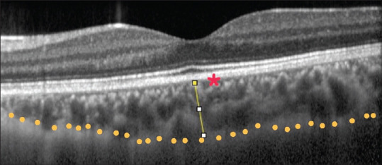
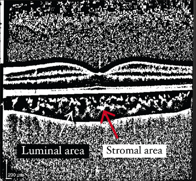
Methods: Sixty-eight eyes from 34 keratoconus (KCN) patients and 72 eyes from 36 healthy subjects were recruited in this prospective, noninterventional, comparative cross-sectional study. EDI-OCT was employed to measure choroidal parameters, including choroidal thickness (CT), total choroidal area (TCA), luminal area, stromal area, and CVI.
Results: Subfoveal CT was 354.6 ± 66.8 μm in the control group and 371 ± 64.5 μm in the KCN group (P = 0.86). There was no significant difference between control and KCN groups in terms of TCA (0.66 ± 0.14 mm2 vs. 0.7 ± 0.12 mm2; P = 0.70), luminal area (0.49 ± 0.10 mm2 vs. 0.53 ± 0.08 mm2; P = 0.67), and stromal area (0.16 ± 0.05 mm2 vs. 0.17 ± 0.05 mm2; P = 0.84). CVI was also comparable in the control group (75.4% ±3.4%) and the KCN group (75.6% ±4.5%; P = 0.43). There was also no significant correlation between other choroidal parameters and KCN severity indices.
Conclusion: It seems that CVI as well as other choroidal biomarkers were not significantly different between patients with KCN and healthy subjects.
Keywords: Choroidal thickness; Choroidal vascularity index; Enhanced depth imaging optical coherence tomography scans; Keratoconus.
Copyright: © 2023 Journal of Current Ophthalmology.
Conflict of interest statement
There are no conflicts of interest.
Figures
References
-
- Mas Tur V, MacGregor C, Jayaswal R, O'Brart D, Maycock N. A review of keratoconus: Diagnosis, pathophysiology, and genetics. Surv Ophthalmol. 2017;62:770–83. - PubMed
-
- Volatier TL, Figueiredo FC, Connon CJ. Keratoconus at a molecular level: A review. Anat Rec (Hoboken) 2020;303:1680–8. - PubMed
-
- Ferrari G, Rama P. The keratoconus enigma: A review with emphasis on pathogenesis. Ocul Surf. 2020;18:363–73. - PubMed
LinkOut - more resources
Full Text Sources