. 2023 Aug 23:14:1248873.
doi: 10.3389/fphar.2023.1248873. eCollection 2023.

Obesity exacerbates influenza-induced respiratory disease via the arachidonic acid-p38 MAPK pathway

Affiliations

Obesity exacerbates influenza-induced respiratory disease via the arachidonic acid-p38 MAPK pathway

Ravishankar Chandrasekaran et al. Front Pharmacol. .

Abstract

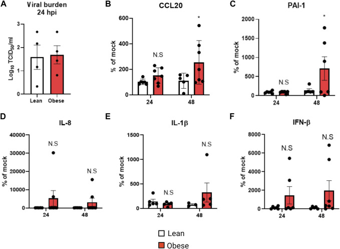
Obesity is a risk factor for severe influenza, and asthma exacerbations caused by respiratory viral infections. We investigated mechanisms that increase the severity of airway disease related to influenza in obesity using cells derived from obese and lean individuals, and in vitro and in vivo models. Primary human nasal epithelial cells (pHNECs) derived from obese compared with lean individuals developed increased inflammation and injury in response to influenza A virus (IAV). Obese mice infected with influenza developed increased airway inflammation, lung injury and elastance, but had a decreased interferon response, compared with lean mice. Lung arachidonic acid (AA) levels increased in obese mice infected with IAV; arachidonic acid increased inflammatory cytokines and injury markers in response to IAV in human bronchial epithelial (HBE) cells. Obesity in mice, and AA in HBE cells, increased activation of p38 MAPK signaling following IAV infection; inhibiting this pathway attenuated inflammation, injury and tissue elastance responses, and improved survival. In summary, obesity increases disease severity in response to influenza infection through activation of the p38 MAPK pathway in response to altered arachidonic acid signaling.

Keywords: arachidonic acid; influenza A virus; lung inflammation; lung injury; obesity; p38 MAPK.


Conflict of interest statement

The authors declare that the research was conducted in the absence of any commercial or financial relationships that could be construed as a potential conflict of interest.

Figures

FIGURE 1
FIGURE 1
Elevated inflammatory and injury marker production in obese pHNECs infected with IAV: (A) TCID50 measurement of viral burden in lean and obese pHNECs (4 samples per group); (B–E): (B) CCL20 (C) PAI-1 (D) IL-1β (E) IL-8 (F) IFN-β protein levels in the supernatant (47 samples per group). * Significant difference between lean and obese pHNECs, N.S, not significant; q values < 0.05 were regarded as discovery or statistically significant. Error bars ±SEM.
FIGURE 2
FIGURE 2
Elevated inflammatory parameters in HFD-IAV mice: (A) Schematic of DIO and IAV-PR8 exposure (B) TCID50 measurement of viral burden in the lungs; (C,D): Cellular infiltration: (C) Total cells and (D) Cell differentials in the BAL; (E–J): Cytokines in BAL (E) CCL20, (F) G-CSF, (G) IL-6 (H) KC (I) IL-1β and (J) CCL2 levels; (K) qRT-PCR for IFN-β. * Significant difference between Mock and IAV, # significant difference between LFD IAV and HFD IAV. q values < 0.05 were regarded as discovery or statistically significant. Error bars ±SEM.
FIGURE 3
FIGURE 3
Elevated lung injury and peribronchial fibrosis in HFD-IAV mice: (A–C): Representative images of Masson’s trichrome staining to measure collagen, Scale bars: 100 µM (A) MOCK (B) LFD IAV (C) HFD IAV; (D) Quantification of Masson’s trichrome staining; (E–H): Lung injury parameters in BAL: (E) LDH activity (F) Dead cell protease activity (G) TGF-β protein level (H) PAI-1 protein level; (I) PAI-1 protein level in serum. * Significant difference between Mock and IAV, # significant difference between LFD IAV and HFD IAV. q values < 0.05 were regarded as discovery or statistically significant. Error bars ± SEM.
FIGURE 4
FIGURE 4
Airway reactivity in lean and obese IAV infected mice: (A) Resistance (Rrs) and (B) Elastance (Ers) (stiffness) as measured using flexivent. *Significant difference between Mock and IAV, # significant difference between LFD IAV and HFD IAV, q values < 0.05 were regarded as discovery or statistically significant. Error bars ±SEM.
FIGURE 5
FIGURE 5
Arachidonic acid mediated inflammation, injury and p38 activation: (A) Arachidonic acid and (B) PGE2 levels in BAL, * significant difference between MOCK and IAV, # significant difference between LFD IAV and HFD IAV; (C–E): Cytokine and injury marker levels in supernatant: (C) CCL20 (D) PAI-1 and (E) IFN-β; (F) Western blot to detect phospho-p38 and phospho-JNK1/2 levels; (G–I): Effect of p38 MAPK inhibitor (DP) treatment on (G) CCL20 (H) PAI-1 and (I) IFNβ levels in supernatant. (J) Western blot to detect phospho and total-p38 MAPK following siRNA treatment; (K) Effect of p38 siRNA treatment on PAI-1 levels. * Significant difference between MOCK and IAV, # significant difference between BSA IAV and AA IAV, $ significant difference between AA IAV and AA IAV/DP or AA IAV/p38si. q values < 0.05 were regarded as discovery or statistically significant. Error bars ±SEM.
FIGURE 6
FIGURE 6
p38 MAPK inhibition reduces IAV-induced inflammation in HFD mice: (A) Western blot to detect phospho-p38 in the lungs; (B) Schematic of DIO, IAV-PR8 exposure and DP-treatment (C) Western blot to detect p38MAPK targets phospho-ATF2 and phospho MAPKAPK2; (D) TCID50 to measure viral burden; (E) Cell differentials in the BAL; F-I: Cytokines in the BAL: (F) CCL20 (G) IL-6 (H) KC; (I) PGE2 level in BAL; (J) IFN-β level in BAL. *Significant difference between HFD mock and HFD IAV, # significant difference between HFD IAV and HFD IAV/DP. q values < 0.05 were regarded as discovery or statistically significant. Error bars ±SEM.
FIGURE 7
FIGURE 7
p38 MAPK inhibition reduces IAV-induced lung injury, lung stiffness, weight change and improves survival in HFD mice: (A–C): Lung injury markers in BAL: (A) Dead cell protease activity (B) TGF-β (C) PAI-1; (D) Resistance (Rrs) and (E) Elastance (Ers) or stiffness as measured by flexivent; (F) Weight change (grams) 12 days post infection; (G) Percent survival 12 days post infection. *Significant difference between HFD mock and HFD IAV, # significant difference between HFD IAV and HFD IAV/DP. q values < 0.05 were regarded as discovery or statistically significant. Error bars ±SEM.

References

    1. Bassetti M., Parisini A., Calzi A., Pallavicini F. M., Cassola G., Artioli S., et al. (2011). Risk factors for severe complications of the novel influenza A (H1N1): analysis of patients hospitalized in Italy. Clin. Microbiol. Infect. 17 (2), 247–250. 10.1111/j.1469-0691.2010.03275.x - DOI - PubMed
    1. Boden G. (2008). Obesity and free fatty acids. Endocrinol. metabolism Clin. N. Am. 37 (3), 635–646. 10.1016/j.ecl.2008.06.007 - DOI - PMC - PubMed
    1. Börgeling Y., Schmolke M., Viemann D., Nordhoff C., Roth J., Ludwig S. (2014). Inhibition of p38 mitogen-activated protein kinase impairs influenza virus-induced primary and secondary host gene responses and protects mice from lethal H5N1 infection. J. Biol. Chem. 289 (1), 13–27. 10.1074/jbc.M113.469239 - DOI - PMC - PubMed
    1. Carey M. A., Bradbury J. A., Seubert J. M., Langenbach R., Zeldin D. C., Germolec D. R. (2005). Contrasting effects of cyclooxygenase-1 (COX-1) and COX-2 deficiency on the host response to influenza A viral infection. J. Immunol. 175 (10), 6878–6884. 10.4049/jimmunol.175.10.6878 - DOI - PubMed
    1. Chandrasekaran R., Bruno S. R., Mark Z. F., Walzer J., Caffry S., Gold C., et al. (2023). Mitoquinone mesylate attenuates pathological features of lean and obese allergic asthma in mice. Am. J. physiology Lung Cell. Mol. physiology 324 (2), L141–L153. 10.1152/ajplung.00249.2022 - DOI - PMC - PubMed

LinkOut - more resources