

Case of lumbar spinal stenosis and chronic tophaceous gout
- PMID: 37680922
- PMCID: PMC10481838
- DOI: 10.25259/SNI_504_2023
Case of lumbar spinal stenosis and chronic tophaceous gout
Abstract
Background: Rarely, chronic tophaceous gout can result in lumbar spinal stenosis and neural compression.
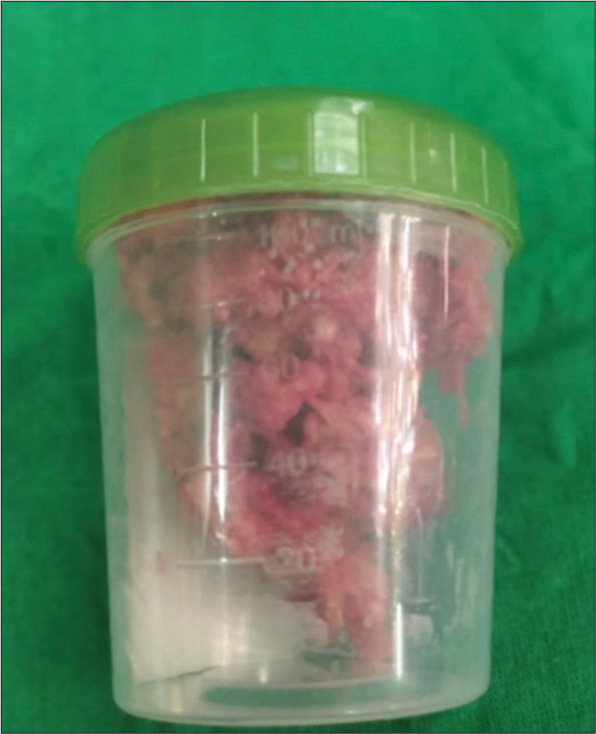
Case description: A 67-year-old male presented with the radiographic and magnetic resonance findings of gout involving and causing compression of the lumbar spine that responded to surgical decompression.
Conclusion: It is difficult to diagnose lumbar spinal stenosis secondary to tophaceous gout. Notably, the treatment, based on the clinical presentation, may include both medication and surgical decompression.
Keywords: Joint inflammation; Lumbar spinal stenosis; Multidisciplinary approach; Tophaceous gouty arthritis; Tophi; Urate crystals.
Copyright: © 2023 Surgical Neurology International.
Conflict of interest statement
There are no conflicts of interest.
Figures


Similar articles
-
A unique presentation of acute tophaceous gout in the lumbar spine causing cauda equina syndrome.Radiol Case Rep. 2023 Jul 14;18(9):3341-3345. doi: 10.1016/j.radcr.2023.06.070. eCollection 2023 Sep. Radiol Case Rep. 2023. PMID: 37520396 Free PMC article.
-
Atypical Cutaneous Presentation of Chronic Tophaceous Gout: A Case Report.Indian Dermatol Online J. 2020 Mar 9;11(2):235-238. doi: 10.4103/idoj.IDOJ_205_19. eCollection 2020 Mar-Apr. Indian Dermatol Online J. 2020. PMID: 32477988 Free PMC article.
-
Lumbar spinal stenosis attributable to tophaceous gout: case report and review of the literature.Ther Clin Risk Manag. 2017 Sep 28;13:1287-1293. doi: 10.2147/TCRM.S145906. eCollection 2017. Ther Clin Risk Manag. 2017. PMID: 29033576 Free PMC article.
-
[Tophaceous gout of the cervical spine, causing cord compression. Case report and review of the literature].Neurochirurgie. 2003 Dec;49(6):600-4. Neurochirurgie. 2003. PMID: 14735005 Review. French.
-
Erosive Tophaceous Gouty Arthropathy of the Hand: A Case Report.S D Med. 2022 May;75(5):216-219. S D Med. 2022. PMID: 35724351 Review.
Cited by
-
Understanding spinal gout: A comprehensive study of 88 cases and their clinical implications.J Craniovertebr Junction Spine. 2024 Apr-Jun;15(2):133-140. doi: 10.4103/jcvjs.jcvjs_166_23. Epub 2024 May 24. J Craniovertebr Junction Spine. 2024. PMID: 38957764 Free PMC article. Review.
References
Publication types
LinkOut - more resources
Full Text Sources