. 2023 Aug 19:33:925-937.
doi: 10.1016/j.omtn.2023.08.020. eCollection 2023 Sep 12.

Novel hybrid silicon-lipid nanoparticles deliver a siRNA to cure autosomal dominant osteopetrosis in mice. Implications for gene therapy in humans

Affiliations

Novel hybrid silicon-lipid nanoparticles deliver a siRNA to cure autosomal dominant osteopetrosis in mice. Implications for gene therapy in humans

Antonio Maurizi et al. Mol Ther Nucleic Acids. .

Abstract

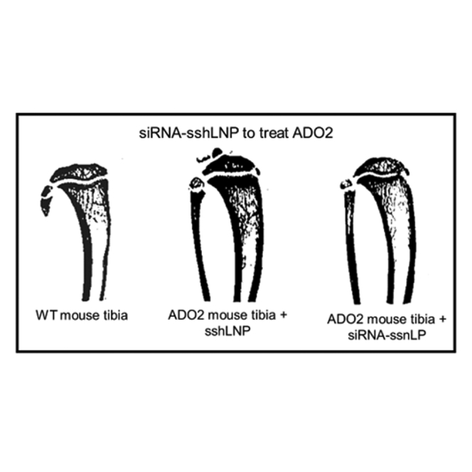
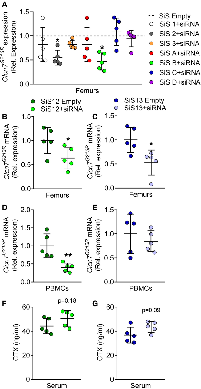
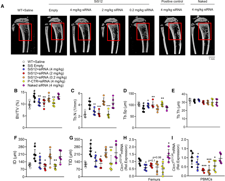
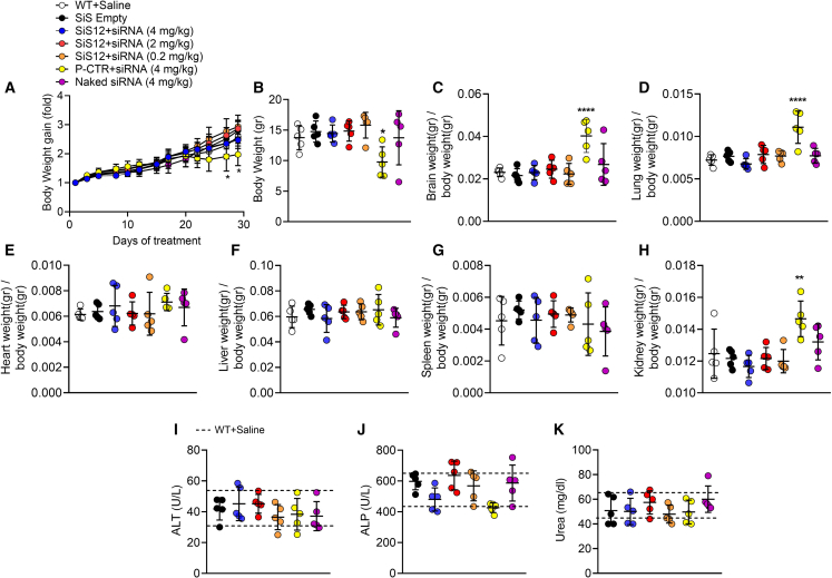
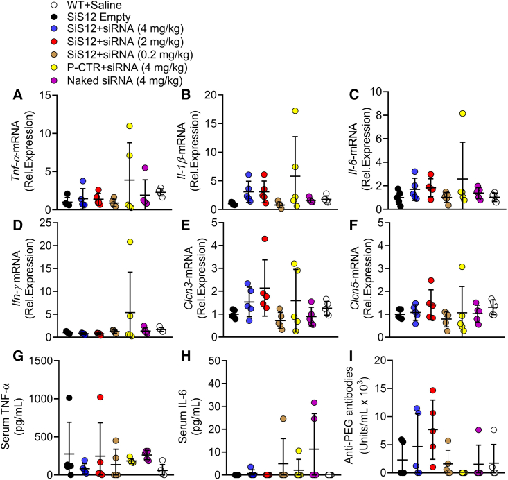
Rare skeletal diseases are still in need of proper clinically available transfection agents as the major challenge for first-in-human translation relates to intrinsic difficulty in targeting bone without exacerbating any inherent toxicity due to used vector. SiSaf's silicon stabilized hybrid lipid nanoparticles (sshLNPs) constitute next-generation non-viral vectors able to retain the integrity and stability of constructs and to accommodate considerable payloads of biologicals, without requiring cold-chain storage. sshLNP was complexed with a small interfering RNA (siRNA) specifically designed against the human CLCN7G215R mRNA. When tested via single intraperitoneal injection in pre-puberal autosomal dominant osteopetrosis type 2 (ADO2) mice, carrying a heterozygous mutation of the Clcn7 gene (Clcn7G213R), sshLNP, this significantly downregulated the Clcn7G213R related mRNA levels in femurs at 48 h. Confirmatory results were observed at 2 weeks and 4 weeks after treatments (3 intraperitoneal injections/week), with rescue of the bone phenotype and demonstrating safety. The pre-clinical results will enable advanced preclinical development of RNA-based therapy for orphan and genetic skeletal disorders by safely and effectively delivering biologicals of interest to cure human systemic conditions.

Keywords: MT: delivery strategies; autosomal dominant osteopetrosis; bone resorption; delivery nanoparticles; genetic bone diseases; osteoclasts; silicon; therapeutic siRNA; therapy.


Conflict of interest statement

A.T. and A.M. served as consultant, P.P had a service agreement, and F.M.S., N.T.-P., P.B.R, C.B., U.N., M.W., and A.D. are scientists working for SiSaf Ltd, a commercial stage biopharmaceutical company. S.S.-S. is founder and CEO of SiSaf Ltd.

Figures

None
Graphical abstract
Figure 1
Figure 1
Clcn7G213R gene expression and serum CTX analysis in ADO2 mice treated with SiS sshLNP formulations Ten-day-old ADO2 male mice were treated i.p. with the indicated SiS sshLNP loaded with 4 mg/kg CLCN7G215R-siRNA or empty. (A) Mice were treated for 48 h and real time RT-PCR was performed using a specific primer pair for the mutant Clcn7G213R (Table S2), normalized by Gapdh. (B and C) Mice were treated for 2 weeks and real time RT-PCR for the mutant Clcn7G213R was performed in femurs and (D and E) in PBMCs normalized by Gapdh. (E and F) Evaluation of serum CTX in ADO2 mice treated for 2 weeks. Data represent the mean ± SD of n = 5 ADO2 mice per group. Student’s t test vs. the SiS empty sshLNP group. ∗p ≤ 0.05; ∗∗p < 0.01.
Figure 2
Figure 2
Treatment with SiS12 and Sis13 formulations for 2 weeks Ten-day-old ADO2 male mice were treated i.p. 3 times a week for 2 weeks with SiS12 or SiS13 sshLNP complexed with 4 mg/kg of CLCN7G215R-siRNA or as such (empty sshLNP). (A and B) At the end of the experiment, the weights of the indicated organs were taken to assess the presence of macroscopic alterations. For each mouse, the organs’ weight was normalized for the body weight. An untreated WT group was examined for comparison. Data are the mean ± SD of n = 5 mice per group. Student’s t test.
Figure 3
Figure 3
Cellular uptake, biodistribution, and pharmacokinetics RAW264.7 cells were incubated with SiS12-18:1PE-CF10%+siRNA at the indicated (A) temperatures (time: 24 h), (B) concentrations (time: 24 h) and (C) times and evaluated for cellular uptake by fluorometry. (D) Cells were flushed out from femurs and allowed to adhere to culture dish for 12 min to separate the adherent fraction (enriched in stromal cells) from the non-adherent fraction (enriched in hematopoietic cells). Five hundred thousand cells per fractions were then incubated with SiS12-18:1PE-CF10%+siRNA for 3 h, washed, and evaluated by fluorometry to detect the green fluorescence. (E) One million of labeled non-adherent cells were sorted by fluorescence-activated cell sorting (FACS) by size and granularity to distinguish the fractions enriched in lymphocytes, monocytes and granulocytes. (F) Image in (E) cleaned from the background to better visualize the three fractions. (G) Mean fluorescence intensity (MFI) in the cell fractions shown in (G). (H) Four-week-old ADO2 mice were injected i.p. and (I) subcutaneously with 4 mg/kg ADO2 siRNA formulated with SiS12-18:PE-CF10%). Mice were sacrificed at the indicated time points, and organs were harvested and analyzed by fluorometry to detect green fluorescence. Results are the mean ± SD of 3 in vitro experiments or 5 mice per group. Statistics: (B and C) multiple comparison ANOVA; (F) Student’s t test.
Figure 4
Figure 4
Bone phenotype analysis Ten-day-old ADO2 male mice were treated with the indicated siRNA and siRNA-SiS sshLNP or positive control (P-CTR) complexes and doses, 3 times a week for 4 weeks. At the end of the experiment, mice were sacrificed, and tibias and femurs were collected and analyzed. (A) Representative μCT reconstructions of proximal tibias. Red squares indicate the region of interest subjected to the analysis. (B) Morphometric analysis of bone volume over total volume (BV/TV%) and (C) trabecular number (Tb.N), (D) separation (Tb.Sp), and (E) thickness (Tb.Th) assessed by μCT. (F) Indentation distance (ID) and (G) total indentation distance (TID) measured in femur midshafts by indentation test using the Biodent device. (H) Downregulation of the mutant Clcn7G213R in femurs and (I) PBMCs, normalized by Gapdh. Results are (A) representative or (B–I) the mean ± SD of n = 5 mice per group. Multiple comparison ANOVA. ∗p < 0.05; ∗∗p < 0.01; ∗∗∗p < 0.001 vs. the SiS empty sshLNP group; #p < 0.01 vs. WT + Saline group.
Figure 5
Figure 5
Safety analysis Ten-day-old ADO2 male mice were treated with the indicated siRNA and siRNA-SiS sshLNP or positive control (P-CTR) complexes and doses, 3 times a week for 4 weeks. (A) Body weights measured at the indicated time points and their gain calculated normalizing the body weights measured during the treatment with the body weights at time = 0 (pre-treatment). (B) Body weights and weights of (C) brains, (D) lungs, (E) hearts, (F) livers, (G) spleens, and (H) kidneys assessed at the end of the experiment. (I and J) Sera collected from the same mice were analyzed by the Reflotron method for the indicated biomarkers of liver and (K) kidney diseases. Results are the mean ± SD of n = 5 mice per group. Multiple comparison ANOVA vs. the WT + Saline group. ∗p < 0.05; ∗∗p < 0.01; ∗∗∗∗p < 0.0001.
Figure 6
Figure 6
Expression of inflammatory cytokines, chloride transporters and anti-PEG antibodies Ten-day-old ADO2 male mice were treated with the indicated siRNA and siRNA-SiS sshLNP or positive control (P-CTR) complexes and doses, 3 times a week for 4 weeks. (A) Real-time RT-PCR of the inflammatory cytokines Tnf-α, (B) IL-1β, (C), IL-6, and (D) INF-γ expressed in femurs, normalized by Gapdh. (E) Real time RT-PCR of the chloride transporters Clcn3 and (F) Clcn5 expressed in femurs, normalized by Gapdh. (G) Serum levels of TNF-α, (H) IL-6 and (I) anti-PEG antibodies. Results are the mean ± SD of n = 5 mice per group. MC-ANOVA vs. the WT+saline group. Differences are statistically not significant.

Similar articles

Cited by

References

    1. Cleiren E., Bénichou O., Van Hul E., Gram J., Bollerslev J., Singer F.R., Beaverson K., Aledo A., Whyte M.P., Yoneyama T., et al. Albers-Schönberg disease (autosomal dominant osteopetrosis, type II) results from mutations in the ClCN7 chloride channel gene. Hum. Mol. Genet. 2001;10:2861–2867. doi: 10.1093/hmg/10.25.2861. - DOI - PubMed
    1. Wu C.C., Econs M.J., Di Meglio L.A., Insogna K.L., Levine M.A., Orchard P.J., Miller W.P., Petryk A., Rush E.T., Shoback D.M., et al. Diagnosis and management of osteopetrosis: consensus guidelines from the osteopetrosis working group. J. Clin. Endocrinol. Metab. 2017;102:3111–3123. doi: 10.1210/jc.2017-01127. - DOI - PubMed
    1. Capulli M., Maurizi A., Ventura L., Rucci N., Teti A. Effective small interfering RNA therapy to treat CLCN7-dependent autosomal dominant osteopetrosis type 2. Mol. Ther. Nucleic Acids. 2015;4:e248. doi: 10.1038/mtna.2015.21. - DOI - PMC - PubMed
    1. Maurizi A., Capulli M., Patel R., Curle A., Rucci N., Teti A. RNA interference therapy for autosomal dominant osteopetrosis type 2. Towards the preclinical development. Bone. 2018;110:343–354. doi: 10.1016/j.bone.2018.02.031. - DOI - PubMed
    1. Zhao Z., Anselmo A.C., Mitragotri S. Viral vector-based gene therapies in the clinic. Bioeng. Transl. Med. 2022;7 doi: 10.1002/btm2.10258. - DOI - PMC - PubMed

LinkOut - more resources