. 2023 Aug 31;12(17):2185.
doi: 10.3390/cells12172185.

Aquaglyceroporins in Human Breast Cancer

Affiliations

Aquaglyceroporins in Human Breast Cancer

Teresa Kirkegaard et al. Cells. .

Abstract

Aquaporins are water channels that facilitate passive water transport across cellular membranes following an osmotic gradient and are essential in the regulation of body water homeostasis. Several aquaporins are overexpressed in breast cancer, and AQP1, AQP3 and AQP5 have been linked to spread to lymph nodes and poor prognosis. The subgroup aquaglyceroporins also facilitate the transport of glycerol and are thus involved in cellular metabolism. Transcriptomic analysis revealed that the three aquaglyceroporins, AQP3, AQP7 and AQP9, but not AQP10, are overexpressed in human breast cancer. It is, however, unknown if they are all expressed in the same cells or have a heterogeneous expression pattern. To investigate this, we employed immunohistochemical analysis of serial sections from human invasive ductal and lobular breast cancers. We found that AQP3, AQP7 and AQP9 are homogeneously expressed in almost all cells in both premalignant in situ lesions and invasive lesions. Thus, potential intervention strategies targeting cellular metabolism via the aquaglyceroporins should consider all three expressed aquaglyceroporins, namely AQP3, AQP7 and AQP9.

Keywords: AQP; aquaglyceroporin; aquaporin; breast cancer; metastasis.


Conflict of interest statement

The authors declare no conflict of interest.

Figures

Figure 1
Figure 1
Graphical illustration of workflow. Formalin-fixed, paraffin-embedded samples from invasive female breast cancer tumors were obtained in fully anonymized form from surgical specimens from the Department of Pathology, Aarhus University Hospital, Denmark. Following retrieval, the anonymized tissue samples were sectioned. For diagnostic assessment, hematoxylin and eosin (H&E) stainings, as well as immunohistochemical stainings for E-cadherin, were performed. Samples representing invasive ductal carcinoma (IDC) and invasive lobular carcinoma (ILC) were further analyzed. Serial sections were processed for immunohistochemical stainings in the following order: #1 AQP7, #2 AQP9, #3 AQP3 and #4 SMMS-1. The SMMS-1 staining identified myoepithelial cells, allowing a distinction between in situ and invasion lesions. Images of all samples were captured by brightfield light microscopy, and select slides for publication figures were scanned on a NanoZoomer 2.0HT (Hamamatsu) with the 40× objective. Part of this figure was generated in BioRender.com; accessed on August 11th 2023.
Figure 2
Figure 2
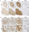
AQP7, AQP9 and AQP3 localize to epithelial cells of lobules and extralobular ducts in benign regions adjacent to tumor tissue. Formalin-fixed, paraffin-embedded samples from invasive female breast cancer tumors were obtained in fully anonymized form from surgical specimens and processed for immunohistochemistry with antibodies against AQP7, AQP9, AQP3 and SMMS-1. The figure depicts representative images from immunohistochemical stainings of serial sections from the normal/benign part of samples from invasive lobular carcinoma. (A) Lobule and (B) extralobular duct. Immune cells in the surrounding connective tissue also stain positive (red asterisks in B). Arrowheads in B point to myoepithelial cells, and the arrows in B point to the apical region of extralobular ducts. Slides were scanned in a NanoZoomer 2.0HT (Hamamatsu) using the 40× objective. White balance was adjusted in Adobe Photoshop CS4. Scale bars are 100 µm.
Figure 3
Figure 3
AQP7, AQP9 and AQP3 localize to neoplastic cells of lobular carcinoma in situ and invasive lobular carcinoma. Formalin-fixed, paraffin-embedded samples from invasive female breast cancer tumors were obtained in fully anonymized form from surgical specimens and processed for immunohistochemistry with antibodies against AQP7, AQP9, AQP3 and SMMS-1. The figure depicts representative images from immunohistochemical stainings of serial sections from patients with invasive lobular carcinoma (ILC) and enlarged images (inserts) of the marked areas. (A) Images are from a region of lobular carcinoma in situ (LCIS) from an ILC patient, and (B) is ILC. Slides were scanned in a NanoZoomer 2.0HT (Hamamatsu) using the 40× objective. White balance was adjusted in Adobe Photoshop CS4. Scale bars are 100 µm (A,B) and 20 µm ((A,B), inserts).
Figure 4
Figure 4
AQP7, AQP9 and AQP3 localize to neoplastic cells of ductal carcinoma in situ and invasive ductal carcinoma. Formalin-fixed, paraffin-embedded samples from invasive female breast cancer tumors were obtained in fully anonymized form from surgical specimens and processed for immunohistochemistry with antibodies against AQP7, AQP9, AQP3 and SMMS-1. The figure depicts representative images from immunohistochemical stainings of serial sections from patients with invasive ductal carcinoma (IDC) and enlarged images (inserts) of the marked areas. (A) Images are from a region of ductal carcinoma in situ (DCIS) from an IDC patient, and (B) is IDC. Slides were scanned in a NanoZoomer 2.0HT (Hamamatsu) using the 40x objective. White balance was adjusted in Adobe Photoshop CS4. Scale bars are 175 µm (A,B) and 50 µm ((A,B), inserts).

References

    1. Login F.H., Nejsum L.N. Aquaporin water channels: Roles beyond renal water handling. Nat. Rev. Nephrol. 2023;19:604–618. doi: 10.1038/s41581-023-00734-9. - DOI - PubMed
    1. Zhang L., Yao D., Xia Y., Zhou F., Zhang Q., Wang Q., Qin A., Zhao J., Li D., Li Y., et al. The structural basis for glycerol permeation by human AQP7. Sci. Bull. 2021;66:1550–1558. doi: 10.1016/j.scib.2020.12.006. - DOI - PubMed
    1. Gotfryd K., Mosca A.F., Missel J.W., Truelsen S.F., Wang K., Spulber M., Krabbe S., Helix-Nielsen C., Laforenza U., Soveral G., et al. Human adipose glycerol flux is regulated by a pH gate in AQP10. Nat. Commun. 2018;9:4749. doi: 10.1038/s41467-018-07176-z. - DOI - PMC - PubMed
    1. Montiel V., Bella R., Michel L.Y.M., Esfahani H., De Mulder D., Robinson E.L., Deglasse J.P., Tiburcy M., Chow P.H., Jonas J.C., et al. Inhibition of aquaporin-1 prevents myocardial remodeling by blocking the transmembrane transport of hydrogen peroxide. Sci. Transl. Med. 2020;12:eaay2176. doi: 10.1126/scitranslmed.aay2176. - DOI - PubMed
    1. Wang H., Schoebel S., Schmitz F., Dong H., Hedfalk K. Characterization of aquaporin-driven hydrogen peroxide transport. Biochim. Biophys. Acta Biomembr. 2020;1862:183065. doi: 10.1016/j.bbamem.2019.183065. - DOI - PubMed

Substances