

Hyperpolarized Carbon 13 MRI: Clinical Applications and Future Directions in Oncology
- PMID: 37682052
- PMCID: PMC10546364
- DOI: 10.1148/rycan.230005
Hyperpolarized Carbon 13 MRI: Clinical Applications and Future Directions in Oncology
Abstract
Hyperpolarized carbon 13 MRI (13C MRI) is a novel imaging approach that can noninvasively probe tissue metabolism in both normal and pathologic tissues. The process of hyperpolarization increases the signal acquired by several orders of magnitude, allowing injected 13C-labeled molecules and their downstream metabolites to be imaged in vivo, thus providing real-time information on kinetics. To date, the most important reaction studied with hyperpolarized 13C MRI is exchange of the hyperpolarized 13C signal from injected [1-13C]pyruvate with the resident tissue lactate pool. Recent preclinical and human studies have shown the role of several biologic factors such as the lactate dehydrogenase enzyme, pyruvate transporter expression, and tissue hypoxia in generating the MRI signal from this reaction. Potential clinical applications of hyperpolarized 13C MRI in oncology include using metabolism to stratify tumors by grade, selecting therapeutic pathways based on tumor metabolic profiles, and detecting early treatment response through the imaging of shifts in metabolism that precede tumor structural changes. This review summarizes the foundations of hyperpolarized 13C MRI, presents key findings from human cancer studies, and explores the future clinical directions of the technique in oncology. Keywords: Hyperpolarized Carbon 13 MRI, Molecular Imaging, Cancer, Tissue Metabolism © RSNA, 2023.
Keywords: Cancer; Hyperpolarized Carbon 13 MRI; Molecular Imaging; Tissue Metabolism.
Conflict of interest statement
Figures
![Simplified schematic of the major metabolic pathways that can be
investigated with hyperpolarized [1–carbon 13]pyruvate MRI. ALT =
alanine transaminase, CA = carbonic anhydrase, CoA = coenzyme A, LDH =
lactate dehydrogenase, PDH = pyruvate dehydrogenase, TCA = tricarboxylic
acid.](https://cdn.ncbi.nlm.nih.gov/pmc/blobs/22d8/10546364/f02df48b0c5c/rycan.230005.fig1.jpg)
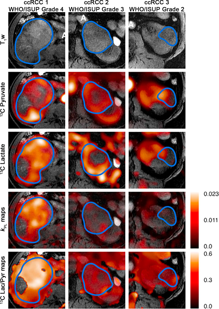
![Hyperpolarized [1–carbon 13]pyruvate MR images in a patient
with triple-negative breast cancer. (A) Coronal T1-weighted
three-dimensional spoiled gradient-echo (SPGR) image. (B) Coronal
reformatted dynamic contrast-enhanced (DCE) image at peak enhancement after
injection of a gadolinium-based contrast agent. (C) Summed hyperpolarized
carbon 13 pyruvate images. (D) Summed hyperpolarized carbon 13 lactate
images. (E) Lactate:pyruvate (LAC/PYR) ratio map. (F, G) Dynamic
hyperpolarized carbon 13 pyruvate and lactate imaging with a 12-second delay
after injection over 15 time points at 4-second intervals. (Reprinted, with
permission, from reference 18.)](https://cdn.ncbi.nlm.nih.gov/pmc/blobs/22d8/10546364/48fb16324d67/rycan.230005.fig3.jpg)
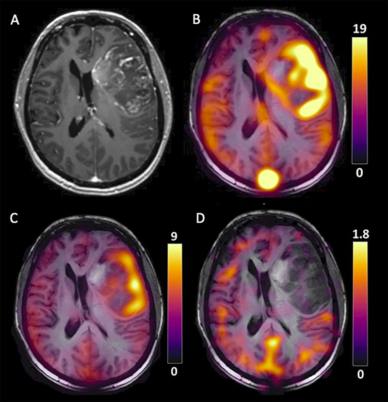
![Images in a 64-year-old patient who underwent robot-assisted radical
prostatectomy. (A) Postsurgical histopathologic assessment confirmed the
diagnosis of adenocarcinoma of the prostate. The red region of interest
represents an International Society of Urological Pathology (ISUP) grade 1
lesion in the right peripheral zone, and the black region of interest
represents a ISUP grade 3 lesion in the left peripheral zone. (B)
T2-weighted MR (T2WI) image demonstrates a single marked area of low signal
intensity corresponding to the target lesion in the left peripheral zone
(yellow arrow). (C) Apparent diffusion coefficient (ADC) map demonstrates a
corresponding focus of markedly restricted diffusion in the left peripheral
zone (blue arrow). (D) Dynamic contrast-enhanced (DCE) MR image demonstrates
the area of early enhancement in the left peripheral zone (green arrow). (E)
Pyruvate signal-to-noise ratio (SNR) map with two areas of high pyruvate
signal intensity, with the red and black arrows corresponding to the grade 1
and grade 3 histopathology-confirmed tumor foci, respectively. (F) Lactate
SNR map demonstrates high [1–carbon 13]lactate signal intensity in
the left peripheral zone lesion. (G) Total carbon SNR map shows higher
signal intensity in the left peripheral zone tumor. (H) The apparent
exchange rate constant for lactate dehydrogenase (kPL) map (presented as
sec-1) shows a higher rate of pyruvate-to-lactate conversion in the more
aggressive left peripheral zone lesion. (Reprinted, under a CC BY 4.0
license, from reference 16.)](https://cdn.ncbi.nlm.nih.gov/pmc/blobs/22d8/10546364/7c6607ca8dfd/rycan.230005.fig5.jpg)

References
-
- Couch MJ , Blasiak B , Tomanek B , et al. . Hyperpolarized and inert gas MRI: the future . Mol Imaging Biol 2015. ; 17 ( 2 ): 149 – 162 . - PubMed
Publication types
MeSH terms
Substances
Grants and funding
LinkOut - more resources
Full Text Sources
Medical
