. 2023 Nov 1;325(5):H1133-H1143.
doi: 10.1152/ajpheart.00306.2023. Epub 2023 Sep 8.

Accelerated atherosclerosis in beta-thalassemia

Affiliations

Accelerated atherosclerosis in beta-thalassemia

Julian Hurtado et al. Am J Physiol Heart Circ Physiol. .

Abstract

Children with beta-thalassemia (BT) present with an increase in carotid intima-medial thickness, an early sign suggestive of premature atherosclerosis. However, it is unknown if there is a direct relationship between BT and atherosclerotic disease. To evaluate this, wild-type (WT, littermates) and BT (Hbbth3/+) mice, both male and female, were placed on a 3-mo high-fat diet with low-density lipoprotein receptor suppression via overexpression of proprotein convertase subtilisin/kexin type 9 (PCSK9) gain-of-function mutation (D377Y). Mechanistically, we hypothesize that heme-mediated oxidative stress creates a proatherogenic environment in BT because BT is a hemolytic anemia that has increased free heme and exhausted hemopexin, heme's endogenous scavenger, in the vasculature. We evaluated the effect of hemopexin (HPX) therapy, mediated via an adeno-associated virus, to the progression of atherosclerosis in BT and a phenylhydrazine-induced model of intravascular hemolysis. In addition, we evaluated the effect of deferiprone (DFP)-mediated iron chelation in the progression of atherosclerosis in BT mice. Aortic en face and aortic root lesion area analysis revealed elevated plaque accumulation in both male and female BT mice compared with WT mice. Hemopexin therapy was able to decrease plaque accumulation in both BT mice and mice on our phenylhydrazine (PHZ)-induced model of hemolysis. DFP decreased atherosclerosis in BT mice but did not provide an additive benefit to HPX therapy. Our data demonstrate for the first time that the underlying pathophysiology of BT leads to accelerated atherosclerosis and shows that heme contributes to atherosclerotic plaque development in BT.NEW & NOTEWORTHY This work definitively shows for the first time that beta-thalassemia leads to accelerated atherosclerosis. We demonstrated that intravascular hemolysis is a prominent feature in beta-thalassemia and the resulting increases in free heme are mechanistically relevant. Adeno-associated virus (AAV)-hemopexin therapy led to decreased free heme and atherosclerotic plaque area in both beta-thalassemia and phenylhydrazine-treated mice. Deferiprone-mediated iron chelation led to deceased plaque accumulation in beta-thalassemia mice but provided no additive benefit to hemopexin therapy.

Keywords: atherosclerosis; beta-thalassemia.


Conflict of interest statement

No conflicts of interest, financial or otherwise, are declared by the authors.

Figures

None
Graphical abstract
Figure 1.
Figure 1.
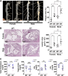
Beta thalassemia (BT) is a hemolytic anemia that causes accelerated atherosclerosis. A: representative aortic en face analysis of plaque accumulation (%) of total luminal area (n = 5–11). B: representative hematoxylin and eosin (H&E) staining of the aortic root and analysis of lesion area (µm2 × 103; n = 5–11). C: serum total cholesterol, triglycerides, LDL, and HDL (n = 10–12). Values represent means ± SE. *P < 0.05; **P < 0.01; ***P < 0.001.
Figure 2.
Figure 2.
Secondary model confirms accelerated atherosclerosis is present in beta thalassemia (BT). Short-term model of accelerated atherosclerosis with angiotensin II infusion, 1 mo high-fat diet (HFD), and proprotein convertase subtilisin/kexin type 9 (PCSK9) gain of function confirms accelerated plaque accumulation in BT mice. A: representative aortic en face analysis (n = 4). B: representative of severe aneurysm formation. C: percent survival in wild-type (WT) and BT mice. Values represent means ± SE. *P < 0.05; **P < 0.01.
Figure 3.
Figure 3.
Baseline hemopexin and free heme levels in beta thalassemia (BT) mice. A: baseline (normal chow diet) serum hemopexin (HPX) levels (n = 6). B: baseline (normal chow diet) serum free heme (n = 6). Values represent means ± SE. **P < 0.01; ***P < 0.001.
Figure 4.
Figure 4.
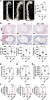
Adeno-associated virus (AAV) overexpression of hemopexin leads to decreased plaque accumulation in beta thalassemia (BT) mice. A: representative aortic en face images and analysis in wild-type (WT), BT, and BT + serum hemopexin (HPX) mice (n = 6–10). B: representative hematoxylin and eosin (H&E) and trichrome staining of the aortic root (n = 6–10). Lesion, necrotic, and collagen area (µm2 × 103) were analyzed. C: serum free heme levels at 1 mo (n = 5–7). D: serum hemopexin (HPX) levels (n = 6) at 3 mo. E: aortic hydrogen peroxide levels at 1 mo (n = 5–7). F: aortic gene expression of IL-6, HOX-1, TNF-α via RT-qPCR at 1 mo (n = 5–7). Values represent means ± SE. *P < 0.05; **P < 0.01; ***P < 0.001.
Figure 5.
Figure 5.
Control serum hemopexin (HPX) vector does not affect atherosclerotic plaque accumulation. A: representative aortic en face images analysis in wild-type (WT) and WT + HPX mice. B: means data (n = 4). Values represent means ± SE.
Figure 6.
Figure 6.
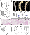
Phenylhydrazine (PHZ)-induced hemolysis increases atherosclerosis and is reversed by serum hemopexin (HPX) expression. A: serum hematocrit at 3 mo (n = 5–7). B: spleen weight at 3 mo (n = 5–7). C: representative aortic en face plaque accumulation in wild-type (WT), PHZ, and PHZ + HPX mice (n = 6–12). D: representative hematoxylin and eosin and trichrome staining of the aortic root (n = 6–10). E: quantitative analysis of aortic root lesion area (n = 6–11). F: serum free heme levels at 3 mo (n = 5–7). G: HPX levels (n = 5–6). Values represent means ± SE. *P < 0.05; **P < 0.01; ***P < 0.001.
Figure 7.
Figure 7.
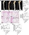
Deferiprone (DFP) decreases atherosclerotic plaque accumulation in beta thalassemia (BT) mice but does not provide an additive benefit over hemopexin therapy. A: representative aortic en face plaque accumulation in WT, BT, and BT + DFP, BT + DFP + HPX mice (n = 5–10). B: representative hematoxylin and eosin and trichrome staining of the aortic root (n = 5–10). C: total serum iron at 3 mo. D: non-transferrin-bound iron at 3 mo. Values represent means ± SE. *P < 0.05; **P < 0.01; ***P < 0.001.

References

    1. Origa R. beta-Thalassemia. Genet Med 19: 609–619, 2017. doi:10.1038/gim.2016.173. - DOI - PubMed
    1. Ansari-Moghaddam A, Adineh HA, Zareban I, Mohammadi M, Maghsoodlu M. The survival rate of patients with beta-thalassemia major and intermedia and its trends in recent years in Iran. Epidemiol Health 40: e2018048, 2018. doi:10.4178/epih.e2018048. - DOI - PMC - PubMed
    1. Borgna-Pignatti C, Cappellini MD, De Stefano P, Del Vecchio GC, Forni GL, Gamberini MR, Ghilardi R, Origa R, Piga A, Romeo MA, Zhao H, Cnaan A. Survival and complications in thalassemia. Ann N Y Acad Sci 1054: 40–47, 2005. doi:10.1196/annals.1345.006. - DOI - PubMed
    1. Morris CR. Mechanisms of vasculopathy in sickle cell disease and thalassemia. Hematology Am Soc Hematol Educ Program : 177–185, 2008. doi:10.1182/asheducation-2008.1.177. - DOI - PubMed
    1. Sherief LM, Dawood O, Ali A, Sherbiny HS, Kamal NM, Elshanshory M, Alazez OA, Alhady MA, Nour M, Mokhtar WA. Premature atherosclerosis in children with beta-thalassemia major: new diagnostic marker. BMC Pediatr 17: 69, 2017. doi:10.1186/s12887-017-0820-1. - DOI - PMC - PubMed

Publication types

MeSH terms

LinkOut - more resources