. 2023 Sep 19;57(37):13901-13911.
doi: 10.1021/acs.est.3c03778. Epub 2023 Sep 8.

Polyethylene Degradation by a Rhodococcous Strain Isolated from Naturally Weathered Plastic Waste Enrichment

Affiliations

Polyethylene Degradation by a Rhodococcous Strain Isolated from Naturally Weathered Plastic Waste Enrichment

Xuanyu Tao et al. Environ Sci Technol. .

Abstract

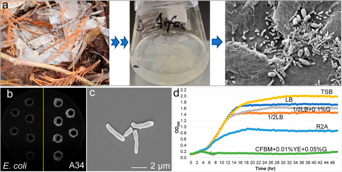
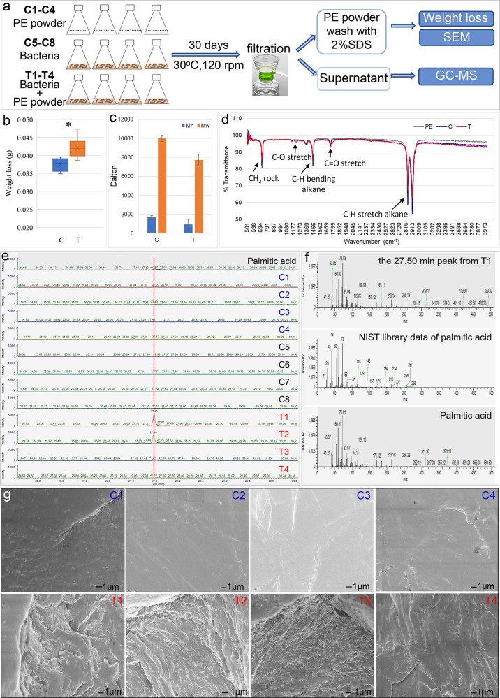
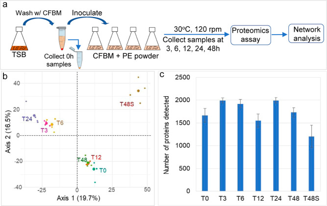
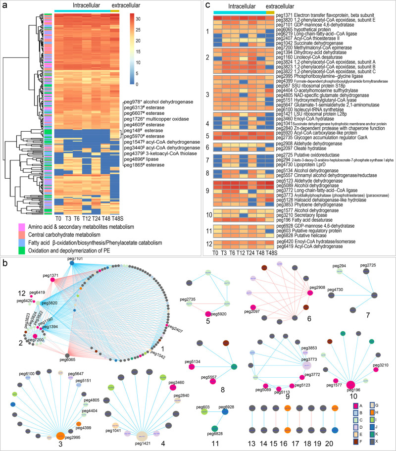
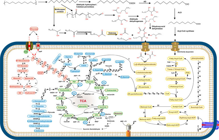
Polyethylene (PE) is the most widely produced synthetic polymer and the most abundant plastic waste worldwide due to its recalcitrance to biodegradation and low recycle rate. Microbial degradation of PE has been reported, but the underlying mechanisms are poorly understood. Here, we isolated a Rhodococcus strain A34 from 609 day enriched cultures derived from naturally weathered plastic waste and identified the potential key PE degradation enzymes. After 30 days incubation with A34, 1% weight loss was achieved. Decreased PE molecular weight, appearance of C-O and C═O on PE, palmitic acid in the culture supernatant, and pits on the PE surface were observed. Proteomics analysis identified multiple key PE oxidation and depolymerization enzymes including one multicopper oxidase, one lipase, six esterase, and a few lipid transporters. Network analysis of proteomics data demonstrated the close relationships between PE degradation and metabolisms of phenylacetate, amino acids, secondary metabolites, and tricarboxylic acid cycles. The metabolic roadmap generated here provides critical insights for optimization of plastic degradation condition and assembly of artificial microbial communities for efficient plastic degradation.

Keywords: Rhodococcus sp.; plastic-degrading enzyme; polyethylene biodegradation; proteomic network analysis; proteomics.


Conflict of interest statement

The authors declare no competing financial interest.

Figures

Figure 1
Figure 1
Isolation and physiological and morphological characterization of Rhodococcus strain A34. (a) Original plastic waste, enrichment culture, and the associated microbial communities on the plastic surface. (b) Lipase activity of strain A34. E. coli was used as negative control. (c) Morphology of A34 cells under a scanning electron microscope. (d) Growth of A34 in liquid media with various levels of nutrients.
Figure 2
Figure 2
Degradation of PE powder by Rhodococcus strain A34. (a) Experiment flowchart. PE weight loss and molecular-weight changes after 30 days of incubation are shown in (b,c), respectively. T, with A34 (T1–T4); C, without A34 (replicates C1–C4). (d) FTIR spectrum showed PE functional group changes in the T group compared to the C group and untreated PE. (e) Unique GC–MS peak was detected in the T group. (f) Unique GC–MS peak was identified as palmitic acid compared to the NIST library data and experimental data of standard palmitic acid. (g) PE surface changes after 30 days of incubation with A34 (T1–T4) or without A34 (C1–C4).
Figure 3
Figure 3
Proteins/enzymes responsive to the change of carbon source to PE powder in medium and the dynamic changes of proteomes over time. (a) General workflow of the time series proteomic incubation experiment. (b) PCA plot of the overall similarities of proteomes at different timepoints. (c) Numbers of proteins detected at each timepoints.
Figure 4
Figure 4
Temporal protein abundance (log 2 transformed) changes of A 34 are shown in the heatmaps [(a) *, PE oxidation; #, PE depolymerization; β, fatty acid β-oxidation]. (b) Networks generated from the temporal proteomics data were partitioned into 20 module. Positive or negative correlations between nodes were shown in blue or red, respectively. Color key for the nodes: A, oxidation of fatty acids or biosynthesis of fatty acids; B, metabolism of amino acid and secondary metabolites; C, metabolism of amino acids; D, metabolism of the secondary metabolites; E, TCA cycle; F, ABC transporter; G, protein degradation; H, purine metabolism; I, regulatory protein; J, sugar metabolism; K, DNA modification; and X, other pathways or hypothetical protein. (c) Protein abundance changes of the interested enzymes in each module (module # is shown in the left of the heatmap).
Figure 5
Figure 5
Conceptual model of metabolic pathways of A34 when grown in defined medium with PE as the carbon source. The pathways are illustrated based on the proteomics data, FTIR, and GC–MS data. Solid arrow represents one-step reaction; broken-line arrow represents the absence of the corresponding enzyme in proteomics data or unknown enzymes. The initial PE depolymerization step indicated by a broken-line arrow might involve multiple enzymes, such as multicopper oxidase, catalase peroxidase, esterase, lipase, and others. PE functional group changes were observed after incubation with crude enzymes of combination of a multicopper oxidase peg 1726 and an esterase peg 6607 (Figure S7). The orange frame highlighted the two types of extracellular key PE degradation enzymes. Color key for intracellular pathways: light brown, fatty acid β-oxidation; fatty acid biosynthesis, phenylacetate metabolism, biosynthesis of pantothenate, and CoA; light green, TCA cycle and the associated pyruvate cycle, glyoxylate cycle, and GABA skanehunt; light red, glycolysis, glycogenolysis, and pentose phosphate pathway; and light blue, biosynthesis of amino acids and secondary metabolites. PE oxidation and depolymerization enzymes: alkane hydroxylase* (genes exist but protein not detected); AD, alcohol dehydrogenase; ALD, aldehyde dehydrogenase; enzymes in fatty acid β-oxidation: 1, acyl-CoA oxidase or acyl-CoA dehydrogenase; 2, enoyl-CoA dehydratase; 3, 3-hydroxyacyl-CoAcdehydrogenase; and 4, β-ketoacyl-CoA thiolase. Enzymes in biosynthesis of pantothenate and CoA: 5, dihydroxy-acid dehydratase; 6, valinepyruvate aminotransferase; 7, ketopantoate hydroxymethyltransferase; 8, ketopantoate reductase PanG or ketol-acid reductoisomerase [NADP(+)]; 9, pantothenate synthetase; 10, pantothenate kinase; 11, phosphopantothenoylcysteine synthetase; 12, phosphopantothenoylcysteine decarboxylase; 13, phosphopantetheine adenylyltransferase; and 14, dephospho-CoA kinase. Enzymes in phenylacetate metabolism: 15, phenylacetate-coenzyme A ligase; 16, 1,2-phenylacetyl-CoA epoxidase; 17, 1,2-epoxyphenylacetyl-CoA isomerase; 18, 2-oxepin-2(3H)-ylideneacetyl-CoA hydrolase; 19, 3-oxo-5,6-dehydrosuberyl-CoA semialdehyde dehydrogenase; 20, 3-hydroxyacyl-CoA dehydrogenase; 21, 3-oxoadipyl-CoA those. Enzymes in fatty acid biosynthesis: 22, acetyl-coenzyme A carboxylase; 23, malonyl CoA-ACP acyltransferase; 24, 3-oxoacyl-ACP synthase of FASI, KASI, or KASII; 25, 3-oxoacyl-ACP reductase; 26, β-hydroxyacyl-ACP dehydratase; and 27, enoyl-ACP reductase. Enzymes in PHA biosynthesis: PhaC, PHA synthase. Enzymes in the TCA cycle and associated pyruvate cycle, glyoxylate cycle, and GABA shunt: PDH, pyruvate dehydrogenase; CS, citrate synthase; ACO, aconitate hydratase; ICDH, isocitrate dehydrogenase; OGDH, 2-oxoglutarate dehydrogenase; SCS, succinyl coenzyme A synthetase; SDH, succinate dehydrogenase; FUM, fumarate hydratase; MDH, malate dehydrogenase; PEPCK, phosphoenolpyruvate carboxykinase; PEPC, phosphoenolpyruvate carboxylase; PK, pyruvate kinase; MAE, NAD-dependent malic enzyme; ICL, isocitrate lyase; MS, malate synthase G; GOGAT, glutamate synthase; GDC, glutamate decarboxylase; GAT, GABA aminotransferase; and SSADH, succinic semialdehyde dehydrogenase. Enzyme in glycolysis, gluconeogenesis, and the pentose phosphate pathway: 28, glucose-6-phosphate isomerase; 29, 6-phosphofructokinase; 30, fructose-bisphosphate aldolase; 31, triosephosphate isomerase; 32, glyceraldehyde-3-phosphate ketol-isomerase; 33, phosphoglycerate kinase; 34, phosphoglycerate mutase; 35, enolase; 36, fructose-1,6-bisphosphatase; 37, glucose-6-phosphate dehydrogenase; 38, 6-phosphogluconolactonase; 39, 6-phosphogluconate dehydrogenase; 40, ribose-5-phosphate isomerase; and 41, transketolase. Enzymes in biosynthesis of amino acids: l-threonine, l-isoleucine, l-valine, and leucine: AST, aspartate aminotransferase; AK, aspartate kinase; ASD, aspartate-semialdehyde dehydrogenase; HD, homoserine dehydrogenase; HK, homoserine kinase; TS, threonine synthase; TD, threonine dehydratase; AHAS, acetolactate synthase; AHAIR, acetohydroxy acid isomeroreductase; DHAD, dihydroxy-acid dehydratase; TA, branched-chain amino acid aminotransferase; IPMS, isopropylmalate synthase; IPMI, α-isopropylmalate isomerase; IPMD, α-isopropylmalate dehydrogenase; tryptophan: 42, anthranilate synthase; 43, anthranilate phosphoribosyltransferase; 44, phosphoribosylanthranilate isomerase 45, tryptophan synthase; l-serine, glycine, and l-cysteine: 46, d-3-phosphoglycerate dehydrogenase; 47, phosphoserine aminotransferase, 48, phosphoserine phosphatase; 49, serine hydroxymethyltransferase; 50, serine acetyltransferase; 51, cysteine synthase; l-lysine: 52, aspartokinase; 53, aspartate-semialdehyde dehydrogenase; 54, 4-hydroxy-tetrahydrodipicolinate synthase; 55, 4-hydroxy-tetrahydrodipicolinate reductase; 56, 2,3,4,5-tetrahydropyridine-2,6-dicarboxylate N-succinyltransferase; 57, N-succinyl-l,l-diaminopimelate aminotransferase; 58, N-succinyl-l,l-diaminopimelate desuccinylase; 59, diaminopimelate epimerase; 60, diaminopimelate decarboxylase; l-valine, 61, alanine dehydrogenase; 62, alanine transaminase; and 63, alanine racemase.

References

    1. Ru J.; Huo Y.; Yang Y. Microbial Degradation and Valorization of Plastic Wastes. Front. Microbiol. 2020, 11, 442. 10.3389/fmicb.2020.00442. - DOI - PMC - PubMed
    1. Ghatge S.; Yang Y.; Ahn J.-H.; Hur H.-G. Biodegradation of polyethylene: a brief review. Appl. Biol. Chem. 2020, 63, 27. 10.1186/s13765-020-00511-3. - DOI
    1. Liu J.; Xu G.; Zhao S.; Chen C.; Rogers M. J.; He J. Mechanistic and microbial ecological insights into the impacts of micro-and nano-plastics on microbial reductive dehalogenation of organohalide pollutants. J. Hazard. Mater. 2023, 448, 130895. 10.1016/j.jhazmat.2023.130895. - DOI - PubMed
    1. Yoshida S.; Hiraga K.; Takehana T.; Taniguchi I.; Yamaji H.; Maeda Y.; Toyohara K.; Miyamoto K.; Kimura Y.; Oda K. A bacterium that degrades and assimilates poly(ethylene terephthalate). Science 2016, 351, 1196–1199. 10.1126/science.aad6359. - DOI - PubMed
    1. Austin H. P.; Allen M. D.; Donohoe B. S.; Rorrer N. A.; Kearns F. L.; Silveira R. L.; Pollard B. C.; Dominick G.; Duman R.; El Omari K.; et al. Characterization and engineering of a plastic-degrading aromatic polyesterase. Proc. Natl. Acad. Sci. U.S.A. 2018, 115, E4350–E4357. 10.1073/pnas.1718804115. - DOI - PMC - PubMed
    2. Lu H.; Diaz D. J.; Czarnecki N. J.; Zhu C.; Kim W.; Shroff R.; Acosta D. J.; Alexander B. R.; Cole H. O.; Zhang Y.; et al. Machine learning-aided engineering of hydrolases for PET depolymerization. Nature 2022, 604, 662–667. 10.1038/s41586-022-04599-z. - DOI - PubMed

Publication types