. 2023 Oct:96:104781.
doi: 10.1016/j.ebiom.2023.104781. Epub 2023 Sep 7.

Biomarker-based risk prediction for the onset of neuroinflammation in X-linked adrenoleukodystrophy

Affiliations

Biomarker-based risk prediction for the onset of neuroinflammation in X-linked adrenoleukodystrophy

Isabelle Weinhofer et al. EBioMedicine. 2023 Oct.

Abstract

Background: X-linked adrenoleukodystrophy (X-ALD) is highly variable, ranging from slowly progressive adrenomyeloneuropathy to severe brain demyelination and inflammation (cerebral ALD, CALD) affecting males with childhood peak onset. Risk models integrating blood-based biomarkers to indicate CALD onset, enabling timely interventions, are lacking. Therefore, we evaluated the prognostic value of blood biomarkers in addition to current neuroimaging predictors for early detection of CALD.

Methods: We measured blood biomarkers in a retrospective, male CALD risk-assessment cohort consisting of 134 X-ALD patients and 66 controls and in a phenotype-blinded validation set (25 X-ALD boys, 4-13 years) using Simoa®and Luminex® technologies.

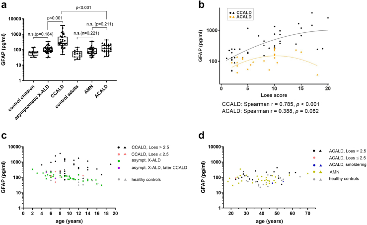
Findings: Among 25 biomarkers indicating axonal damage, astrocye/microglia activation, or immune-cell recruitment, neurofilament light chain (NfL) had the highest prognostic value for early indication of childhood/adolescent CALD. A plasma NfL cut-off level of 8.33 pg/mL, determined in the assessment cohort, correctly discriminated CALD with an accuracy of 96% [95% CI: 80-100] in the validation group. Multivariable logistic regression models revealed that combining NfL with GFAP or cytokines/chemokines (IL-15, IL-12p40, CXCL8, CCL11, CCL22, and IL-4) that were significantly elevated in CALD vs healthy controls had no additional benefit for detecting neuroinflammation. Some cytokines/chemokines were elevated only in childhood/adolescent CALD and already upregulated in asymptomatic X-ALD children (IL-15, IL-12p40, and CCL7). In adults, NfL levels distinguished CALD but were lower than in childhood/adolescent CALD patients with similar (MRI) lesion severity. Blood GFAP did not differentiate CALD from non-inflammatory X-ALD.

Interpretation: Biomarker-based risk prediction with a plasma NfL cut-off value of 8.33 pg/mL, determined by ROC analysis, indicates CALD onset with high sensitivity and specificity in childhood X-ALD patients. A specific pro-inflammatory cytokine/chemokine profile in asymptomatic X-ALD boys may indicate a primed, immanent inflammatory state aligning with peak onset of CALD. Age-related differences in biomarker levels in adult vs childhood CALD patients warrants caution in predicting onset and progression of CALD in adults. Further evaluations are needed to assess clinical utility of the NfL cut-off for risk prognosis of CALD onset.

Funding: Austrian Science Fund, European Leukodystrophy Association.

Keywords: Biomarker; Cytokines; GFAP; Neurodegeneration; Neurofilament light chain; X-ALD.


Conflict of interest statement

Declaration of interests MP received support from Amicus, Merck, Novartis and Sanofi-Genzyme; BZ received support from ACTRIMS 2022 and 2023 endMS SPRINT; JG received support from Quanterix; HAY was supported by an emerging investigator grant from ALD connect; CGB received grants from the German Research Foundation and the Ministry for Science and Culture of Lower Saxony; ME received support from Minoryx and is member of the advisory board of Minoryx, Poxel and SwanBio Therapeutics; FE is holding a license for “Intrathecal delivery of nucleic acid sequences encoding ABCD1 for treatment of Adrenomyeloneuropathy” (NO. 29539-021PCT), received consulting fees from SwanBio Therapeutics and UpToDate, is founder of SwanBio Therapeutics, ALD Connect and organizer of trial sites for ASPA, Bluebird Bio Therapeutics, Ionis Pharmaceuticals and Sanofi; AP received consulting fees from Swanbio Therapeutics and Sanofi and is member of the Advisory Board of Bluebird Bio Therapeutics and MedDay Therapeutics. JSK is member of the advisory board for Krabbe Disease of PassageBio. MCR received a grant from Novartis. EM has received funding from the National Institutes of Health (K23NS118044). All remaining authors declare no competing interests.

Figures

Fig. 1
Fig. 1
NfL has a higher capacity to discriminate onset of CALD in children than in adults with X-ALD. (a) NfL in plasma and serum samples of childhood/adolescent X-ALD patients (asymptomatic X-ALD, n = 20, median age = 9.4 years, total sample number = 40; inflammatory CALD [CCALD], n = 41, median age = 9 years, total sample number = 45), adult X-ALD patients (non-inflammatory AMN, n = 58, median age = 39 years, total sample number = 84; adult CALD [ACALD], n = 22, median age = 37 years, total sample number = 25) and healthy controls (childhood/adolescents: n = 15, with additional NfL levels of 138 boys measured using the same methodology and described in, median age = 11 years, total sample number = 153; adults: n = 49, median age = 39 years, total sample number = 49). The data are depicted as boxplots (median ± IQR). Total sample numbers include samples collected longitudinally for some patients during disease progression. Comparison of log (NfL) levels was done using a linear mixed model adjusted for sample type (serum vs plasma) with the addition of a random ID factor to account for the longitudinal sampling. Multiple testing was corrected by the Bonferroni-Holm method. Adjustment of the group comparisons for age differences did not change the significance or the reported p-values. (b) Comparison of MRI severity (Loes score, indicating location and activity of the inflammatory myelin destruction) in groups of CCALD (n = 38, median age = 9 years, total sample number = 40) and adult ACALD (n = 19, median age = 37 years, total sample number = 22) patients. The data are depicted as boxplots (median ± IQR). (c) Association between plasma/serum NfL levels and Loes scores in CCALD (n = 38, median age = 6 years, total sample number = 42, black symbols) and ACALD patients (n = 22, median age = 37 years, total sample number = 24, orange symbols). The Linear mixed model included a fixed factor for sample type and a random ID factor as well as a linear and quadratic term for the Loes score. (de) Association of NfL and age in samples from (d) CCALD (Loes >2.5, black symbols; early onset CCALD, Loes ≤2.5, pink symbols) and asymptomatic X-ALD patients (asympt. X-ALD, dark green symbols; patients who later converted to CALD, lilac symbols). For curve fitting using linear regression including a quadratic term of log (NfL) on age in healthy control children/adolescents, data described by was used. The cut-off value of 8.33 pg/mL to discriminate plasma samples of CCALD from asymtomatic X-ALD children is indicated by a dotted line. 1Asymptomatic X-ALD patient 1 (asympt. X-ALD 1). (e) Association of NfL and age in samples from ACALD (Loes >2.5, black symbols; early onset ACALD, Loes ≤2.5, pink symbols; smouldering ACALD, blue symbols), non-inflammatory AMN, light green symbols, and adult healthy controls (grey symbols). Serum samples are indicated by triangles and plasma samples by circles.
Fig. 2
Fig. 2
Blood GFAP indicative of astrocyte damage is associated with CALD only in childhood/adolescent X-ALD. (a) GFAP levels in samples of childhood/adolescent X-ALD patients (asymptomatic X-ALD, n = 20, median age = 9.4 years, total sample number = 40; CCALD, n = 38, median age = 9 years, total sample number = 42), adult X-ALD patients (non-inflammatory AMN, n = 23, median age = 40 years, total sample number = 27; ACALD, n = 22, median age = 37 years, total sample number = 25), and healthy controls (childhood/adolescents: n = 15, median age = 12 years, total sample number = 15; adults: n = 14, median age = 42 years, total sample number = 14). The data are depicted as boxplots (median ± IQR). Total sample numbers include samples collected longitudinally from some patients during disease progression. Comparison of log (GFAP) levels was done using a linear mixed model adjusted for sample type (serum vs plasma) with the addition of a random ID factor to account for the longitudinal sampling. Multiple testing was corrected by the Bonferroni-Holm method. Adjustment of the group comparisons for age differences did not change the significance or the reported p-values. (b) Association between plasma/serum GFAP levels and brain lesion severity (Loes score) in CCALD (n = 38, median age = 6 years, total sample number = 42, black symbols) and ACALD patients (n = 22, median age = 37 years, total sample number = 24, orange symbols). The Linear mixed model included a fixed factor for sample type and a random ID factor as well as a linear and quadratic term for the Loes score. (cd) Association of GFAP and age in samples from (c) CCALD (Loes >2.5, black symbols; early onset CALD, Loes ≤2.5, pink symbols) and asymptomatic X-ALD patients (asympt. X-ALD, dark green symbols; patients who later converted to CALD, lilac symbols; 1Asymptomatic X-ALD patient 1, asympt. X-ALD 1) and (d) ACALD (Loes >2.5, black symbols; early stage ACALD, Loes ≤2.5, pink symbols; smouldering ACALD, blue symbols), non-inflammatory AMN (light green symbols), and adult healthy controls (grey symbols). Serum samples are indicated by triangles and plasma samples by circles (a–d).
Fig. 3
Fig. 3
Increased blood IL-15, CCL7 and IL-12p40 levels indicate an X-ALD immanent pro-inflammatory status in childhood that does not persist in adulthood. Plasma and serum samples derived from childhood/adolescent X-ALD patients (asymptomatic X-ALD, n = 20, median age = 9 years, total sample number = 21; inflammatory CALD, n = 37, median age = 9 years, total sample number = 41), adult X-ALD patients (non-inflammatory AMN, n = 24, median age = 40 years, total sample number = 27; adult CALD, n = 24, median age = 40 years, total sample number = 24), and healthy controls (childhood/adolescents: n = 16, median age = 12 years, total sample number = 16; adults: n = 14, median age = 42 years, total sample number = 14) were used to determine blood levels of and their association with Loes scores for MRI-severity for (a) IL-15, (b) CCL7, (c) IL-12p40, (d) CXCL8, (e) CCL4, and (f) CCL11 (linear mixed models). The median is indicated by a horizontal line in the dot plots. Total sample numbers include several samples collected longitudinally from the same patients during disease progression. Reported Spearman's r have been partialized for the sample type serum or plasma. Serum samples are indicated by grey triangles and plasma samples by black circles. Childhood X-ALD patients before and after conversion to CALD are indicated by lilac circles. The number of data points that were below the detection limit are indicated in brackets on the x-axis above the respective group.
Fig. 4
Fig. 4
Evaluation of plasma NfL to indicate cerebral involvement in X-ALD patients independent of clinical neuroimaging-based assessment. (a) Association of plasma NfL and age in samples from the development cohort of childhood/adolescent X-ALD patients (CALD, n = 38, median age = 9 years, total sample number = 40, black symbols; and asymptomatic X-ALD, n = 20, median age = 9.4 years, total sample number = 40, green symbols; asymptomatic X-ALD patients who later converted to CALD, lilac symbols) and the phenotype-blinded childhood/adolescent validation cohort (n = 25, median age = 6 years, total sample number = 25). (b) Differentiation of CALD onset in the validation cohort based on the plasma NfL cut-off level of 8.33 pg/mL. Blue symbols indicate correctly and red incorrectly assigned phenotypes. Numbers in brackets indicate the MRI brain lesion severity (Loes score), disclosed after NfL–based designations.

References

    1. Turk B.R., Theda C., Fatemi A., Moser A.B. X-linked adrenoleukodystrophy: pathology, pathophysiology, diagnostic testing, newborn screening and therapies. Int J Dev Neurosci. 2020;80(1):52–72. - PMC - PubMed
    1. Mosser J., Douar A.M., Sarde C.O., et al. Putative X-linked adrenoleukodystrophy gene shares unexpected homology with ABC transporters. Nature. 1993;361(6414):726–730. - PubMed
    1. Weinhofer I., Zierfuss B., Hametner S., et al. Impaired plasticity of macrophages in X-linked adrenoleukodystrophy. Brain. 2018;141:2329–2342. - PMC - PubMed
    1. Zierfuss B., Buda A., Villoria-Gonzalez A., et al. Saturated very long-chain fatty acids regulate macrophage plasticity and invasiveness. J Neuroinflammation. 2022;19(1):305. - PMC - PubMed
    1. Weinhofer I., Rommer P., Zierfuss B., et al. Neurofilament light chain as a potential biomarker for monitoring neurodegeneration in X-linked adrenoleukodystrophy. Nat Commun. 2021;12(1):1816. - PMC - PubMed