. 2023 Dec 1:240:109705.
doi: 10.1016/j.neuropharm.2023.109705. Epub 2023 Sep 6.

Volatile anesthetics inhibit presynaptic cGMP signaling to depress presynaptic excitability in rat hippocampal neurons

Affiliations

Volatile anesthetics inhibit presynaptic cGMP signaling to depress presynaptic excitability in rat hippocampal neurons

Iris Speigel et al. Neuropharmacology. .

Abstract

Volatile anesthetics alter presynaptic function through effects on Ca2+ influx and neurotransmitter release. These actions are proposed to play important roles in their pleiotropic neurophysiological effects including immobility, unconsciousness and amnesia. Nitric oxide and cyclic guanosine monophosphate (NO/cGMP) signaling has been implicated in presynaptic mechanisms, and disruption of NO/cGMP signaling has been shown to alter sensitivity to volatile anesthetics in vivo. We investigated volatile anesthetic actions NO/cGMP signaling in relation to presynaptic function in cultured rat hippocampal neurons using pharmacological tools and genetically encoded biosensors and sequestering probes of cGMP levels. Using the fluorescent cGMP biosensor cGull, we found that electrical stimulation-evoked NMDA-type glutamate receptor-independent presynaptic cGMP transients were inhibited 33.2% by isoflurane (0.51 mM) and 26.4% by sevoflurane (0.57 mM) (p < 0.0001) compared to control stimulation without anesthetic. Stimulation-evoked cGMP transients were blocked by the nonselective inhibitor of nitric oxide synthase N-ω-nitro-l-arginine, but not by the selective neuronal nitric oxide synthase inhibitor N5-(1-imino-3-butenyl)-l-ornithine. Isoflurane and sevoflurane inhibition of stimulation-evoked increases in presynaptic Ca2+ concentration, measured with synaptophysin-GCaMP6f, and of synaptic vesicle exocytosis, measured with synaptophysin-pHlourin, was attenuated in neurons expressing the cGMP scavenger protein sponge (inhibition of exocytosis reduced by 54% for isoflurane and by 53% for sevoflurane). The anesthetic-induced reduction in presynaptic excitability was partially occluded by inhibition of HCN channels, a cGMP-modulated excitatory ion channel that can facilitate glutamate release. We propose that volatile anesthetics depress presynaptic cGMP signaling and downstream effectors like HCN channels that are essential to presynaptic function and excitability. These findings identify novel mechanisms by which volatile anesthetics depress synaptic transmission via second messenger signaling involving the NO/cGMP pathway in hippocampal neurons.

Keywords: Cyclic GMP; HCN channel; Isoflurane; Mechanisms of anesthesia; Neurotransmitter release; Nitric oxide; Sevoflurane; Synaptic transmission.


Conflict of interest statement

Declaration of competing interest Funding for the project was provided by USNational Institutes of Health Grant R01 GM058055-21 (to HCH) and F31 GM133115 (to VO). IAS, KP, and VO have no competing interests to declare. HCH is editor-in-chief of the British Journal of Anesthesia and has received consulting fees from Elsevier unrelated to this work.

Figures

Figure 1
Figure 1. cGull reports basal and electrically-stimulated cGMP concentrations in hippocampal presynaptic boutons.
(A) At rest, cGull fluorescence is evident in somata and proximal dendrites (rest, left), and increases in all compartments following electrical stimulation (20 AP, middle), especially within presynaptic boutons co-expressing the presynaptic marker VAMP-mCherry (right). (B) Presynaptic boutons expressing VAMP-mCherry (VAMP) and cGull (far left and left), kymograph of evoked cGull fluorescence changes (middle), and regions of interest (ROIs) drawn over responsive boutons (right). (C) Boutons from one cell analyzed together as an ensemble averaged time series. Peak amplitude: ΔF= Fmax-F0. Traces show mean over time per cell. (D) Representative cGMP transients from boutons measured using cGull compared to signals from the soma. (E) Transient waveform is slowed and prolonged in the presence of the phosphodiesterase inhibitor IBMX. (F) Presynaptic cGull transient peak amplitudes increase with increases in external Ca2+ concentration. Each line represents data from a single cell (n=5). (G) Presynaptic cGMP transients are resistant to the selective nNOS inhibitor L-VNIO, and completely abolished by the combined eNOS/nNOS inhibitor L-NNA. Calculated as percent of a control stimulation: control= 97.2 ± 8.9%, n=6; L-VNIO = 88.6 ± 6.3%, n= 5, L-NNA = 0.64 ± 0.60%, n=4. One-way ANOVA F(2, 12)= 272.3, p<0.0001 with Tukey’s post hoc test: control vs L-VNIO p=0.135, 95%CI [−2.41, 19.65], control vs L-NNA p<0.0001, 95%CI [84.8,108.3]; L-VNIO vs L-NNA p<0.0001, 95%CI [75.8, 100.2]. ns, not significant; ****, p<0.0001.
Figure 2:
Figure 2:. Volatile anesthetics inhibit stimulus-evoked presynaptic cGMP increases.
(A) Live-cell imaging and drug perfusion paradigm. (B) Representative traces of presynaptic cGull transients in the presence of isoflurane (0.51 ± 0.09 mM) or sevoflurane (0.57 ± 0.13 mM) compared to control without volatile anesthetic. (C) Isoflurane or sevoflurane inhibits the evoked increase in presynaptic cGMP. Peak cGMP transient amplitude was reduced by each anesthetic compared to stimulation without anesthetic. Calculated as a percentage of control stimulation: no anesthetic=103.6 ± 8.8%, n=11; isoflurane=69.2 ± 11.3%, n=13, sevoflurane = 76.2 ± 10.2%, n=6. One-way ANOVA F(2, 27) = 35.3, p<0.0001, with Tukey’s post hoc test: no anesthetic vs isoflurane p<0.0001, 95%CI [23.9, 44.7], no anesthetic vs sevoflurane p<0.0001, 95%CI [14.5, 40.3]; isoflurane vs sevoflurane p=0.373, 95%CI [−19.4, −5.62]. *, p<0.05.
Figure 3:
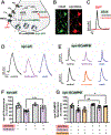
Figure 3:. Sequestration of cGMP by sponGee blunts volatile anesthetic inhibition of presynaptic function.
(A) Schematic diagram showing probes used to investigate anesthetic effects on cGMP generation, Ca2+ influx, and synaptic vesicle (SV) exocytosis. NMDA receptors are blocked by APV included in the perfusion buffers to prevent recurrent excitation. NOS is shown in the bouton, but might also function as a retrograde messenger from postsynaptic neurons. Abbreviations: Cav (voltage-gated Ca2+ channel), cGMP (cyclic guanosine monophosphate), eNOS and nNOS (endothelial and neuronal nitric oxide synthase), Nav (voltage-gated Na+ channel), NMDA (N-methyl-D-aspartate), NO (nitric oxide), sGC (soluble gyanylyl cyclase), SV (synaptic vesicle), syn-GCaMP6f (synaptophysin-GCaMP6f), syn-pH (synaptophysin-pHluorin). (B) Overlapping red and green somatic fluorescence confirms sponGee-mRFP and cGull co-expression. (C) Co-transfection of cGull with sponGee blocks activity-dependent cGMP increase with electrical stimulation. (D) Isoflurane reversibly inhibits (by −31.7%) synaptic vesicle exocytosis evoked by electrical stimulation as shown by representative traces from a syn-pH transfected neuron. (E) Isoflurane and sevoflurane reversibly inhibit presynaptic Ca2+ entry as shown by representative traces from syn-GCaMP6f transfected neurons (−23.3 and −21.9%, respectively). (F) Isoflurane inhibition of synaptic vesicle exocytosis is reduced by co-expression of syn-pH with the cGMP binding protein sponGee. Calculated as % control stimulation (left-right): syn-pH, 92.9 ± 10.6%, n=8; syn-pH plus isoflurane, 64.3 ± 7.1%, n=11; syn-pH and sponGee, 99.0 ± 7.2%, n=6; syn-pH and sponGee, plus isoflurane, 85.4 ± 11.1%. Two-way ANOVA F(3,14)=12.87, p<0.0001, with Tukey’s post hoc test: syn-pH plus isoflurane vs syn-pH and sponGee plus isoflurane p=0.0004, 95%CI [13.33, 43.57]. (G) Inhibition by isoflurane (or sevoflurane; data not shown) of presynaptic Ca2+ influx was reduced by co-expressing syn-GCaMP6f with sponGee. Calculated as % control stimulation (left-right): syn-GCaMP6f = 92.4 ± 8.0%, n=8; syn-GCaMP6f plus isoflurane 71.8 ± 10.4%, n=11; syn-GCaMP6f plus sevoflurane 72.9 ± 12.4%, n=5; syn-GCaMP6f and sponGee 99.7 ± 4.5%, n=10; syn-GCaMP6f and sponGee plus isoflurane 86.3 ± 8.0%, n=11; syn-GCaMP6f and sponGee plus sevoflurane 89.9 ± 3.0%, n=7. Two-way ANOVA F(5,33)=14.99, p<0.0001: Isoflurane: syn-GCaMP6f vs syn-GCaMP6f plus sponGee; p=0.0020, 95%CI [4.74, 27.86]. Sevoflurane: syn-GCaMP6f vs syn-GCaMP6f plus sponGee p=0.0311, 95%CI [−30.45, −0.98]. *, p<0.05; **, p<0.01; ***, p<0.001.
Figure 4:
Figure 4:. Isoflurane inhibits presynaptic Ca2+ influx through cGMP modulation of HCN channels independent of cGMP-dependent protein kinase.
(A) Live-cell imaging paradigm to identify the contribution of cGMP-dependent protein kinase (PKG) to isoflurane actions using the selective PKG inhibitor KT-5823. (B) Live-cell imaging paradigm to identify the contribution of HCN channels to isoflurane actions, using the HCN inhibitor ivabradine. (C) KT-5823 had no significant effect on isoflurane inhibition of presynaptic Ca2+ influx, while ivabradine diminished isoflurane inhibition of presynaptic Ca2+ influx Calculated as % control stimulation (left-right): control (no isoflurane), 92.4 ± 8.0%, n=8; 0.51 ± 0.09 mM isoflurane, 71.8 ± 10.4%, n=11; 500 nM KT-5823, no isoflurane, 102.3 ± 8.4%, n=7; KT-5823 plus isoflurane, 75.7 ± 7.2%, n=10; 30 μM ivabradine, no isoflurane, 97.6 ± 6.9%, n=10; ivabradine plus isoflurane, 84.6 ± 10.8%, n=14. Two-way ANOVA, F(5,41)=18.76, with Tukey’s post hoc test: no isoflurane vs isoflurane, p=0.00002, 95%CI [10.54, 35.74]; isoflurane vs isoflurane plus KT-5823, p=0.732, 95%CI [−16.6, 6.72]; no isoflurane plus ivabradine vs isoflurane plus ivabradine, p=0.03, 95%CI [0.61, 23.3]; isoflurane vs isoflurane plus ivabradine, p=0.03, 95% CI [−25.4, −3.6]. ns, not significant; *, p<0.05; **, p<0.01; ****, p<0.0001.

Similar articles

Cited by

References

    1. Kim SH, Ryan TA. Balance of Calcineurin Aα and CDK5 Activities Sets Release Probability at Nerve Terminals. The Journal of Neuroscience 2013; 33: 8937–50 - PMC - PubMed
    1. Hemmings HC Jr., Akabas MH, Goldstein PA, Trudell JR, Orser BA, Harrison NL. Emerging molecular mechanisms of general anesthetic action. Trends Pharmacol Sci 2005; 26: 503–10 - PubMed
    1. Baumgart JP, Zhou ZY, Hara M, et al. Isoflurane inhibits synaptic vesicle exocytosis through reduced Ca2+ influx, not Ca2+-exocytosis coupling. Proc Natl Acad Sci U S A 2015; 112: 11959–64 - PMC - PubMed
    1. MacIver MB. Anesthetic agent-specific effects on synaptic inhibition. Anesth Analg 2014; 119: 558–69 - PMC - PubMed
    1. Study RE. Isoflurane inhibits multiple voltage-gated calcium currents in hippocampal pyramidal neurons. Anesthesiology 1994; 81: 104–16 - PubMed