Case Reports
. 2023 Sep;51(9):3000605231193929.
doi: 10.1177/03000605231193929.

Low-grade malignant myofibroblastic sarcoma of the larynx: a case report

Affiliations
Case Reports

Low-grade malignant myofibroblastic sarcoma of the larynx: a case report

Yu Feng et al. J Int Med Res. 2023 Sep.

Abstract

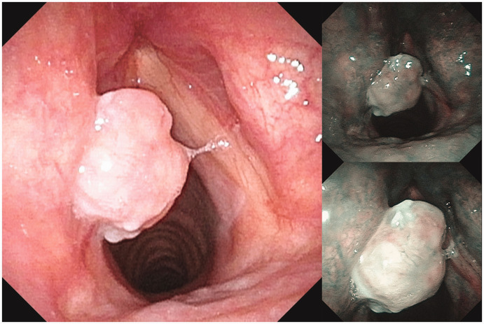
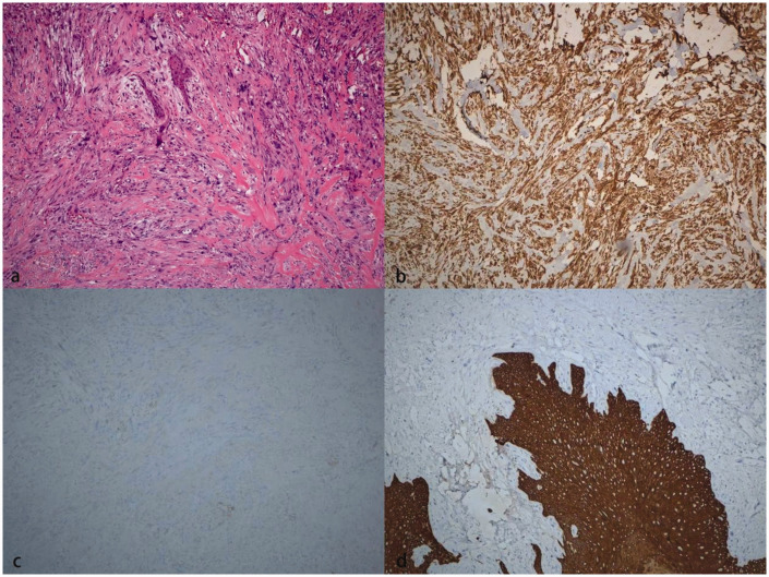
Low-grade myofibroblastic sarcoma (LGMS) is a rare malignant mesenchymal tumor derived from myofibroblasts. It is commonly identified in the head and neck, and particularly in the oral cavity, but rarely in the larynx. In this case report, we describe a patient who presented with hoarseness and underwent electronic fiber laryngoscopy, which revealed a neoplasm on the surface of his left vocal cord. The vocal cord tumor was resected under general anesthesia, and a malignant LGMS was diagnosed on postoperative pathologic examination. The results of immunohistochemical staining of the sections for vimentin (diffuse +), actin (partial +), and desmin (-) were consistent with this diagnosis. The patient recovered well after the surgery, and there was no recurrence of the neoplasm.

Keywords: Low-grade myofibroblastic sarcoma; case report; laryngeal tumor; malignancy; sarcoma; surgery; vocal cord.


Conflict of interest statement

The authors declare that there is no conflict of interest.

Figures

Figure 1.
Figure 1.
Laryngoscopic image, showing a pink neoplasm in the middle and posterior thirds of the left vocal cord. Thick submucosal blood vessels were visible on the surface of the neoplasm on narrow-band imaging, and no abnormal intrapapillary capillary loop dilatation was observed.
Figure 2.
Figure 2.
A 5-mm nodular shadow was visible in the left glottic region on transverse computed tomography. The shapes of the bilateral pre-epiglottic space and epiglottic vallecula were acceptable and all the laryngeal cartilages were normal.
Figure 3.
Figure 3.
Results of the pathologic examination of the resected tumor. (a) Hematoxylin and eosin-stained section, showing spindle-shaped tumor cells at ×100 magnification, some of which have prominent cell atypia, with scattered dividing nuclei. Interstitial fibers are stained red. (b) Immunohistochemical staining of a section for vimentin, showing strong, diffuse expression of vimentin. (c) Immunohistochemical staining of a section for actin, showing weak expression in some tumor cells and (d) Immunohistochemical staining of a section for cytokeratin, showing no expression in tumor cells. Normal squamous epithelium acts as the positive control.

Similar articles

References

    1. Jemal A, Siegel R, Ward E, et al.. Cancer statistics, 2009. CA Cancer J Clin 2009; 59: 225–249. doi: 10.3322/caac.20006. PMID: 19474385. - PubMed
    1. Velez Torres JM, Martinez Duarte E, Diaz-Perez JA, et al.. Primary sarcomas of the larynx: a clinicopathologic study of 27 cases. Head Neck Pathol 2021; 15: 905–916. doi: 10.1007/s12105-021-01314-8. PMID: 33686585; PMCID: PMC8384992. - PMC - PubMed
    1. Astl J, Holy R, Tuckova I, et al.. Sarcomas of the larynx: one institution’s experience and treatment protocol analyses. Medicina (Kaunas) 2021; 57: 192. doi: 10.3390/medicina57030192. PMID: 33668739; PMCID: PMC7996352. - PMC - PubMed
    1. Mentzel T, Dry S, Katenkamp D, et al.. Low-grade myofibroblastic sarcoma: analysis of 18 cases in the spectrum of myofibroblastic tumors. Am J Surg Pathol 1998; 22: 1228–1238. doi: 10.1097/00000478-199810000-00008. PMID: 9777985. - PubMed
    1. Li J, Huang XY, Zhang B. Low-grade myofibroblastic sarcoma of the liver misdiagnosed as cystadenoma: a case report. World J Gastroenterol 2022; 28: 4456–4462. doi: 10.3748/wjg.v28.i31.4456. PMID: 36159015; PMCID: PMC9453762. - PMC - PubMed

Publication types