. 2023 Sep 8;13(1):14831.
doi: 10.1038/s41598-023-41248-5.

Aspacochioside C from Asparagus cochinchinensis attenuates eumelanin synthesis via inhibition of TRP2 expression

Affiliations

Aspacochioside C from Asparagus cochinchinensis attenuates eumelanin synthesis via inhibition of TRP2 expression

Silvia Yunmam et al. Sci Rep. .

Abstract

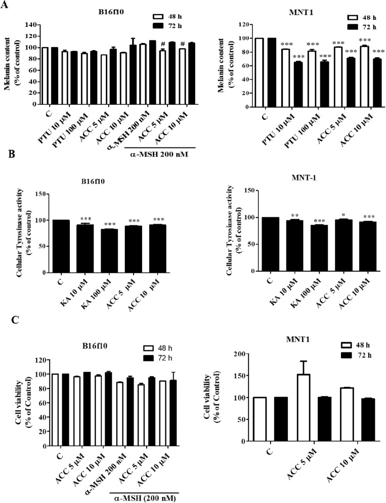
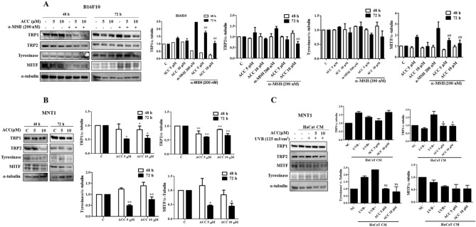
Aspacochioside C (ACC) is a steroidal saponin isolated from Asparagus cochinchinensis. Steroidal saponins, such as pseudoprotodioscin and dioscin, are known to inhibit melanogenesis, but the role of ACC in melanogenesis remains unknown. Due to the toxic effect of the commonly used skin whitening agents like arbutin, kojic acid and α-lipoic acid alternative plant products are recentlybeen studied for their anti-hypergmentation effect. This study explores the role of ACC in melanogenesis in both in vivo and in vitro models. Here, we for the first time demonstrate that ACC attenuated α-MSH- and UVB-induced eumelanin production by inhibiting tyrosinase-related protein (TRP)-2 protein expression in both murine B16F10 and human melanoma MNT1 cells. However, ACC had no significant effect on pheomelanin concentration. ACC also decreased the pigmentation density in zebrafish embryos, which indicates that ACC targets TRP2 and inhibits eumelanin synthesis. Our results demonstrate that ACC inhibits TRP2, thereby attenuating eumelanin synthesis both in in vitro and in vivo zebrafish model. Therefore, ACC can potentially be used as an anti-melanogenic agent for both aesthetic and pharmaceutical purposes.


Conflict of interest statement

The authors declare no competing interests.

Figures

Figure 1
Figure 1
Effect of aspacochioside C (ACC) on zebrafish pigmentation. (A) Chemical structure of ACC. Zebrafish embryos were treated with or without ACC (1 or 5 μM) and phenylthiourea (PTU, 25 μM). PTU was used as a positive control. (B) ACC inhibited zebrafish pigmentation. Pigmentation in zebrafish embryos was observed under a IX7 microscope at 72 hpf. Scale bar: 0.5 mm. (C) Pigmentation area density was determined using the ImageJ software (n = 5). The values represent the mean ± standard error of the mean (SEM) of three independent experiments. ***p < 0.001 versus C.
Figure 2
Figure 2
Effect of aspacochioside C (ACC) on melanin production. B16F10 cells were treated with or without ACC (5 or 10 μM) and 200 nM alpha-melanocyte-stimulating hormone (α-MSH), while MNT1 cells were treated with or without ACC (5 or 10 μM) for 48 and 72 h. (A) ACC inhibited, while α-MSH induced melanin production in MNT1cells. PTU was used as a positive control. (B) ACC decreased the cellular tyrosinase activity in B16F10 and MNT1cells. Kojic acid (KA) was used as a positive control. (C) ACC had no cytotoxic effects on B16F10 and MNT1 cells. The values represent the mean ± (SD) of three independent experiments. *p < 0.05, ***p < 0.001 versus C; #p < 0.05 versus α-MSH.
Figure 3
Figure 3
Effects of aspacochioside C (ACC) on melanogenesis-related proteins. (A) ACC inhibited α-MSH-induced tyrosinase-related protein (TRP)-2 and melanocyte-inducing transcription factor (MITF) protein expression in B16F10 cells. B16F10 cells were treated with or without ACC (5 or 10 μM) and 200 nM α-MSH for 48 and 72 h. Cells were lysed and proteins were separated according to size via sodium dodecyl sulfate–polyacrylamide gel electrophoresis (SDS-PAGE). (B) ACC inhibited TRP1, TRP2, tyrosinase, and MITF protein expression in MNT1 cells. Cells were treated with or without ACC (5 or 10 μM) 48 and 72 h. Cells were lysed and proteins were separated according to size via SDS-PAGE. (C) ACC inhibited UVB-induced TRP2 and tyrosinase protein expression in MNT1 cells. HaCaT cells were irradiated with 125 mJ/cm2 UVB and treated with or without ACC (5 or 10 μM). After 24 h, conditioned media (CM) of HaCaT cells were collected and used to treat MNT1 cells. After 48 h, proteins were collected and separated according to size via SDS-PAGE. NC: MNT1 cells grown in 2% fetal bovine serum (FBS) containing Dulbecco's Modified Eagle’s Medium (DMEM), UVB: MNT1 cells grown in HaCaT CM, UVB+: MNT1 cells grown in UVB irradiated HaCaT CM. The values represent the mean ± SD of three independent experiments. *p < 0.05, ***p < 0.001 versus C; ##< 0.01 versus α-MSH; $$p << 0.01 versus UVB+.
Figure 4
Figure 4
Effect of aspacochioside C (ACC) on eumelanin and pheomelanin production. Cells were treated with ACC as described. Eumelanin and pheomelanin concentrations were measured using Eumelanin and Pheomelanin enzyme-linked immunosorbent assay (ELISA) kit, according to the manufacturer’s protocol. (A) ACC inhibited α-MSH-induced eumelanin production in B16F10 cells, with no significant change in pheomelanin production. (B) ACC inhibited eumelanin production in MNT1 cells, with no significant change in pheomelanin production. (C) ACC inhibited UVB-induced eumelanin production in MNT1 cells, with no significant change in pheomelanin production. NC: MNT1 cells grown in 2% FBS containing DMEM, UVB: MNT1 cells grown in HaCaT CM, UVB+: MNT1 cells grown in UVB irradiated HaCaT CM. The values represent the mean ± SD of three independent experiments. ***p < 0.001 versus C; ##p < 0.01, ###p < 0.001 versus α-MSH; $p < 0.05, $$p < 0.01, $$$p < 0.001 versus UVB.
Figure 5
Figure 5
Effect of aspacochioside C (ACC) on ratio of pheomelanin (TTCA)/eumelanin (PTCA) production. Cells were treated with ACC as described. The ratio of pheomelanin (TTCA)/eumelanin (PTCA) was measured using HPLC analysis. (A) ACC inhibited α-MSH-induced eumelanin (PTCA) production in B16F10 cells, with no significant change in pheomelanin (TTCA) production. (B) ACC inhibited α-MSH-induced eumelanin (PTCA) production in MNT1 cells, no significant change in pheomelanin (TTCA) production. The values represent the mean ± SD of three independent experiments. ***p < 0.001 versus C; ##p < 0.01, $p < 0.05, $$p < 0.01, $$$p < 0.001 versus α-MSH.

Similar articles

References

    1. Brenner M, Hearing VJ. The protective role of melanin against UV damage in human skin. Photochem. Photobiol. 2008;84:539–549. doi: 10.1111/j.1751-1097.2007.00226.x. - DOI - PMC - PubMed
    1. Narayanan DL, Saladi RN, Fox JL. Ultraviolet radiation and skin cancer. Int. J. Dermatol. 2010;49:978–986. doi: 10.1111/j.1365-4632.2010.04474.x. - DOI - PubMed
    1. D'Mello SA, Finlay GJ, Baguley BC, Askarian-Amiri ME. Signaling pathways in melanogenesis. Int. J. Mol. Sci. 2016 doi: 10.3390/ijms17071144. - DOI - PMC - PubMed
    1. Brozyna AA, VanMiddlesworth L, Slominski AT. Inhibition of melanogenesis as a radiation sensitizer for melanoma therapy. Int. J. Cancer. 2008;123:1448–1456. doi: 10.1002/ijc.23664. - DOI - PubMed
    1. Brozyna AA, Jozwicki W, Roszkowski K, Filipiak J, Slominski AT. Melanin content in melanoma metastases affects the outcome of radiotherapy. Oncotarget. 2016;7:17844–17853. doi: 10.18632/oncotarget.7528. - DOI - PMC - PubMed

Publication types