. 2023 Sep 8;22(1):151.
doi: 10.1186/s12943-023-01856-1.

CircXRN2 suppresses tumor progression driven by histone lactylation through activating the Hippo pathway in human bladder cancer

Affiliations

CircXRN2 suppresses tumor progression driven by histone lactylation through activating the Hippo pathway in human bladder cancer

Bo Xie et al. Mol Cancer. .

Abstract

Background: Bladder cancer (BCa) is the fourth most common malignant tumor with a poor prognosis worldwide. Further exploration and research are needed to unmask the underlying roles and molecular mechanisms of circular RNAs. In the current study, our findings showed that circXRN2 suppresses tumor progression driven by histone lactylation by activating the Hippo pathway in human bladder cancer.

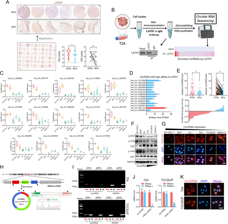
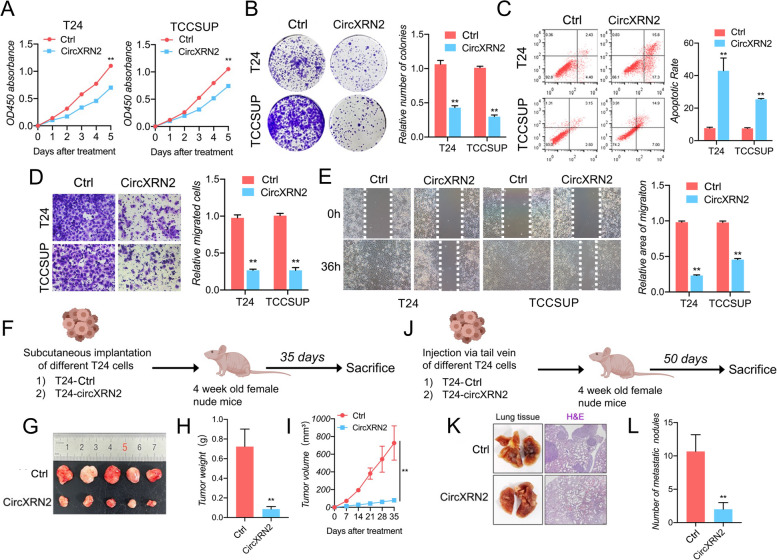
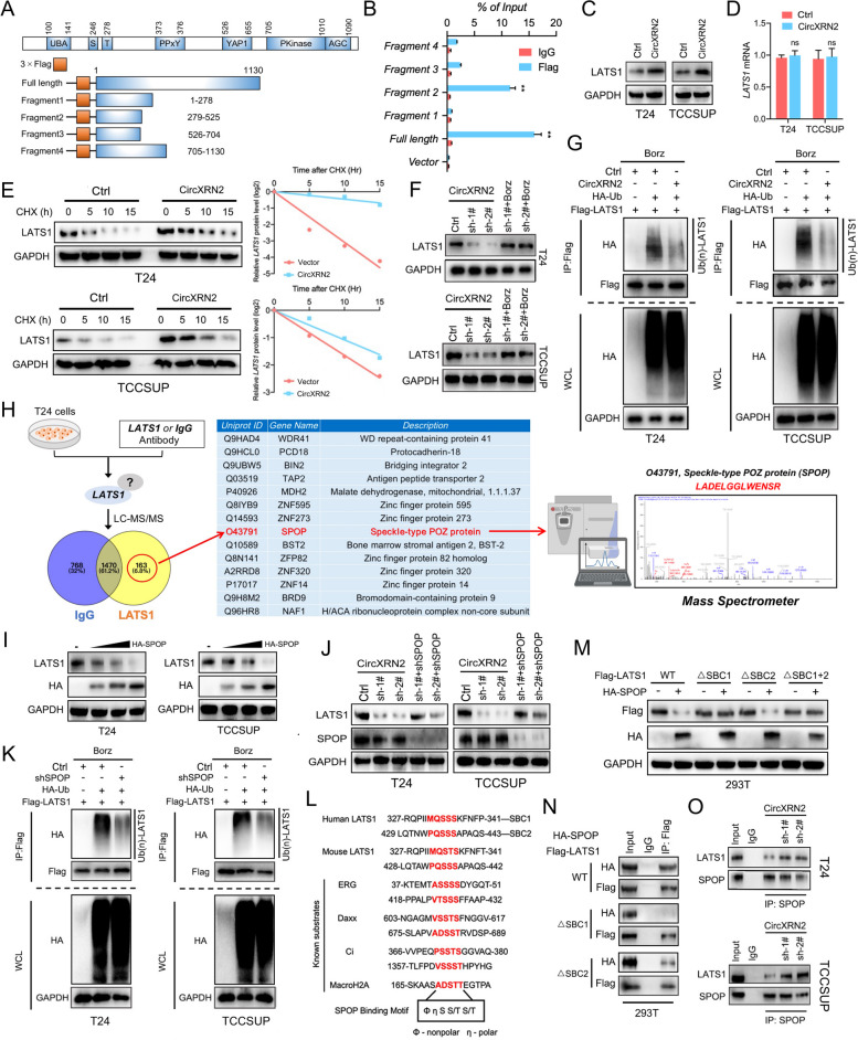
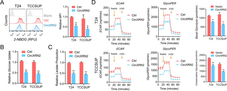
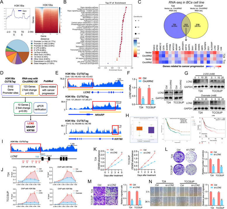
Methods: RNA immunoprecipitation (RIP) followed by circRNA sequencing confirmed circXRN2 as the research object. Overexpression of circXRN2 and knockdown of TAZ/YAP further verified the biological functions in T24 and TCCSUP cells. RIP, immunoprecipitation and coimmunoprecipitation were used to elucidate the interaction between circXRN2 and LATS1. A Seahorse metabolic analyzer was used to determine the glycolytic rate. Cleavage under targets and Tagmentation (CUT&Tag) and chromatin immunoprecipitation (ChIP) were employed to ensure the regulatory roles of H3K18 lactylation in the transcriptional activity of LCN2.

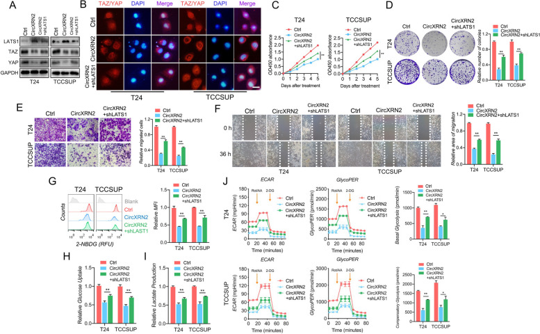
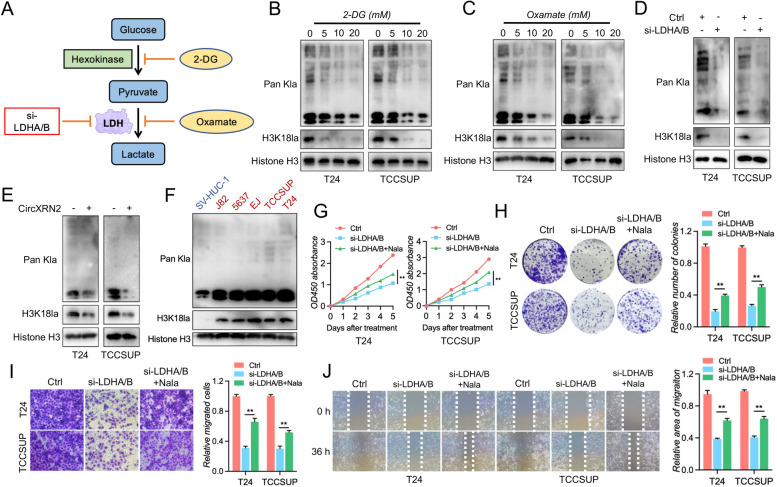
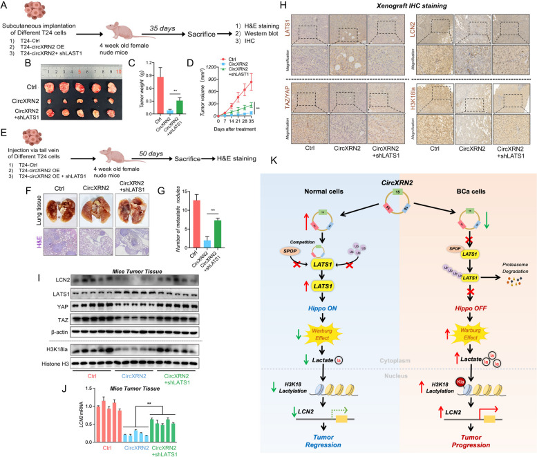
Results: CircXRN2 is aberrantly downregulated in bladder cancer tissues and cell lines. CircXRN2 inhibits the proliferation and migration of tumor cells both in vitro and in vivo. In addition, circXRN2 serves as a negative regulator of glycolysis and lactate production. Mechanistically, circXRN2 prevents LATS1 from SPOP-mediated degradation by binding to the SPOP degron and then activates the Hippo signaling pathway to exert various biological functions. The circXRN2-Hippo pathway regulatory axis further modulates tumor progression by inhibiting H3K18 lactylation and LCN2 expression in human bladder cancer.

Conclusions: CircXRN2 suppresses tumor progression driven by H3K18 lactylation by activating the Hippo signaling pathway in human bladder cancer. Our results indicated novel therapeutic targets and provided promising strategies for clinical intervention in human bladder cancer.

Keywords: Bladder cancer; Hippo pathway; Histone lactylation; Tumor progression; circXRN2.


Conflict of interest statement

The authors declare no competing interests.

Figures

Fig. 1
Fig. 1
LATS1 is downregulated and interacts with circXRN2 in bladder cancer. a LATS1 was downregulated in surgical tissue samples (n = 30). Scale bar: 200 μm. b RIP assay followed by RNA sequencing revealed that LATS1 protein could interact with numerous circRNAs. c CircRNAs binding with LATS1 were validated by qRT‒PCR in different cell lines (SV-HUC-1, 5637, EJ, T24, TCCSUP and UM-UC-3). d The affinities of dysregulated circRNAs to the LATS1 protein were evaluated by RIP assay, and the results showed that circXRN2 had the highest affinity for LATS1. e Total RNA was extracted from clinical specimens of our own cohort (n = 40). Dysregulation of circXRN2 in surgical tissue samples was confirmed by qRT‒PCR. f The expression levels of LATS1, p-TAZ, TAZ, p-YAP and YAP in cell lines were determined by western blotting. g Immunofluorescence indicated the localization of TAZ/YAP in cell lines with various abundances of circXRN2. Scale bar: 50 μm. h The biological structure of circXRN2 is represented by a graphic illustration, and the back-splicing site was verified by PCR and Sanger sequencing. i CircXRN2 was only amplified by divergent primers in cDNA but not in gDNA of T24 and TCCSUP cells. GAPDH was used as a negative control. j Cells were incubated in the absence or presence of RNase R, and the expression levels of circXRN2 and linear XRN2 were determined by qRT‒PCR. k RNA FISH indicated that CircXRN2 was mainly located in the cytoplasm. The nuclei were stained with DAPI, while circXRN2 was red. Scale bar: 50 μm. All the data are presented as the mean ± standard deviation (n = 3). *P < 0.05, **P < 0.01, compared with the control group
Fig. 2
Fig. 2
CircXRN2 inhibits the proliferation and migration of bladder cancer cells in vitro and in vivo. a A CCK-8 assay was conducted to evaluate cell viability in control and circXRN2-overexpressing cells. b CircXRN2 suppressed the colony-formation ability of bladder cancer cells. c CircXRN2-induced cell apoptosis in T24 and EJ cells. de Transwell migration assays and wound healing assays were conducted to evaluate cell migratory abilities. f Schematic illustration of the subcutaneous tumorigenesis model in nude mice with negative control or circXRN2-overexpressing cells. g-i CircXRN2 inhibited tumor growth of human bladder cancer in vivo (n = 5). j Schematic illustration of the lung metastatic model in nude mice with negative control or circXRN2-overexpressing cells. k-l Overexpressing circXRN2 led to fewer lung metastatic nodules than in the control group. All the data are presented as the mean ± standard deviation (n = 5). *P < 0.05, **P < 0.01, compared with the control group
Fig. 3
Fig. 3
CircXRN2 prevents LATS1 from SPOP-mediated degradation by binding to its degron sequence. a We contrasted different fragments of the LATS1 protein to test the exact region interacting with circXRN2. A schematic illustration of the depletion map of LATS1 protein. b Fragment 2 and full-length LATS1 protein interacted with circXRN2, as determined by RIP assay in 293T cells. c, d CircXRN2 enhanced the expression level of LATS1 at the protein level but not the mRNA level. e Cycloheximide (CHX) was used to block protein synthesis. LATS1 protein levels were detected by western blotting in control and circXRN2-overexpressing bladder cancer cells. f Downregulation of LATS1 protein induced by circXRN2 knockdown was reversed by treatment with bortezomib (500 nM, an inhibitor of the proteasome). g Cells were pretreated with bortezomib (500 nM) and NEM (10 μM, an inhibitor of deubiquitinating enzyme) for 8 h. The ubiquitinated LATS1 protein levels in the control group and circXRN2-overexpressing group were detected by IP assay. h A mass spectrometer was used to detect the proteins interacting with LATS1. i LATS1 protein levels were detected by immunoblot analysis in cells transfected with incremental doses of SPOP plasmid. j Downregulation of LATS1 protein could be rescued by depletion of SPOP. k SPOP increased the ubiquitination of LATS1 protein in T24 and TCCSUP cells. l Schematic illustration of the sequence alignment of the LATS1 protein with the SPOP-binding degron among known substrates. m Flag-LATS1-containing wild-type or mutant SBCs and HA-SPOP were transfected into 293T cells. Western blotting indicated that mutation of SBC1 led to remarkable blockade of LATS1 degradation mediated by SPOP, while depletion of SBC2 had little effect. n Co-IP results showed that wild-type LATS1 could bind to SPOP, but the interaction of SBC1-mutant LATS1 with SPOP was almost completely diminished. Western blotting was performed to determine the expression levels of Flag-LATS1 and HA-SPOP. o The interactions between LATS1 and SPOP in control and circXRN2 knockdown cells were verified by Co-IP assay. All the data are presented as the mean ± standard deviation (n = 3). *P < 0.05, **P < 0.01
Fig. 4
Fig. 4
CircXRN2 is a negative regulator of glycolysis in human bladder cancer cells. a Control or circXRN2-overexpressing bladder cancer cells were incubated with 2-NBDG (a fluorescent D-glucose analog) and subjected to flow cytometry to monitor glucose uptake. b Overexpression of circXRN2 decreased the uptake of glucose in T24 and TCCSUP cells. c Lactate production was suppressed in the presence of circXRN2. d Seahorse metabolic analysis showed that circXRN2 significantly inhibited glycolysis in bladder cancer cells, which was marked by a decreased glycoPER (glycolytic proton efflux rate), basal glycolysis rate and compensatory glycolysis rate. All the data are presented as the mean ± standard deviation (n = 3). *P < 0.05, **P < 0.01
Fig. 5
Fig. 5
CircXRN2 activates the Hippo pathway through LATS1 to regulate cell proliferation, migration and glycolysis in human bladder cancer. a The expression levels of LATS1, TAZ and YAP were determined in different cells. b CircXRN2 altered the location of TAZ/YAP in T24 and TCCSUP cells, and knockdown of LATS1 reversed the effect of circXRN2 on TAZ/YAP. Scale bar: 50 μm. c CCK-8 assay showed that shLATS1 alleviated the suppression of cell viability induced by circXRN2. d Control, circXRN2-overexpressing and circXRN2 + shLATS1 cells were used for colony formation assays. e, f Cell migratory ability was impaired by circXRN2 and rescued by depletion of LATS1, as validated by Transwell migration and wound healing assays. g-i CircXRN2-induced inhibition of glucose uptake and lactate production was reversed by elimination of LATS1. j Cells were subjected to a Seahorse metabolic analyzer to determine the glycolytic rate. The glycoPER, basal glycolytic rate and compensatory glycolytic rate were higher in the circXRN2 overexpression group along with the shLATS1 group than in the circXRN2 overexpression group. All the data are presented as the mean ± standard deviation (n = 3). *P < 0.05, **P < 0.01
Fig. 6
Fig. 6
CircXRN2-mediated H3K18 lactylation promotes tumor progression in human bladder cancer. a A schematic illustration of glycolysis is presented, and the methods used to inhibit the production of lactate and histone lactylation in this research are indicated. b, c Cells were treated with 2-DG or oxamate (glycolysis inhibitors) at different concentrations for 24 h. Cells were collected for measuring the level of Pan Kla and H3K18 lactylation. d Immunoblots were carried out to determine the effect of LDH depletion on Pan Kla and H3K18 lactylation. e CircXRN2 suppressed intracellular Pan Kla and H3K18 lactylation in T24 and TCCSUP cells. f Pan Kla and H3K18 lactylation were evaluated in SV-HUC-1 and bladder cancer cell lines. g A CCK-8 assay was used to determine the viability of LDH-deficient bladder cancer cells. h Cell proliferation and colony formation ability were measured by colony formation assay. i, j The migratory ability of LDH-deficient cells was evaluated by Transwell migration assay and wound healing assay. All the data are presented as the mean ± standard deviation (n = 3). *P < 0.05, **P < 0.01, compared with the control group
Fig. 7
Fig. 7
LCN2 is a target of H3K18 lactylation and acts as an oncogene. a CUT&Tag was performed with H3K18la antibodies in T24 cells. H3K18la could be enriched in the promoter region of numerous genes. b KEGG database analysis was carried out in H3K18la-related genes. c Transcriptome sequencing was performed in control and circXRN2-overexpressing T24 and TCCSUP cells. d Flow diagram confirming the downstream target of H3K18la. e H3K18la peaks in the promoter regions of LCN2, NRARP and KRT80. f LCN2 mRNA and protein levels were measured in circXRN2-overexpressing cells. g Different concentrations of 2-DG and oxamate were added to T24 and TCCSUP cells, and LCN2 protein levels were measured by immunoblotting. h LCN2 mRNA was upregulated in bladder cancer and was closely related to the overall survival time of bladder cancer patients in the TCGA database and K‒M plotter. i Primers targeting different fragments of the LCN2 gene are indicated, and the c, d, and e sites are H3K18la peaks of the LCN2 promoter region in CUT&Tag. j ChIP assay following qPCR was used to detect the binding status of H3K18la in the LCN2 promoter region in bladder cancer cells treated with or without glycolysis inhibitors. k-n The roles of LCN2 in cell proliferation and migration were verified by CCK-8 assay, colony formation assay, Transwell migration assay and wound healing assay. All the data are presented as the mean ± standard deviation (n = 3). *P < 0.05, **P < 0.01, compared with the control group
Fig. 8
Fig. 8
CircXRN2/LATS1 axis inhibits tumor growth and metastasis of bladder cancer in vivo. a T24 cells (6 × 106) transfected with control, circXRN2 or circXRN2 and shLATS1 were injected subcutaneously to establish a tumorigenesis model in nude mice. b, c At Day 35 after treatment, all mice were sacrificed, and tumors were dissected and photographed. Tumor weight was also recorded. d The parameters of subcutaneous tumors were measured and recorded every 7 days. We calculated the volume of the tumor according to the formula below: tumor volume = 0.5 × length × width × width (mm3). e We also established a lung metastasis model by injecting 5 × 106 different T24 cells into the tail vein to evaluate the ability of cells to metastasize in vivo. f, g Fifty days later, mice were sacrificed, and lung tissues were photographed and subjected to H&E staining. The number of lung metastatic nodules was counted. h Immunohistochemistry was used to visualize and compare the protein levels of LATS1, TAZ/YAP, LCN2 and H3K18la in tumors collected from the control, circXRN2 overexpression and circXRN2 with shLATS1 groups. Scale bar: 20 μm. i Immunoblotting was used to determine the protein levels of LATS1, TAZ/YAP, LCN2 and H3K18la in tumors collected from the control, circXRN2 overexpression and circXRN2 with shLATS1 groups. j The expression levels of LCN2 mRNA were measured by qRT‒PCR. k Schematic illustration of the current study: circXRN2 suppresses tumor progression driven by H3K18 lactylation in human bladder cancer by activating the Hippo signaling pathway. All the data are presented as the mean ± standard deviation (n = 5 in the tumorigenesis model, n = 3 in the metastatic model). *P < 0.05, **P < 0.01, compared with the circXRN2 overexpression group

References

    1. Lenis AT, Lec PM, Chamie K, Mshs MD. Bladder cancer: a review. JAMA. 2020;324:1980–1991. doi: 10.1001/jama.2020.17598. - DOI - PubMed
    1. Antoni S, et al. Bladder cancer incidence and mortality: a global overview and recent trends. Eur Urol. 2017;71:96–108. doi: 10.1016/j.eururo.2016.06.010. - DOI - PubMed
    1. Li HZ, et al. Bladder cancer incidence, mortality and temporal trends in China. Zhonghua Zhong Liu Za Zhi. 2021;43:293–298. doi: 10.3760/cma.j.cn112152-20200421-00362. - DOI - PubMed
    1. Siegel RL, Miller KD, Fuchs HE, Jemal A. Cancer statistics, 2021. CA Cancer J Clin. 2021;71:7–33. doi: 10.3322/caac.21654. - DOI - PubMed
    1. Patel VG, Oh WK, Galsky MD. Treatment of muscle-invasive and advanced bladder cancer in 2020. CA Cancer J Clin. 2020;70:404–423. doi: 10.3322/caac.21631. - DOI - PubMed

Publication types