. 2023 Aug 28;13(17):2740.
doi: 10.3390/ani13172740.

Reduced Nitric Oxide Synthase Involvement in Aigamo Duck Basilar Arterial Relaxation

Affiliations

Reduced Nitric Oxide Synthase Involvement in Aigamo Duck Basilar Arterial Relaxation

Siyuan Wu et al. Animals (Basel). .

Abstract

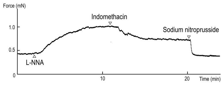
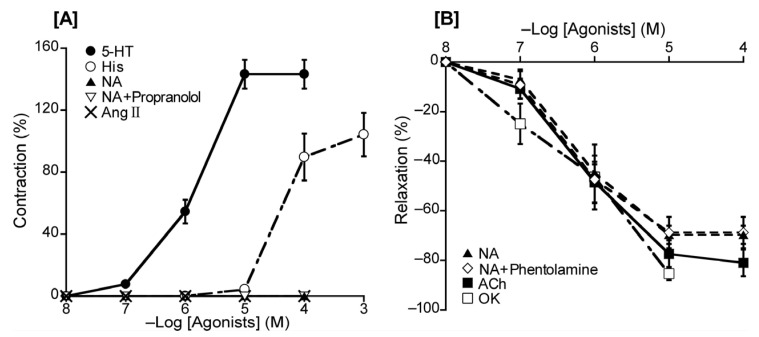
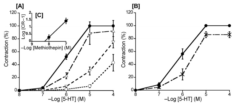
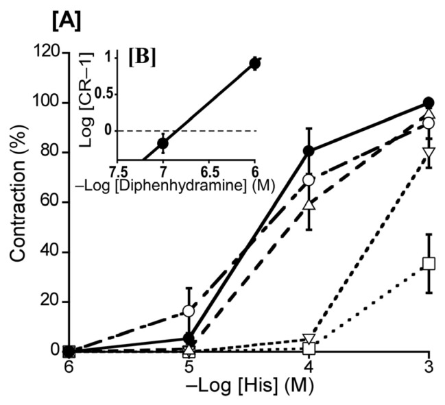
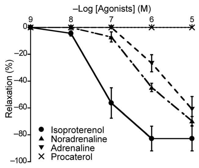
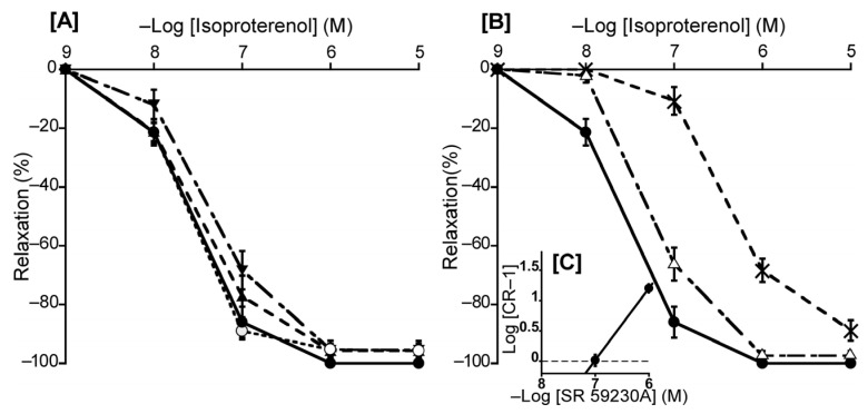
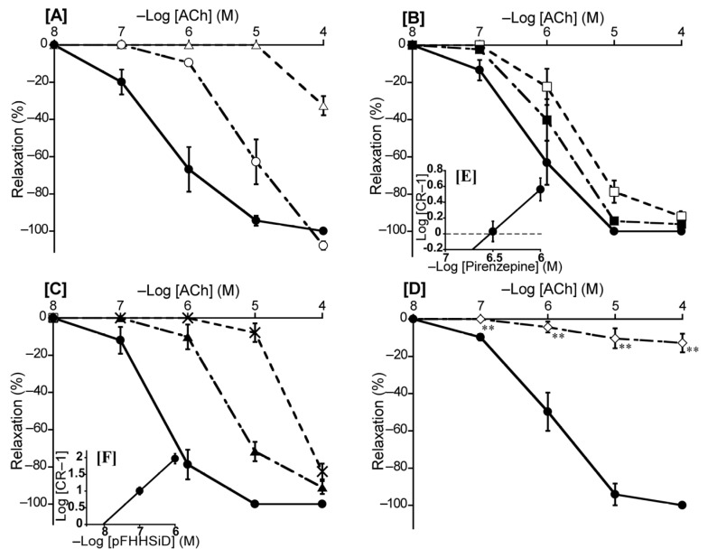
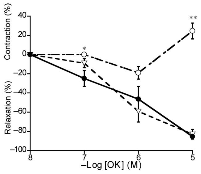
The basilar arterial endothelium mediates blood vessel relaxation partly through the release of nitric oxide (NO). Apoptosis of cerebrovascular endothelial cells is linked to a high mortality rate in chickens infected with the highly pathogenic avian influenza virus, but interestingly, ducks exhibit a greater resistance to this virus. In this study, we examined the responsiveness of duck basilar arteries (BAs) to various vasoactive substances, including 5-hydroxytryptamine (5-HT), histamine (His), angiotensin (Ang) II, noradrenaline (NA), acetylcholine (ACh), and avian bradykinin ornithokinin (OK), aiming to characterize the receptor subtypes involved and the role of endothelial NO in vitro. Our findings suggest that arterial contraction is mediated with 5-HT1 and H1 receptors, while relaxation is induced with β3-adrenergic and M3 receptors. Additionally, OK elicited a biphasic response in duck BAs, and Ang II had no effect. Endothelial NO appears to be crucial in relaxation mediated with M3 and OK receptors but not β3-adrenergic receptors in the duck BA. The reduced endothelial NO involvement in the receptor-mediated relaxation response in duck BAs represents a clear difference from the corresponding response reported in chicken BAs. This physiological difference may explain the differences in lethality between ducks and chickens when vascular endothelial cells are infected with the virus.

Keywords: cerebral artery; duck; endothelium; highly pathogenic avian influenza; nitric oxide.


Conflict of interest statement

The authors declare no conflicts of interest.

Figures

Figure 1
Figure 1
Typical contraction induced with Nω-nitro-L-arginine (L-NNA, 10−4 M) and relaxation induced with indomethacin (10−5 M) under precontracted conditions induced with L-NNA.
Figure 2
Figure 2
Responsiveness to 5-hydroxytriptamine (5-HT), histamine (His), noradrenaline (NA), and angiotensin (Ang) II under resting tension (A) and to NA, acetylcholine (ACh), and ornithokinin (OK) under a precontracted condition induced with 5-HT (B). The contraction induced with 60 mM KCl (A) and the relaxation induced with 10−4 M sodium nitroprusside (B) was taken as 100% contraction and relaxation, respectively. Each point represents the mean ± SEM of 4–6 ducks. The percentage of reacting vessels is 100%.
Figure 3
Figure 3
Effects of 5-hydroxytriptamine (5-HT) antagonists on 5-HT-induced contraction in the duck basilar artery. The antagonists were methiothepin (Δ: 10−8 M, ▽: 10−7 M, ○: 10−6 M) (A) and ketanserin (×: 10−6 M) (B). The Schild plot for methiothepin is shown in (C). The maximal contraction induced with 5-HT was taken as 100%. Each point represents the mean ± SEM of 6 ducks.
Figure 4
Figure 4
Effects of histamine (His) antagonists on His-induced contraction in the duck basilar artery. The antagonists were diphenhydramine (Δ: 10−7 M, ▽: 10−6 M, □: 10−5 M) and cimetidine (○: 10−5 M) (A). The Schild plot for diphenhydramine is shown in (B). The maximal contraction induced with His was taken as 100%. Each point represents the mean ± SEM of 5 ducks.
Figure 5
Figure 5
Responsiveness of duck basilar arteries to isoproterenol (●), noradrenaline (▲), adrenaline (▼), and procaterol (×) under precontracted conditions. The relaxation induced with 10−4 M sodium nitroprusside was taken as 100%. Each point represents the mean ± SEM of 4–6 ducks.
Figure 6
Figure 6
Effects of β-adrenergic receptor antagonists and a NOS inhibitor (L-NNA) on isoproterenol-induced relaxation in the duck basilar artery. The antagonists or inhibitor were atenolol (▲: 10−6 M), butoxamine (▼: 10−6 M), L-NNA (○: 10−4 M) (A), and SR 59230A (Δ: 10−7 M, ×: 10−6 M) (B). The Schild plot for SR 59230A is shown in (C). The maximal relaxation induced with isoproterenol was taken as 100%. Each point represents the mean ± SEM of 4 or 5 ducks.
Figure 7
Figure 7
Effects of acetylcholine (ACh) antagonists and a NOS inhibitor (L-NNA) on ACh-induced relaxation in the duck basilar artery. The antagonists or inhibitor were atropine (○: 10−8 M, Δ: 10−7 M) (A), pirenzepine (■: 10−6.5 M, □: 10−6 M) (B), hexahydro-sila-difenidol hydrochloride, p-fluoroanalog (pFHHSiD; ▲: 10−7 M, ×: 10−6 M) (C), and L-NNA (◇:10−4 M) (D). The Schild plots for pirenzepine and pFHHSiD were shown in (E,F), respectively. The maximal relaxation induced with ACh was taken as 100%. Each point represents the mean ± SEM of 5 ducks. (** p < 0.01 vs. control).
Figure 8
Figure 8
Effects of L-NNA (○: 10−4 M) and L-NNA plus indomethacin (10−5 M) (▽) on ornithokinin (OK)-induced relaxation (●) in duck basilar arteries. The contraction induced with 60 mM KCl and the relaxation induced with 10−4 M sodium nitroprusside were taken as 100% contraction and relaxation, respectively. Each point represents the mean ± SEM of 4 ducks. (* p < 0.05, ** p < 0.01 vs. control).

Similar articles

References

    1. Davis R.L., Choi G., Kuiken T., Quéré P., Trapp S., Short K.R., Richard M. The culture of primary duck endothelial cells for the study of avian influenza. BMC Microbiol. 2018;18:138. doi: 10.1186/s12866-018-1307-4. - DOI - PMC - PubMed
    1. de Bruin A.C., Spronken M.I., Bestebroer T.M., Fouchier R.A., Richard M. Reduced replication of highly pathogenic avian influenza virus in duck endothelial cells compared to chicken endothelial cells is associated with stronger antiviral responses. Viruses. 2022;14:165. doi: 10.3390/v14010165. - DOI - PMC - PubMed
    1. Keawcharoen J., van Riel D., van Amerongen G., Bestebroer T., Beyer W.E., van Lavieren R., Osterhaus A.D., Fouchier R.A., Kuiken T. Wild ducks as long-distance vectors of highly pathogenic avian influenza virus (H5N1) Emerg. Infect. Dis. 2008;14:600–607. doi: 10.3201/eid1404.071016. - DOI - PMC - PubMed
    1. Ito T., Kobayashi Y., Morita T., Horimoto T., Kawaoka Y. Virulent influenza A viruses induce apoptosis in chickens. Virus Res. 2002;84:27–35. doi: 10.1016/S0168-1702(01)00414-2. - DOI - PubMed
    1. Vreman S., Bergervoet S.A., Zwart R., Stockhofe-Zurwieden N., Beerens N. Tissue tropism and pathology of highly pathogenic avian influenza H5N6 virus in chickens and Pekin ducks. Res. Vet. Sci. 2022;146:1–4. doi: 10.1016/j.rvsc.2022.03.010. - DOI - PubMed

LinkOut - more resources