

Study of Root Transparency in Different Postmortem Intervals Using Scanning Electron Microscopy
- PMID: 37685346
- PMCID: PMC10487109
- DOI: 10.3390/diagnostics13172808
Study of Root Transparency in Different Postmortem Intervals Using Scanning Electron Microscopy
Abstract
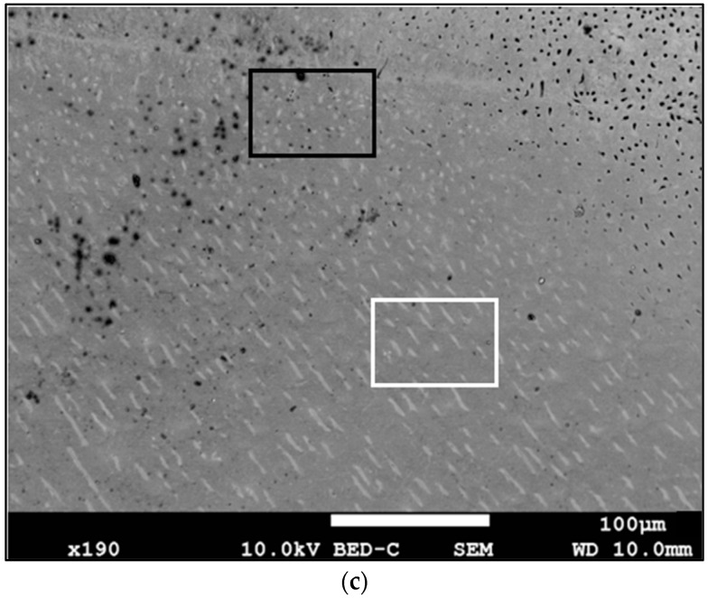
In the fields of forensics, the identification of human remains is a recurrent problem. The estimated age at death is one of the copious criteria to be evaluated. In adult teeth, the height of the root dentin transparency is used to estimate age. However, in archaeological material, this phenomenon appears inconstant. The aim of this work was to observe the structural modifications of the sclerotic dentin in the teeth for different postmortem intervals. The study included two parts (retrospective and prospective study) with 21 human monoradicular teeth, from bodies donated to medical science with postmortem intervals (PMIs) of 0, 1, 2 and 5 years and archeological excavation. After inclusion based on resin, section and polishing, the samples were analyzed with a scanning electron microscope (SEM) JSM-7800F®, and the procedure was completed via a semiquantitative analysis of calcium and phosphorus using EDX microanalysis. The analysis showed the existence of tubular and chemical modifications of sclerotic dentin at different PMIs. Our SEM study allowed us to observe a difference in tubule aspects linked to an increased PMI: the loss of peritubular collar and the lumen obstruction of tubules with a hyperdense material. Microanalysis highlighted variations in phosphocalcic ratios among the different groups, especially in the pulp area and the canine. Our hypotheses that explain these differences are based on the postmortem modifications of the crystals of the mineral phase of sclerotic dentin under the influence of chemical and/or bacterial action.
Keywords: estimated age at death; forensic science; postmortem changes; root transparency; scanning electron microscopy; sclerotic dentin; taphonomy.
Conflict of interest statement
The authors declare no conflict of interest.
Figures


Similar articles
-
A scanning electron microscopy and electron probe X-ray microanalysis (SEM-EPMA) of pink teeth.J Forensic Sci. 1988 Nov;33(6):1328-31. J Forensic Sci. 1988. PMID: 3204342
-
Bonding of a self-etching primer to non-carious cervical sclerotic dentin: interfacial ultrastructure and microtensile bond strength evaluation.J Adhes Dent. 2000 Spring;2(1):9-28. J Adhes Dent. 2000. PMID: 11317411
-
Novel Management of Hypersensitive Dentin Using Propolis-based Herbal Desensitizing Agents: An In Vitro Scanning Electron Microscopic Study.J Contemp Dent Pract. 2021 Sep 1;22(9):1030-1034. J Contemp Dent Pract. 2021. PMID: 35000948
-
[Sclerotic dentin: aetio-pathogenetic hypotheses].Minerva Stomatol. 2002 Jul-Aug;51(7-8):285-92. Minerva Stomatol. 2002. PMID: 12434123 Review. Italian.
-
[Micromorphological aspects of dentin].Minerva Stomatol. 1995 Sep;44(9):377-87. Minerva Stomatol. 1995. PMID: 8668111 Review. Italian.
Cited by
-
Decoding Time of Death: Histopathological Dynamics of Intervertebral Discs as a Novel Marker for Postmortem Interval Estimation.Diagnostics (Basel). 2025 Mar 2;15(5):605. doi: 10.3390/diagnostics15050605. Diagnostics (Basel). 2025. PMID: 40075852 Free PMC article.
-
Diagnostic Utility of Podoplanin Immunohistochemistry Combined with the NanoSuit-Correlative Light and Electron Microscopy Method for Thoracic Malignant Tumors.Diagnostics (Basel). 2025 May 21;15(10):1298. doi: 10.3390/diagnostics15101298. Diagnostics (Basel). 2025. PMID: 40428290 Free PMC article.
References
-
- Porter A.E., Nalla R.K., Minor A., Jinschek J.R., Kisielowski C., Radmilovic V., Kinney J.H., Tomsia A.P., Ritchie R. A transmission electron microscopy study of mineralization in age-induced transparent dentin. Biomaterials. 2005;26:7650–7660. doi: 10.1016/j.biomaterials.2005.05.059. - DOI - PubMed
LinkOut - more resources
Full Text Sources
