

Integrating Artificial Intelligence Tools in the Clinical Research Setting: The Ovarian Cancer Use Case
- PMID: 37685352
- PMCID: PMC10486639
- DOI: 10.3390/diagnostics13172813
Integrating Artificial Intelligence Tools in the Clinical Research Setting: The Ovarian Cancer Use Case
Abstract
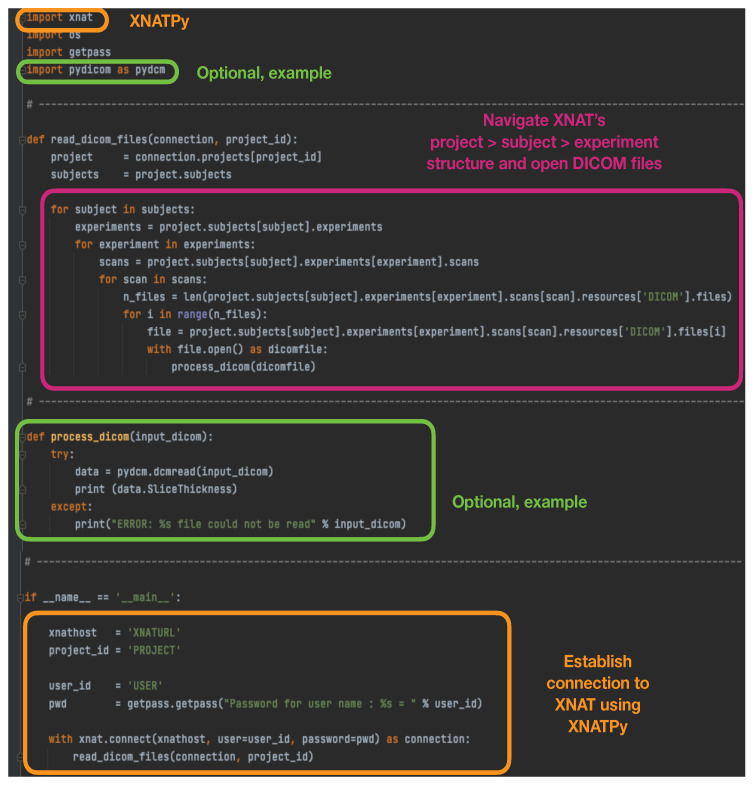
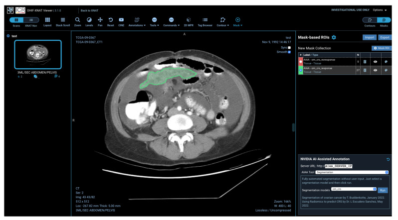
Artificial intelligence (AI) methods applied to healthcare problems have shown enormous potential to alleviate the burden of health services worldwide and to improve the accuracy and reproducibility of predictions. In particular, developments in computer vision are creating a paradigm shift in the analysis of radiological images, where AI tools are already capable of automatically detecting and precisely delineating tumours. However, such tools are generally developed in technical departments that continue to be siloed from where the real benefit would be achieved with their usage. Significant effort still needs to be made to make these advancements available, first in academic clinical research and ultimately in the clinical setting. In this paper, we demonstrate a prototype pipeline based entirely on open-source software and free of cost to bridge this gap, simplifying the integration of tools and models developed within the AI community into the clinical research setting, ensuring an accessible platform with visualisation applications that allow end-users such as radiologists to view and interact with the outcome of these AI tools.
Keywords: artificial intelligence; cancer research; clinical integration; imaging; radiomics.
Conflict of interest statement
E.S: Lucida Medical (co-founder and shareholder), GE HealthCare (research support, speakers’ bureau), Canon (research support, speakers’ bureau). The rest of the authors declare no potential conflict of interest.
Figures


References
-
- Aerts H., Rios Velazquez E., Leijenaar R., Parmar C., Grossmann P., Cavalho S., Bussink J., Monshouwer R., Haibe-Kains B., Rietveld D., et al. Decoding tumour phenotype by noninvasive imaging using a quantitative radiomics approach. Nat. Commun. 2014;5:4006. doi: 10.1038/ncomms5006. - DOI - PMC - PubMed
-
- Lambin P., Leijenaar R.T., Deist T.M., Peerlings J., De Jong E.E., Van Timmeren J., Sanduleanu S., Larue R.T., Even A.J., Jochems A., et al. Radiomics: The bridge between medical imaging and personalized medicine. Nat. Rev. Clin. Oncol. 2017;14:749–762. doi: 10.1038/nrclinonc.2017.141. - DOI - PubMed
-
- Rundo L., Beer L., Escudero Sanchez L., Crispin-Ortuzar M., Reinius M., McCague C., Sahin H., Bura V., Pintican R., Zerunian M., et al. linically Interpretable Radiomics-Based Prediction of Histopathologic Response to Neoadjuvant Chemotherapy in High-Grade Serous Ovarian Carcinoma. Front Oncol. 2022 doi: 10.3389/fonc.2022.868265. - DOI - PMC - PubMed
Grants and funding
LinkOut - more resources
Full Text Sources
Miscellaneous
