. 2023 Aug 27;11(17):2404.
doi: 10.3390/healthcare11172404.

Technical Note: Tibial Spine Avulsion Treatment with Arthroscopic Reduction and Internal Fixation with Kirschner Wires in Skeletally Immature Patients

Affiliations

Technical Note: Tibial Spine Avulsion Treatment with Arthroscopic Reduction and Internal Fixation with Kirschner Wires in Skeletally Immature Patients

Vittorio Calvisi et al. Healthcare (Basel). .

Abstract

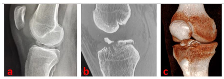
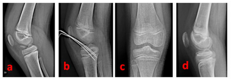
Introduction: Tibial spine avulsion injury, tibial eminence injury, tibial spine fracture, and anterior cruciate ligament (ACL) avulsion are multiple terms that express the same pathological condition. It can be encountered both in the pediatric and adult population. A wide array of surgical techniques have been proposed to manage displaced tibial spine avulsions. Anyway, insufficient evidence is currently available to prefer one fixation technique over another, and a gold-standard arthroscopy-based technique is still missing. In this article, we describe a mini-invasive, safe and user-friendly technique for arthroscopic reduction and internal fixation of displaced tibial eminence fractures.

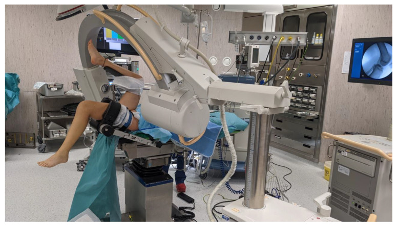
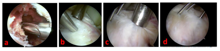
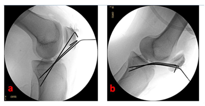
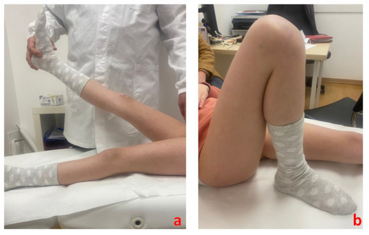
Materials and methods: Standard and patient-specific accessory arthroscopic portals allow for full access to knee visualization and management of concomitant intraarticular lesions. After performing the debridement of the inflammatory tissue and the release of eventual interposed tissues in the fracture site, the tibial eminence avulsion can be reduced by using a less-invasive bone impactor. With the knee flexed to 90°, the fracture fragments are then synthesized (under fluoroscopic control) with three thin Kirschner wires inserted in a proximal-distal direction in a cross-shaped geometry.

Results: This technique allows a fast surgical and hospitalization time, a punctiform arthrotomy, proximal tibial physis preservation, and an early rehabilitation program.

Conclusions: This novel technique seems attractive and very promising since it is respectful of the epiphyseal growth plates and is thus suitable for children and adolescents.

Keywords: anterior cruciate ligament; arthroscopy; osteosynthesis; tibial eminence fracture; tibial spine fracture.


Conflict of interest statement

Each author declares that they have no commercial associations (e.g., consultancies, stock ownership, equity interest, patent/licensing arrangement, etc.) that might pose a conflict of interest in connection with the submitted article.

Figures

Figure 1
Figure 1
A true lateral knee radiographic view of the right knee of a 16-year-old male patient of our series shows a Type 3 tibial avulsion fracture (a); CT scans are useful to define the pattern of the fracture and drive the management: CT sagittal view scan (b) and 3D CT reconstruction (c) of the same patient.
Figure 2
Figure 2
Operating theatre setup: the patient is in a supine position with the affected leg immobilized in a leg holder and the opposite leg held with a gynecologic leg holder to grant adequate workspace. C-arm fluoroscopy is set to radiographically guide fracture reduction and osteosynthesis with Kirschner wires.
Figure 3
Figure 3
Arthroscopic view of tibial spine avulsion management of the left knee of a 16-year-old female patient—(a): light fracture site debridement is necessary to grant adequate visualization; (b): the reduction of the fracture is facilitated by using a less-invasive bone impactor; (c): a cannulated aimer is a useful tool to drive the Kirschner wires positioning; (d): the osteosynthesis is performed and completed by positioning three thin Kirschner wires in a cross-shaped geometry.
Figure 4
Figure 4
Fluoroscopic control of fracture reduction and osteosynthesis devices positioning of the left knee of a 16-year-old female patient with the knee flexed to 90° (a) and the knee completely extended (b).
Figure 5
Figure 5
A radiographic summary overview of the right knee of a 9-year-old female patient—(a): pre-operative lateral view of a 3-type TSA; (b): post-operative radiographic control showing the cross-shaped geometry of the Kirschner wires; (c): antero-posterior radiographic control 15 months after surgery; (d): lateral radiographic control 15 months after surgery (complete restoration).
Figure 6
Figure 6
Three-month post-surgery clinical follow-up of the right knee of a 9-year-old female patient shows complete restoration of (a) full knee extension and (b) full knee flexion.

References

    1. Coyle C., Jagernauth S., Ramachandran M. Tibial eminence fractures in the paediatric population: A systematic review. J. Child. Orthop. Surg. 2010;18:395–405. doi: 10.1007/s11832-014-0571-6. - DOI - PMC - PubMed
    1. Wyatt J.D., Treme G., Veitch A.J., Johnson A.C. Incidence of Associated Knee Injury in Pediatric Tibial Eminence Fractures. J. Knee Surg. 2014;27:215–219. doi: 10.1055/s-0033-1360656. - DOI - PubMed
    1. Salvato D., Green D.W., Accadbled F., Tuca M. Tibial spine fractures: State of the art. J. ISAKOS Jt. Disord. Orthop. Sport. Med. 2023;13:S2059-7754(23)00518-7. doi: 10.1016/j.jisako.2023.06.001. - DOI - PubMed
    1. Patterson S.P., Christiansen G.B., Daffner R.H. Avulsion fracture of the tibial eminence in an adult with a unique mechanism of injury. Radiol. Case Rep. 2018;13:843–847. doi: 10.1016/j.radcr.2018.05.015. - DOI - PMC - PubMed
    1. Yu D., Yu R., Zhang J., Chen T., Zhang B. Arthroscopic treatment of adult displaced tibial eminence fractures with anchor and pushlock fixation. Medicine. 2020;99:e21237. doi: 10.1097/MD.0000000000021237. - DOI - PMC - PubMed

LinkOut - more resources