Review
. 2023 Sep 1;12(17):5720.
doi: 10.3390/jcm12175720.

Acute-Onset Retinal Conditions Mimicking Acute Optic Neuritis: Overview and Differential Diagnosis

Affiliations
Review

Acute-Onset Retinal Conditions Mimicking Acute Optic Neuritis: Overview and Differential Diagnosis

Emanuela Interlandi et al. J Clin Med. .

Abstract

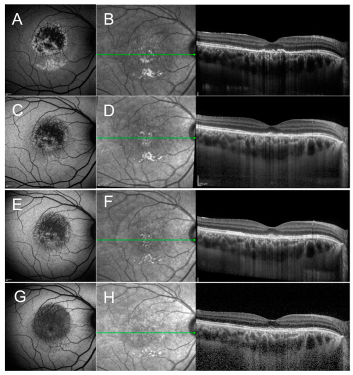
Acute optic neuritis (AON) is a common cause of sudden visual loss in young patients. Because of the risk of demyelinating disease, patients affected by unilateral or bilateral optic neuritis should be evaluated and treated accordingly. Despite advancements in imaging of the brain and retina, misdiagnosis of AON is not uncommon. Indeed, some acute disorders of the retina have the potential to mimic AON and their prompt diagnosis may avoid unnecessary neurologic investigation, psychological stress to the patient, and delays in treatment. This review describes uncommon retinal disorders presenting with sudden-onset visual loss and absent or subtle funduscopic manifestation that can mimic AON. Multimodal retinal imaging is essential in detecting these conditions and in their differential diagnosis. It behooves neurologists and general ophthalmologists to be aware of these entities and be familiar with multimodal imaging of the retina.

Keywords: acute-onset visual loss; differential diagnosis; multimodal imaging; optic neuritis; retinal conditions.


Conflict of interest statement

The authors declare no conflict of interest.

Figures

Figure 1
Figure 1
Fundus autofluorescence (FAF) and optical coherence tomography (OCT) scan of a case of acute idiopathic maculopathy of the right eye at presentation (A,B), 4 weeks after presentation (C,D), 8 weeks after presentation (E,F), and 12 weeks (G,H) after presentation. (A) FAF shows a predominant hypoautofluorescent signal surrounded by a hyperautofluorescent signal (Pattern 1 of the proposed disease Stage 1); (B) OCT shows disruption of the retinal pigment epithelium (RPE) and outer retinal layers with increased choroidal transmission; (CH) Subsequent FAF and OCT scan during follow-up show decreasing intensity of the hypoautofluorescent signal and gradual improvement of the integrity of RPE and outer retinal layers, respectively. (G) Two years after presentation, FAF shows homogeneously decreased FAF (Pattern 4 of the proposed disease Stage 4).
Figure 2
Figure 2
Multimodal retinal imaging of a case of acute retinal pigment epitheliitis of the left eye at presentation (AG) and after 6 weeks (HJ). (A) Color fundus photograph at presentation shows abnormal foveal reflex; (B) Fundus autofluorescence (AF) shows absence of AF abnormalities; (C) Optical coherence tomography (OCT) scan shows subfoveal retinal pigment epithelium and outer retinal disruption; (DG) Fluorescein angiography (D,E) and indocyanine green angiography (F,G) in the early and late frames do not show obvious abnormalities of the dyes circulation. (H) Color fundus photograph six weeks after presentation shows improvement of the foveal reflex; (I) AF findings were stable; (J) OCT scan shows recovery of the reflectivity of the subfoveal RPE and outer retinal layers.
Figure 3
Figure 3
Near-infrared reflectance imaging (NIR) and simultaneous optical coherence tomography (OCT) scan of a case of acute macular neuroretinopathy (AMN) of the right eye at presentation (A) and 4 weeks after presentation (B). (A) OCT scan passing through the tear-shaped AMN lesions visible on NIR reveals increased reflectivity of the outer plexiform layer and outer nuclear layer with disruption of the ellipsoid zone. (B) Follow-up OCT scan shows attenuation of the ellipsoid zone and overlying thinning of the outer nuclear layer.
Figure 4
Figure 4
Acute macular neuroretinopathy (AMN) of the right eye at presentation (A,B) and 2 weeks after presentation (C,D). (A) Color fundus photography is unremarkable, (B) OCT scan shows a small juxtafoveal hyperreflectivity in the outer retina. (C) Microperimetry reveals a focal depression of the macular sensitivity; (D) OCT scan shows attenuation of the ellipsoid zone in correspondence of a tear-shaped hyporeflective area visible in NIR (white arrow).
Figure 5
Figure 5
Near-infrared reflectance (NIR) imaging and simultaneous optical coherence tomography (OCT) scan of a case of paracentral acute middle maculopathy (PAMM) of the left eye at presentation (A) 2 weeks after presentation (B) and 8 weeks after presentation (C). (A) OCT scan shows hyperreflective band-like lesions involving the middle layers of the retina at the level of the inner nuclear layer (INL). (B,C) Follow-up OCT scan shows decreased reflectivity and subsequent thinning of the INL.
Figure 6
Figure 6
Multimodal imaging of acute zonal occult outer retinopathy, variant acute annular outer retinopathy of the right eye. (A) Pseudocolor fundus photography shows peripapillary atrophy and pigmentary changes in the superior periphery. (B) Ultra-widefield autofluorescence shows a hyperautofluorescent ring around the optic nerve and hypofluorescent lesions in the superior periphery. (C) Structural optical coherence tomography scan centered on the fovea (top image) and on the optic nerve (bottom image) shows the loss of the peripapillary photoreceptor layers (white curly brackets). (D) Fluorescein angiography shows a hyperfluorescent annular lesion around the optic nerve and hypo/hyperfluorescent lesions in the superior periphery. (E) Indocyanine green angiography shows mild hypofluorescence around the optic nerve and hypofluorescent lesions in the superior periphery.
Figure 7
Figure 7
Multimodal imaging of acute idiopathic blind spot enlargement of the left eye, at presentation and during follow-up. (A) Baseline row shows in order: blue autofluorescence with hyperautofluorescent spots centered on the optic nerve resembling multiple evanescent white dot syndrome and peripapillary hypoautofluorescent pigmentary changes; infrared image with corresponding retinal nerve fiber layer (RNFL) optical coherence tomography (OCT) showing disruption of the outer retina and increased thickness of RNFL; macular OCT showing disruption of the outer retina. (B) Two-week follow-up row shows in order: faint hyperautofluorescent lesions; infrared image with corresponding RNFL OCT showing decreased thickness of RNFL and improved outer retina; macular OCT showing improvement of the outer retina. (C) One-month follow-up row shows in order: disappearance of the lesions on autofluorescence; almost resolved appearance of the peripapillary outer retina; resolved appearance of outer retina at the macula. (D) Visual field test one year before the appearance of the spots (left image) and at disease onset (right image) showing a peripapillary scotoma.
Figure 8
Figure 8
Multimodal imaging of multiple evanescent white dot syndrome of the right eye at presentation. (A) Pseudocolor fundus photography shows some faint yellowish lesions at the posterior pole. (B) Green autofluorescence shows hyperautofluorescent spots spread beyond the vascular arcades. (C) Fluorescein angiography shows hyperfluorescent lesions in a wreath-like pattern and hot disc. (D) Near-infrared image shows the same lesions with enhanced granularity at the fovea. (E) Structural optical coherence tomography scan corresponding to the green line in panel (D) showing outer retina disruption.

References

    1. Peng P.H., Lee T.S., Cheng C.K., Peng C.H., Chan W.C. Acute zonal occult outer retinopathy presenting as optic neuritis. Taiwan J. Ophthalmol. 2020;10:222–226. doi: 10.4103/tjo.tjo_11_20. - DOI - PMC - PubMed
    1. Shindo A., Kokubo Y., Taniguchi A., Kuze M., Kuzuhara S. Case of acute zonal occult outer retinopathy (AZOOR): A 15 years’ mislabeling as retrobulbar optic neuritis. Rinsho Shinkeigaku. 2007;47:116–118. - PubMed
    1. Pellegrini F., Interlandi E. A case of multiple evanescent white dot syndrome misdiagnosed as optic neuritis: Differential diagnosis for the neurologist. J. Neurosci. Rural Pract. 2016;7:283–285. doi: 10.4103/0976-3147.178658. - DOI - PMC - PubMed
    1. Yannuzzi L.A., Jampol L.M., Rabb M.F., Sorenson J.A., Beyrer C., Wilcox L.M. Unilateral acute idiopathic maculopathy. Arc. Ophthalmol. 1991;109:1411–1416. doi: 10.1001/archopht.1991.01080100091049. - DOI - PubMed
    1. Freund K.B., Yannuzzi L.A., Barile G.R., Spaide R.F., Milewski S.A., Guyer D.R. The expanding clinical spectrum of unilateral acute idiopathic maculopathy. Arch. Ophthalmol. 1996;114:555–559. - PubMed

LinkOut - more resources