. 2023 Sep 2;12(17):5727.
doi: 10.3390/jcm12175727.

Intra-Articular Ultrasonography Probe for Minimally Invasive Upper Extremity Arthroscopic Surgery: A Phantom Study

Affiliations

Intra-Articular Ultrasonography Probe for Minimally Invasive Upper Extremity Arthroscopic Surgery: A Phantom Study

Shintaro Oyama et al. J Clin Med. .

Abstract

Background: Upper extremity arthroscopic surgery is a highly technique-dependent procedure that requires the surgeon to assess difficult cartilage conditions and manage the risk of iatrogenic damage to nerves and vessels adjacent to the joint capsule in a confined joint space, and a device that can safely assist in this procedure has been in demand.

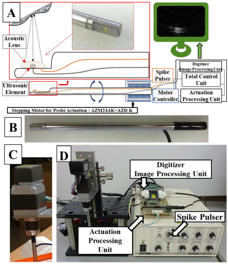
Methods: In this study, we developed a small intra-articular ultrasound (AUS) probe for upper extremity joint surgery, evaluated its safety using underwater sound field measurement, and tested its visualization with a phantom in which nerves and blood vessels were embedded.

Results: Sound field measurement experiments confirmed the biological safety of the AUS probe's output, while confirming that sufficient output power level performance was obtained as an ultrasound measurement probe. In addition, images of blood vessels and nerves were reconstructed discriminatively using A-mode imaging of the agar phantom.

Conclusions: This study provides proof-of-concept of the AUS probe in upper extremity surgery. Further studies are needed to obtain approval for use in future medical devices.

Keywords: arthroscopic ultrasonography; intra-articular examination probe; medical image processing; phantom study.


Conflict of interest statement

The authors declare no conflict of interest.

Figures

Figure 1
Figure 1
A system schema (A) of the manufactured probe (orange line: acoustic pulse line, black line: signal line from the ultrasonic element to the digitizer), a photograph (B) of the actual probe, a stepping motor (C) for controlling rotation of the probe, and an overall mechanism diagram (D).
Figure 2
Figure 2
A detailed system block diagram. The AZD-KD motor controller can execute motions with multiple motion modes, origins, and speeds set in advance by inputting signals to M0, M1, and M2. M0 and M1 were controlled from the APX-5040 digital output channel, D03 and D04, and M2 were fixed at a high level. START/READY on AZD-KD are a signal to notify the start/completion of motion. In this experiment, the registered motion settings of AZD-KD were set to reciprocate between angles −45° and 45° at a speed of 5000 r/min.
Figure 3
Figure 3
A computer aided design (CAD) model of the mold for the agar phantom. Lids have a width of 142 mm, a length of 142 mm, and a height of 100 mm, each having a diameter of 7 mm and having 14 holes each for passing tissue on a diagonal line at intervals of 10 mm on the lid. A hole of 15 mm diameter is made in the center of the top lid for inserting the probe (A); an actual molded plastic mold. Nerves and blood vessels can be fixed here. A tube is connected from a syringe to maintain the lumen of the blood vessel, and the vessel is filled with physiological saline (B); and the agar dissolved therein is cooled to 40 °C and poured and solidified (C); and finally physiological saline is put into the central hole, and an ultrasonic probe is inserted and observed (D). Please find the Supplementary Video S1.
Figure 4
Figure 4
Setting of underwater acoustic intensity measuring device (A). It is measured in pure water at room temperature. The hydrophone moves as it measures around the probe (B). The image below shows the measured amplitude overlaid on a photo. Graph plotting ultrasonic intensity around the probe (for the first measurement) (C); the waveform remains almost the same for all three measurements. The intensity distribution is uniform around the probe, and the strongest point shows sufficient intensity for an ultrasonic measurement probe.
Figure 5
Figure 5
Ultrasonography images of blood vessel (A) and ulnar nerve (B) in 2D and 3D reconstruction. Intensity represented in grayscale (high intensity: white, low intensity: black) in 2D, heatmap (high intensity: yellow, low intensity: red) in 3D.

References

    1. Randelli P., Dejour D., van Dijk C.N., Denti M., Seil R. Arthroscopy: Basic to Advanced. Springer; Berlin/Heidelberg, Germany: 2016.
    1. Ahmed A.F., Alzobi O.Z., Hantouly A.T., Toubasi A., Farsakoury R., Alkhelaifi K., Zikria B. Complications of Elbow Arthroscopic Surgery: A Systematic Review and Meta-analysis. Orthop. J. Sports Med. 2022;10:23259671221137863. doi: 10.1177/23259671221137863. - DOI - PMC - PubMed
    1. Atesok K., Doral M.N., Whipple T., Mann G., Mei-Dan O., Atay O.A., Beer Y., Lowe J., Soudry M., Schemitsch E.H. Arthroscopy-assisted fracture fixation. Knee Surg. Sports Traumatol. Arthrosc. 2011;19:320–329. doi: 10.1007/s00167-010-1298-7. - DOI - PubMed
    1. Beredjiklian P.K., Bozentka D.J., Leung Y.L., Monaghan B.A. Complications of wrist arthroscopy. J. Hand Surg. Am. 2004;29:406–411. doi: 10.1016/j.jhsa.2003.12.020. - DOI - PubMed
    1. Stetson W.B., Vogeli K., Chung B., Hung N.J., Stevanovic M., Morgan S. Avoiding Neurological Complications of Elbow Arthroscopy. Arthrosc. Tech. 2018;7:e717–e724. doi: 10.1016/j.eats.2018.03.010. - DOI - PMC - PubMed

LinkOut - more resources