. 2023 Aug 25;24(17):13221.
doi: 10.3390/ijms241713221.

Structures of the Insecticidal Toxin Complex Subunit XptA2 Highlight Roles for Flexible Domains

Affiliations

Structures of the Insecticidal Toxin Complex Subunit XptA2 Highlight Roles for Flexible Domains

Cole L Martin et al. Int J Mol Sci. .

Abstract

The Toxin Complex (Tc) superfamily consists of toxin translocases that contribute to the targeting, delivery, and cytotoxicity of certain pathogenic Gram-negative bacteria. Membrane receptor targeting is driven by the A-subunit (TcA), which comprises IgG-like receptor binding domains (RBDs) at the surface. To better understand XptA2, an insect specific TcA secreted by the symbiont X. nematophilus from the intestine of entomopathogenic nematodes, we determined structures by X-ray crystallography and cryo-EM. Contrary to a previous report, XptA2 is pentameric. RBD-B exhibits an indentation from crystal packing that indicates loose association with the shell and a hotspot for possible receptor binding or a trigger for conformational dynamics. A two-fragment XptA2 lacking an intact linker achieved the folded pre-pore state like wild type (wt), revealing no requirement of the linker for protein folding. The linker is disordered in all structures, and we propose it plays a role in dynamics downstream of the initial pre-pore state.

Keywords: Cryo-EM; TcA; X-ray crystallography; toxin translocase.


Conflict of interest statement

The authors declare no conflict of interest.

Figures
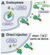
Figure 1
Figure 1
Proposed mechanisms of ABC Toxin Complex (Tc) cell recognition and cytotoxicity. (A) Central mechanism. Step 1: TcA and TcB-TcC subunits are secreted from bacteria (e.g., Xenorhabdus, Photorhabdus, Yersinia, etc., see Figure S2 for phylogenetic analysis); The structure presented here is an insect specific TcA designated XptA2 secreted by Xenorhabdus nematophila. Step 2: holotoxin formation by complex assembly; Step 3: recognition, binding to cell surface membrane receptor(s), endocytosis, and transport to lysosome; Step 4: pH-dependent change to pore state and injection of the toxin peptide into the cytoplasm where it folds into an enzyme that is toxic for cell division [26]. (B) Alternative mechanism. Steps 1 and 2 as with the central mechanism; Step 3: recognition and binding of Tcs to cell surface membrane receptors; Step 4: the high pH of the insect gut may allow the transition to the pore state at the plasma membrane and direct injection of the toxin into the cytoplasm [23].
Figure 2
Figure 2
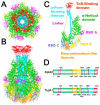
Structure of XptA2 from Xenorhabdus nematophila. (A) Bottom view of XptA2 colored by domain. (B) Side view of XptA2 colored by domain. (C) A single chain of XptA2 with each domain color-coded and labeled. (D) Domain comparison of XptA2 and TcdA1 according to the color schematic in A, B, and C.
Figure 3
Figure 3
RMSD comparison of the structures of XptA2 by X-ray crystallography and Cryo-EM. (A) Side view of XptA2 with RBD-B (residues 1365–1508) of chain D encircled. (B) An end-on view of XptA2 with RBD-B (residues 1365–1508) of chain D encircled. (C) Zoom of XptA2 chain D shows the indentation of RBD-B due to lattice packing of this specific X-ray structure (See Figure S3 for 2mFo-DFc and cryo-EM map comparisons). RBD-B chain D flexibility is likely a property of this domain in all five pentamers and highlights a potential hotspot for conformational dynamics and/or receptor binding. The scale bar shows the color gradient of the RMSD schema used for all three panels. The panels were created within PyMOL using the Python module colorbyrmsd.py written by Shandilya, Vertrees and Holder.
Figure 4
Figure 4
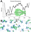
XptA2 pore dimensions and constriction analysis. (A) Width constrictions over the 250 Å translocation channel. The tightest dimension (1.5 Å) occurs at the transmembrane (TM) region, followed by a constriction at R2190 (~2.5 Å) and another at Q2204 (~3 Å). (B) Constriction of the XptA2 pore from the five-fold arrangement of R2190 side chains in the X-ray crystal structure. 2mFo-DFc map is colored in blue mesh contoured to 1.5 σ, and the mFo-DFc map is colored orange contoured to 7 σ. A strong unknown density appears at the five-fold axis. (C) Same view of R2190 for the cryo-EM structure as panel (B). Cryo-EM density is colored in blue mesh contoured to 5 σ.
Figure 5
Figure 5
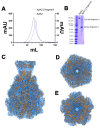
Pre-pore state of two-fragment XptA2 at 8.2 Å resolution. (A) Overlay of size exclusion chromatographs (SEC) of wt XptA2 and the XptA2 2-fragment construct. (B) SDS-PAGE comparing wtXptA2 and 2-fragment XptA2 after SEC. (C) Side view of the refined model for 2-fragment XptA2 (orange cartoon) fit into the experimental cryo-EM density (blue mesh). (D) 2-fragment XptA2 viewed from the toxin-binding face of the pentamer. (E) View of 2-fragment XptA2 from the transmembrane piercing face.
Figure 6
Figure 6
Molecular Dynamics and Differential Scanning Fluorometry of wt vs. 2-fragment XptA2. (A) RMSD comparison of wt XptA2 to 2-fragment XptA2 over 100 ns. (B) Solvent-accessible surface area (SASA) comparison of wtXptA2 to 2-fragment XptA2 over 100 ns. (C) Fluorescence emission at 330 nm as a function of temperature for the wt XptA2. (D) Fluorescence emission at 330 nm as a function of temperature for the 2-fragment XptA2.

References

    1. Sheets J.J., Hey T.D., Fencil K.J., Burton S.L., Ni W., Lang A.E., Benz R., Aktories K. Insecticidal toxin complex proteins from Xenorhabdus nematophilus: Structure and pore formation. J. Biol. Chem. 2011;286:22742–22749. doi: 10.1074/jbc.M111.227009. - DOI - PMC - PubMed
    1. Chaston J.M., Suen G., Tucker S.L., Andersen A.W., Bhasin A., Bode E., Bode H.B., Brachmann A.O., Cowles C.E., Cowles K.N., et al. The entomopathogenic bacterial endosymbionts Xenorhabdus and Photorhabdus: Convergent lifestyles from divergent genomes. PLoS ONE. 2011;6:e27909. doi: 10.1371/journal.pone.0027909. - DOI - PMC - PubMed
    1. Sicard M., Hinsinger J., Le Brun N., Pages S., Boemare N., Moulia C. Interspecific competition between entomopathogenic nematodes (Steinernema) is modified by their bacterial symbionts (Xenorhabdus) BMC Evol Biol. 2006;6:68. doi: 10.1186/1471-2148-6-68. - DOI - PMC - PubMed
    1. Shawer R., Donati I., Cellini A., Spinelli F., Mori N. Insecticidal Activity of Photorhabdus luminescens against Drosophila suzukii. Insects. 2018;9:148. doi: 10.3390/insects9040148. - DOI - PMC - PubMed
    1. Blackburn M.B., Domek J.M., Gelman D.B., Hu J.S. The broadly insecticidal Photorhabdus luminescens toxin complex a (Tca): Activity against the Colorado potato beetle, Leptinotarsa decemlineata, and sweet potato whitefly, Bemisia tabaci. J Insect Sci. 2005;5:32. doi: 10.1093/jis/5.1.32. - DOI - PMC - PubMed

LinkOut - more resources