Review
. 2023 Aug 28;24(17):13316.
doi: 10.3390/ijms241713316.

Significance of the cGAS-STING Pathway in Health and Disease

Affiliations
Review

Significance of the cGAS-STING Pathway in Health and Disease

Jinglin Zhou et al. Int J Mol Sci. .

Abstract

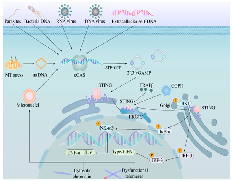
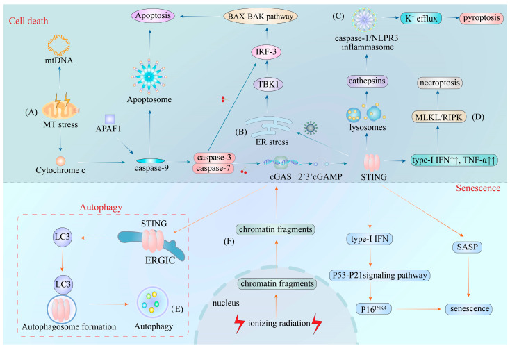
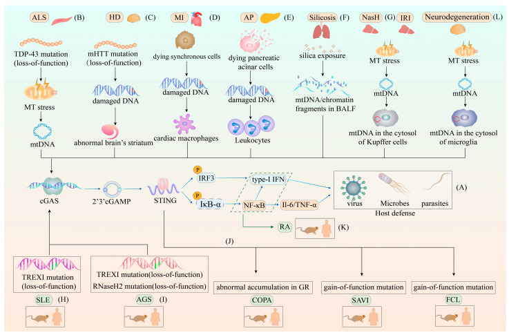
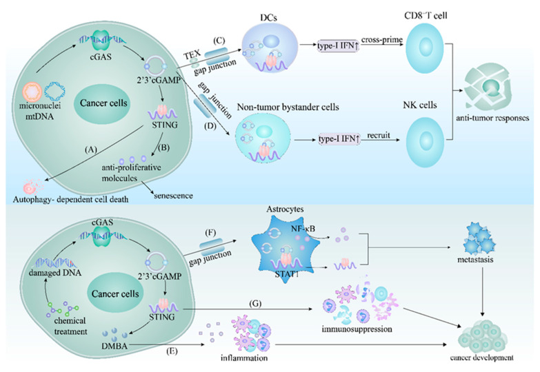
The cyclic GMP-AMP synthase (cGAS)-stimulator of interferon genes (STING) pathway plays a significant role in health and disease. In this pathway, cGAS, one of the major cytosolic DNA sensors in mammalian cells, regulates innate immunity and the STING-dependent production of pro-inflammatory cytokines, including type-I interferon. Moreover, the cGAS-STING pathway is integral to other cellular processes, such as cell death, cell senescence, and autophagy. Activation of the cGAS-STING pathway by "self" DNA is also attributed to various infectious diseases and autoimmune or inflammatory conditions. In addition, the cGAS-STING pathway activation functions as a link between innate and adaptive immunity, leading to the inhibition or facilitation of tumorigenesis; therefore, research targeting this pathway can provide novel clues for clinical applications to treat infectious, inflammatory, and autoimmune diseases and even cancer. In this review, we focus on the cGAS-STING pathway and its corresponding cellular and molecular mechanisms in health and disease.

Keywords: anti-pathogen immunity; autoimmune disorder; cGAS–STING pathway; cancer; inflammation.


Conflict of interest statement

The authors declare no conflict of interest.

Figures

Figure 1
Figure 1
Activation of the cGAS–STING pathway during innate immune response. The DNA from intracellular and extracellular sources is sensed by cGAS. After DNA recognition, cGAS catalyzes its substrates, GTP and ATP, into cGAMP. cGAMP is then sensed by STING located at the ER. Upon cGAMP binding, STING translocates from the ER to the ERGIC and recruits TBK1, followed by which STING and TBK1 are trafficked to the Golgi with the assistance of COPII and TRAPβ. Finally, STING and TBK1 interact with IRF3 and IκBα, culminating in type-I IFN and cytokine production. Abbreviations: mtDNA, mitochondrial DNA; cGAS, cyclic GMP-AMP synthase; 2′3′-cGAMP, 2′3′-cGMP-AMP; STING, stimulator of interferon genes; ER, endoplasmic reticulum; ERGIC, ER–Golgi intermediate compartment; TRAPβ, translocon-associated protein β; COPII, cytoplasmic coat protein complex-II; TBK1, TANK-binding kinase 1; IRF-3, interferon regulatory factor-3; IκBα: inhibitors of transcription factor NF-κB; type-I IFN, type-I interferon; IL-6, interleukin 6; TNF-α, tumor necrosis factor-α; GTP, guanosine triphosphate; ATP, adenosine triphosphate.
Figure 2
Figure 2
The cGAS–STING pathway in cell death, autophagy, and senescence. (A) The cGAS–STING pathway in MT stress-derived apoptosis. Permeabilization of the mitochondrial outer membrane induces the release of mtDNA and cytochrome c into the cytosol. The mtDNA then triggers the activation of the cGAS–STING pathway, leading to the production of type-I IFN and the association of cytochrome c with APAF1 to form the caspase-9-induced apoptosome. This structure further drives apoptosis and promotes caspase-3 and -7 production, which in turn causes the cleavage of cGAS and IRF-3. (B) The cGAS–STING pathway in virus-derived apoptosis. During infection with extracellular pathogens, the pathogen-related ER stress results in the direct recruitment of phosphorylated TBK1 by STING to trigger apoptosis via a BAX–BAK-dependent pathway. (C) The cGAS–STING pathway in pyroptosis. In specific cell types, STING is sorted into lysosomes after the activation of the cGAS–STING pathway, which leads to the permeabilization of the lysosome membrane and the release of cathepsins into the cytosol. Next, the cytosolic cathepsins trigger the activation of caspase-1 and NLRP3 inflammasome to drive K+ efflux-dependent pyroptosis. (D) The cGAS–STING pathway in necroptosis. The activation of the cGAS–STING pathway by mtDNA leads to the overexpression of type-I IFN and TNF-α. This overexpression further stimulates the activation of MLKL and RIPK to execute necroptosis. (E) The cGAS–STING pathway in autophagy. To prevent the overexpression of type-I IFN, STING traffics to the ERGIC after binding to cGAMP, independently of the recruitment of TBK1. This STING-containing ERGIC then acts as a source of LC3B, which is essential to autophagosome formation to induce autophagy. (F) The cGAS–STING pathway in senescence. When cells undergo ionizing radiation, the NL cannot maintain the NE structure, leading to the leakage of chromatin fragments (created by senescence-associated DNA damage) from the nucleus to the cytosol to initiate the overproduction of type-I IFN and SASP via the cGAS–STING pathway. Consequently, the overproduction of type-I IFN activates the p53-p21 signaling pathway to increase p16INK4 levels, a hallmark of cellular senescence. Abbreviations: MT stress, mitochondrial stress; mt DNA, mitochondrial DNA; APAF1, adapter apoptotic protease activating factor-1; BAX, BCL-2 associated X protein; BAK, BCL-2 homologous killer; K+ efflux, potassium efflux; type-I IFN, type-I interferon; TNF-α, tumor necrosis factor-α; MLKL, mixed lineage kinase domain-like protein; RIPK3, receptor-interacting serine/threonine protein kinase 3; LC3B, microtubule-associated proteins 1A/1B light chain 3B; SASP, senescence-associated secretory phenotype; cGAS–STING, cyclic GMP-AMP synthase-stimulator of interferon genes; IRF-3, interferon regulatory factor-3; ER, endoplasmic reticulum; TBK1, TANK-binding kinase 1; ERGIC, ER–Golgi intermediate compartment; NL, nuclear lamina; NE, nuclear envelope.
Figure 3
Figure 3
The different roles of the cGAS–STING pathway in host defense and various diseases. (A) The role of the cGAS–STING pathway in host defense. The genome of invading pathogens, such as viruses, bacteria and parasites, in the cytosol can be sensed by cGAS, triggering the cGAS–STING pathway-induced production of type-I IFN and other cytokines to eliminate the genomes of invading pathogens. (B) The role of the cGAS–STING pathway in ALS. The missense mutation in the TDP-43 protein enables its absorption into the mitochondria and causes mtDNA leakage into the cytosol, prompting overproduction of type-I IFN and NF-κB to initiate hyperinflammatory responses. (C) The role of the cGAS–STING pathway in HD. The mutation of the N-terminal polyglutamine in the huntingtin protein (mHTT) leads to the abnormal accumulation of damaged DNA in the brain’s striatum, initiating the STING-dependent type-I IFN production and autophagy. (D) The role of the cGAS–STING pathway in MI. Synchronously dying cells are sensed by cardiac macrophages, and damaged DNA from these cells is internalized by cardiac macrophages. Furthermore, this internalization process leads to the inhibition of the transformation of cardiac macrophages from inflammatory cells to a reparative phenotype via the activation of the cGAS–STING pathway. (E) The role of the cGAS–STING pathway in AP. Damaged DNA released by the dying pancreatic acinar cells is internalized by leukocytes, resulting in severe inflammation due to the cGAS–STING pathway-induced overproduction of type-I IFN and other cytokines. (F) The role of the cGAS–STING pathway in silicosis. Silica microparticles invade the lungs, causing an increased release of chromatin fragments and mtDNA from dying cells into the BALF. These chromatin fragments and mtDNA then engage the cGAS–STING pathway to increase type-I IFN and other cytokine levels, causing chronic progressive fibrotic inflammation in the lungs. (G) The role of the cGAS–STING pathway in NASH and liver IRI. Lipotoxicity in NASH and ROS in IRI both lead to mitochondrial stress and the release of mtDNA into the cytosol of Kupffer cells. Furthermore, these changes result in adipose tissue inflammation via the cGAS–STING pathway and cGAS–STING–NLRP3 inflammasome-driven cell death in NASH and IRI, respectively. (H) The role of the cGAS–STING pathway in SLE. The loss-of-function mutation in TREX1 is suggested as the main cause of SLE. TREXI mutations lead to the abnormal accumulation of cytosolic DNA, which continuously activates the type-I IFN signaling cascade and amplifies inflammation via cGAS–STING. (I) The role of the cGAS–STING pathway in AGS. The loss-of-function mutation in TREX1 and the RNaseH2 complex are considered the major components of AGS. In this context, mutations in TREX1 and the RNaseH2 complex lead to the failure to eliminate genomic fragments in the cytosol and maintain genome stability, causing increased cGAS–STING pathway-dependent expression of type-I IFN. (J) The role of the cGAS–STING pathway in COPA, SAVI, and FCL. COPA is caused by the abnormal accumulation of STING in the GR. SAVI is attributed to the gain-of-function mutation in the STING-encoding gene, which leads to the spontaneous translocation of STING from the ER to the GR. This abnormal translocation activates intensive type-I IFN signals independently of cGAMP stimulation. Finally, FCL is caused by the heterozygous gain-of-function mutation in STING, resulting in the spontaneous dimerization of STING and constitutive activation of the type-I IFN signature. (K) The role of the cGAS–STING pathway in RA. The abnormal accumulation of cytosolic DNA leads to the expression of cytokines and chemokines via cGAS–STING activation, contributing to RA development. (L) The role of the cGAS–STING pathway in neurodegeneration. In microglia, mtDNA in the cytosol of microglia contributes to the chronic inflammatory response, which leads to neurodegeneration. Abbreviations: ALS, amyotrophic lateral sclerosis; MT stress, mitochondrial stress; mt DNA, mitochondrial DNA; TDP-43 protein, TAR DNA-binding protein 43; HD, Huntington disease; mHTT, mutation of the N-terminal polyglutamine in huntingtin protein; MI, myocardial infarction; AP, acute pancreatitis; BALF, bronchoalveolar lavage fluid; NASH, nonalcoholic steatohepatitis; IRI, ischemia-reperfusion injury; SLE, systemic lupus erythematosus; AGS, Aicardi–Goutières syndrome; COPA, COPA syndrome; SAVI, STING-associated vasculopathy with infantile-onset; FCL, familial chilblain lupus; RA, rheumatoid arthritis; cGAS–STING, cyclic GMP-AMP synthase-stimulator of interferon genes; type-I IFN, type-I interferon.
Figure 4
Figure 4
The cGAS–STING pathway in cancer. (A) In certain cancers, the cGAS–STING pathway induces autophagy-dependent cell death to limit the transformation of normal cells into cancerous cells. (B) The cGAS–STING pathway inhibits the growth of certain cancer cells by inducing the secretion of anti-proliferative molecules that promote senescence. (C) In specific cancers, 2′3′-cGAMP from the cancer cells enters the cytosol of DCs through the gap junction or TEX, activating STING to produce type-I IFN. The type-I IFN from the DCs then cross-primes CD8+ T cells to eliminate tumorigenic cells. (D) Within some cancers, 2′3′-cGAMP from the cancer cells is transferred into non-tumor bystander cells via the gap junction and activates STING, resulting in the recruitment of NK cells to restrain tumor growth. (E) In the case of certain cancer cells, chemical treatments cause the introduction of damaged DNA into the cytosol. This damaged DNA further engages the cGAS–STING pathway, inducing the secretion of DMBA to facilitate inflammation and maintain the tumor microenvironment. (F) STING activation also occurs after 2′3′-cGAMP from specific cancer cells are trafficked to astrocytes via the gap junction. The activated STING in the astrocytes promotes STAT1 and NF-κB production, further facilitating the development of metastatic cancer cells. (G) The loss of function of cGAS or STING leads to immunosuppression in cancer cells due to the hindered production of type-I IFN and other cytokines. Abbreviations: TEX, tumor-derived exosomes; DCs, infiltrating dendritic cells; NK cells, natural killer cells; DMBA, 7, 12-dimethylbenz(a)anthracene; cGAS–STING, cyclic GMP-AMP synthase-stimulator of interferon genes; 2′3′-cGAMP, 2′3′-cGMP-AMP; type-I IFN, type-I interferon.

References

    1. Roers A., Hiller B., Hornung V. Recognition of Endogenous Nucleic Acids by the Innate Immune System. Immunity. 2016;44:739–754. doi: 10.1016/j.immuni.2016.04.002. - DOI - PubMed
    1. Gong T., Liu L., Jiang W., Zhou R. DAMP-sensing receptors in sterile inflammation and inflammatory diseases. Nat. Rev. Immunol. 2020;20:95–112. doi: 10.1038/s41577-019-0215-7. - DOI - PubMed
    1. Hopfner K.P., Hornung V. Molecular mechanisms and cellular functions of cGAS-STING signalling. Nat. Rev. Mol. Cell Biol. 2020;21:501–521. doi: 10.1038/s41580-020-0244-x. - DOI - PubMed
    1. Ishikawa H., Barber G.N. STING is an endoplasmic reticulum adaptor that facilitates innate immune signalling. Nature. 2008;455:674–678. doi: 10.1038/nature07317. - DOI - PMC - PubMed
    1. Burdette D.L., Monroe K.M., Sotelo-Troha K., Iwig J.S., Eckert B., Hyodo M., Hayakawa Y., Vance R.E. STING is a direct innate immune sensor of cyclic di-GMP. Nature. 2011;478:515–518. doi: 10.1038/nature10429. - DOI - PMC - PubMed