. 2023 Aug 28;24(17):13334.
doi: 10.3390/ijms241713334.

Striking Neurochemical and Behavioral Differences in the Mode of Action of Selegiline and Rasagiline

Affiliations

Striking Neurochemical and Behavioral Differences in the Mode of Action of Selegiline and Rasagiline

Laszlo G Harsing Jr et al. Int J Mol Sci. .

Abstract

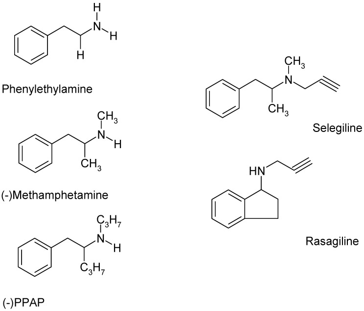
Selegiline and rasagiline are two selective monoamine oxidase B (MAO-B) inhibitors used in the treatment of Parkinson's disease. In their clinical application, however, differences in L-dopa-sparing potencies have been observed. The aim of this study was to find neurochemical and behavioral explanations for the antiparkinsonian effects of these drugs. We found that selegiline possesses a dopaminergic enhancer effect: it stimulated the electrically induced [3H]dopamine release without influencing the resting [3H]dopamine release from rat striatal slices in 10-10-10-9 mol/L concentrations. Rasagiline added in 10-13 to 10-5 mol/L concentrations did not alter the resting or electrically stimulated [3H]dopamine release. Rasagiline (10-9 mol/L), however, suspended the stimulatory effect of selegiline on the electrically induced [3H]dopamine release. The trace amine-associated receptor 1 (TAAR1) antagonist EPPTB (10-8-10-7 mol/L) also inhibited the stimulatory effect of selegiline on [3H]dopamine release. The effect of selegiline in its enhancer dose (5.33 nmol/kg) against tetrabenazine-induced learning deficit measured in a shuttle box apparatus was abolished by a 5.84 nmol/kg dose of rasagiline. The selegiline metabolite (-)methamphetamine (10-9 mol/L) also exhibited enhancer activity on [3H]dopamine release. We have concluded that selegiline acts as an MAO-B inhibitor and a dopaminergic enhancer drug, and the latter relates to an agonist effect on TAAR1. In contrast, rasagiline is devoid of enhancer activity but may act as an antagonist on TAAR1.

Keywords: [3H]dopamine release; conditioned avoidance response; dopaminergic activity enhancer effect; monoamine oxidase inhibition; rasagiline; rat striatum; selegiline; trace amine-associated receptor 1.


Conflict of interest statement

We declare this research work was partly supported by the Fujimoto Pharmaceutical Corporation, Osaka, Japan.

Figures

Figure 1
Figure 1
The chemical structures of the trace amine β-phenylethylamine, the releaser compound methamphetamine, the selective catecholamine activity enhancer (−)PPAP ((−)-1-phenyl-2-propylaminopentane), and the MAO-B inhibitors selegiline ((−)deprenyl) and rasagiline. Although the compounds shown here can be derived from β-phenylethylamine, there are definitive differences in the attached alkylamine side chains. In addition, a phenyl ring is present in the structures of β-phenylethylamine, methamphetamine, (−)PPAP, and selegiline, and there is an indane ring in rasagiline. (−)PPAP exerts a catecholamine activity enhancer effect, primarily on catecholaminergic transmission, but leaves MAO activity unchanged [10]. Selegiline and rasagiline, which possess a propargyl group attached to the nitrogen atom in the side chain, proved to be potent MAO-B inhibitors.
Figure 2
Figure 2
The time course of resting and electrical stimulation-induced [3H]dopamine release from rat striatum. Slices from rat striatum were prepared, incubated in the presence of [3H]dopamine, and superfused with oxygenated and preheated Krebs bicarbonate buffer. The [3H]dopamine release was expressed as the percent of content released, i.e., a percentage of the amount of [3H]dopamine in the tissue at the time of the release. The [3H]dopamine release was stimulated electrically (40 V, 10 Hz, 2 msec for 3 min) in fractions 4 (S1) and 18 (S2). The electrically stimulated fractional release S2 (2nd stimulation) over fractional release S1 (1st stimulation) (S2/S1 ratio) was 0.85 ± 0.06, representing a [3H]dopamine release with a vesicular origin. The resting fractional release B2 (fraction 17) over fractional release B1 (fraction 3) (B2/B1 ratio) was 0.77 ± 0.06. The tissue [3H]dopamine content approached a value of 232.60 ± 58.03 kBq/g at the end of the superfusion, mean ± S.E.M., n = 4. When the drug effect was studied, the drugs were added to the superfusion buffer between the 1st and 2nd electrical stimulations and maintained throughout the experiment.
Figure 3
Figure 3
Figure 3 shows the time course of resting [3H]dopamine release in the presence and absence of drugs (β-phenylethylamine, selegiline, rasagiline) measured from the rat striatum. Striatal slices from a rat brain were prepared, loaded with [3H]dopamine, and superfused with aerated and preheated Krebs bicarbonate buffer. The release of [3H]dopamine was expressed as a fractional rate calculated as the percent of content released. (A) Non-vesicular [3H]dopamine release was evoked by β-phenylethylamine (PEA) added to the superfusion buffer from fraction 10 and maintained throughout the experiment. A quantity of 10−5 mol/L of β-phenylethylamine increased non-vesicular [3H]dopamine release from 1.89 ± 0.21 to 3.27 ± 0.20 percent of the content released in 3 min (fractions 9 and 10), mean ± S.E.M., n = 4–5. (B) The effects of the MAO B inhibitors selegiline and rasagiline on resting [3H]dopamine release from rat striatum. For control, see Figure 3A. Selegiline and rasagiline were added to striatal slices from fraction 10 in a concentration of 10−5 mol/L and kept present throughout the experiment. In contrast to β-phenylethylamine, neither selegiline nor rasagiline altered the resting [3H]dopamine release in striatal slices, mean ± S.E.M., n = 4.
Figure 4
Figure 4
Concentration-dependent effects of selegiline on resting and electrical stimulation-induced [3H]dopamine release from rat striatum. For experimental procedure, see Figure 2. The resting and the electrical stimulation-induced [3H]dopamine release were determined as fractional rates. Selegiline was added in a concentration range from 10−13 to 10−5 mol/L to striatal slices. (A) The effect of selegiline on the electrical stimulation-induced [3H]dopamine release determined in the 1st (absence of drug, S1) and 2nd (presence of drug, S2) stimulations carried out in fractions 4 and 18. In the control experiments (c), the S2/S1 value was 0.80 ± 0.06. Selegiline exerted a dual effect: it increased electrical stimulation-induced [3H]dopamine release in 10−10 to 10−9 and 10−6 to 10−5 mol/L concentrations. ANOVA followed by Dunnett’s test, F(9,36) = 3.622, p = 0.002, * p < 0.05, mean ± S.E.M., n = 4–6. (B) The B2/B1 ratio indicates the effect of selegiline on the resting fractional [3H]dopamine release determined in fraction 3 (absence of drug, B1) and in fraction 17 (presence of drug, B2). The B2/B1 value was 0.92 ± 0.14 in the control experiments (c). Selegiline added in a concentration range of 10−13 to 10−5 mol/L was without effect on the resting [3H]dopamine release from the rat striatum. ANOVA followed by Dunnett’s test, F(9,36) = 0.581, p = 0.803, mean ± S.E.M., n = 4–6.
Figure 5
Figure 5
Reversal by EPPTB of the selegiline-induced [3H]dopamine release in rat striatum. For experimental procedure, see Figure 2. The resting and the electrical stimulation-induced [3H]dopamine release were determined as fractional rates. In these experiments, selegiline was added from fraction 8 to striatal slices in a concentration of 10−9 mol/L and maintained throughout the experiment in the presence and absence of EPPTB. When used, EPPTB was added to striatal slices from fraction 1 in a concentration of either 10−8 or 10−7 mol/L and was present throughout the experiment. One-way ANOVA was followed by Dunnett’s test, F(5,32) = 2.977, p < 0.05. Student t-statistics for two-means, control vs. selegiline effect, # p < 0.01, selegiline vs. selegiline plus EPPTB effects, (EPPTB 10−8 mol/L, * p < 0.05 and EPPTB 10−7 mol/L, * p < 0.01), mean ± S.E.M., n = 4–8.
Figure 6
Figure 6
The lack of dopaminergic enhancer effect of rasagiline on the resting and electrical stimulation-induced [3H]dopamine release from rat striatum. For experimental procedure, see Figure 2. The resting and electrical stimulation-induced [3H]dopamine release was determined as a fractional rate. Rasagiline was added in a concentration range from 10−13 to 10−5 mol/L to the superfusion buffer from fraction 8 and maintained throughout the experiment. (A) The S2/S1 ratio indicates the effect of rasagiline on the electrical stimulation-induced [3H]dopamine release determined in the 1st (absence of drug, S1) and 2nd (presence of drug, S2) stimulations carried out in fractions 4 and 18. The S2/S1 ratio in the control experiments (c) was found to be 0.83 ± 0.04. ANOVA followed by Dunnett’s test, F(9,30) = 0.197, p = 0.992, mean ± S.E.M., n = 4. (B) The B2/B1 ratio indicates the effect of rasagiline on the resting fractional [3H]dopamine release determined in fraction 3 (absence of drug, B1) and in fraction 17 (presence of drug, B2). The B2/B1 value was 0.88 ± 0.05 in the control experiments (c). ANOVA followed by Dunnett’s test, F(9,30) = 1.357, p = 0.250, mean ± S.E.M., n = 4.
Figure 7
Figure 7
Rasagiline reversed the selegiline-induced [3H]dopamine release in rat striatum. For experimental procedure, see Figure 2. The resting and electrical stimulation-induced [3H]dopamine release was determined as a fractional rate. Selegiline was added to striatal slices from fraction 8 in a concentration of 10−9 mol/L and maintained throughout the experiment in the presence and absence of rasagiline. When used, rasagiline was added to the striatal slices from fraction 1 in a concentration of 10−9 mol/L and maintained throughout the experiment. ANOVA followed by Dunnett’s test, F(3,28) = 7.228, p < 0.001. Student’s t-statistics for two-means, control vs. selegiline effect, # p < 0.01, selegiline vs. selegiline plus rasagiline effects, * p < 0.05; rasagiline vs. rasagiline plus selegiline effects did not differ significantly, mean ± S.E.M., n = 8.
Figure 8
Figure 8
The effects of the despropargyl metabolite of selegiline (−)methamphetamine ((±)/(−)methamph) on [3H]dopamine release from rat striatum. For experimental procedure, see Figure 3 and Figure 4. The resting and the electrical stimulation-induced [3H]dopamine release were determined as fractional rates. (A) (−)Methamphetamine increased vesicular (electrically induced, S2/S1) but not the non-vesicular (resting, B2/B1) [3H]dopamine release when added to the striatal slices in a concentration of 10−9 mol/L, Student’s t-statistics for two-means, * p < 0.01, mean ± S.E.M., n = 4–4. (B) The effects of (−) and (±)methamphetamine in 10−5 mol/L concentration on resting [3H]dopamine release in rat striatum. As expected, the [3H]dopamine-releasing effect of (−)methamphetamine was substantially less than that of the racemic form. Striatal [3H]dopamine release was 4.49 ± 0.60 and 13.05 ± 1.65 percent of the content released in response to 10−5 mol/L (−)methamphetamine and (±)methamphetamine, respectively. Student’s t-statistics for two means, p < 0.01, mean ± S.E.M., n = 4. (−)Methamphetamine added in a concentration of 10−9 mol/L was without effect on [3H]dopamine release.
Figure 9
Figure 9
Effect of selegiline, rasagiline, and their combination against tetrabenazine-induced memory loss, measured in a shuttle box. (A) panel—number of conditioned avoidance responses (CAR), (B) panel—number of escape failures (EF). Tetrabenazine (1 mg/kg) abolished the positive CARs ((A) panel) and the escape responses, resulting in about 80% EF ((B) panel). This effect was reduced by selegiline (0.001 mg/kg equivalent to 5.33 nmol/kg, sc.) but not rasagiline. The combination of rasagiline with selegiline, however, terminated selegiline’s effect. TBZ, tetrabenazine; SEL, selegiline; RAS, rasagiline. Statistical analysis—CAR - F = 53.86 *** p < 0.001, EF F = 73.78 *** p < 0.001. No of animals, 8 per group.
Figure 10
Figure 10
A model for the role of the trace amine associated receptor 1 (TAAR1) and signaling in the mechanisms of selegiline and rasagiline to influence dopaminergic neurotransmission in rat striatum. We propose that the dopaminergic enhancer activity effect of selegiline is related to the stimulation of TAAR1 and its signal transduction pathway. Selegiline is taken up by the plasma membrane dopamine transporter (DAT) and activates TAAR1, a Gs protein-coupled, intracellularly located metabotropic receptor [33,34]. Stimulation of this receptor will result in adenylyl cyclase activation, followed by an increase in cAMP production and augmented intracellular phosphorylation. Protein kinase C (PKC) phosphorylates a series of proteins involved in the exocytotic processes [35,36,37], and the stimulation-evoked vesicular release of dopamine increases. Classical amines, trace amines, and the releaser amphetamines also act as agonists on TAAR1 [19]. Our experiments strongly suggest that the MAO-B inhibitor selegiline might act as an agonist on TAAR1, whereas the MAO-B inhibitor rasagiline suspends selegiline–TAAR1 interaction. Enhanced TAAR1 and signaling may have a beneficial role in the abnormalities in dopaminergic neurochemical transmission observed both in presynaptic and postsynaptic levels [38].
Figure 11
Figure 11
Schematic chart displaying the behavioral experiment design.

Similar articles

Cited by

References

    1. Knoll J., Magyar K. Some puzzling effects of monoamine oxidase inhibitors. Adv. Biochem. Psychopharmacol. 1972;5:393–408. - PubMed
    1. Birkmayer W., Riederer P., Ambrozi L., Youdim M. Implications of combined treatment with ‘madopar’ and l-deprenil in parkinson’s disease: A Long-term Study. Lancet. 1977;309:439–443. doi: 10.1016/S0140-6736(77)91940-7. - DOI - PubMed
    1. Lancet Editorial Deprenyl in Parkinson’s Disease. Lancet. 1982;2:695–696. - PubMed
    1. Morelli M., Pinna A. Neurobiology of Parkinson’s Disease. Int. J. Mol. Sci. 2023;24:9933. doi: 10.3390/ijms24129933. - DOI - PMC - PubMed
    1. Glover V., Sandler M., Owen F., Riley G.J. Dopamine is a monoamine oxidase B substrate in man. Nature. 1977;268:80–81. doi: 10.1038/265080a0. - DOI - PubMed

LinkOut - more resources