. 2023 Aug 30;24(17):13437.
doi: 10.3390/ijms241713437.

Insight into Tetramolecular DNA G-Quadruplexes Associated with ALS and FTLD: Cation Interactions and Formation of Higher-Ordered Structure

Affiliations

Insight into Tetramolecular DNA G-Quadruplexes Associated with ALS and FTLD: Cation Interactions and Formation of Higher-Ordered Structure

Matja Zalar et al. Int J Mol Sci. .

Abstract

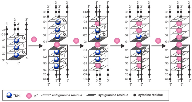
The G4C2 hexanucleotide repeat expansion in the c9orf72 gene is a major genetic cause of familial amyotrophic lateral sclerosis (ALS) and frontotemporal lobar degeneration (FTLD), with the formation of G-quadruplexes directly linked to the development of these diseases. Cations play a crucial role in the formation and structure of G-quadruplexes. In this study, we investigated the impact of biologically relevant potassium ions on G-quadruplex structures and utilized 15N-labeled ammonium cations as a substitute for K+ ions to gain further insights into cation binding and exchange dynamics. Through nuclear magnetic resonance spectroscopy and molecular dynamics simulations, we demonstrate that the single d(G4C2) repeat, in the presence of 15NH4+ ions, adopts a tetramolecular G-quadruplex with an all-syn quartet at the 5'-end. The movement of 15NH4+ ions through the central channel of the G-quadruplex, as well as to the bulk solution, is governed by the vacant cation binding site, in addition to the all-syn quartet at the 5'-end. Furthermore, the addition of K+ ions to G-quadruplexes folded in the presence of 15NH4+ ions induces stacking of G-quadruplexes via their 5'-end G-quartets, leading to the formation of stable higher-ordered species.

Keywords: ALS; DNA; FTLD; NMR; c9orf7; cations; quadruplex.


Conflict of interest statement

The authors declare no conflict of interest.

Figures

Figure 1
Figure 1
Imino and aromatic regions of the 1H NMR spectrum of d(G4C2) in the absence (A) and the presence (B) of 100 mM of 15NH4Cl. Spectra were recorded in 90% H2O and 10% 2H2O at 25 °C, pH 6.0, on a 600 MHz NMR spectrometer. The oligonucleotide concentration was 2.0 mM per strand. The doublet signal (J = 74 Hz) denoted with asterisks corresponds to ammonium ions bound to binding site O5 within the G-quadruplex.
Figure 2
Figure 2
(A) Folding topology and cation binding sites within G-quadruplex adopted by d(G4C2) in the presence of 15NH4+ ions, with the three ion binding sites shown. Syn- and anti-conformations of guanines are represented with grey and white rectangles, respectively. The arrows represent the observed movements between cation binding sites, with apparent rate constants depicted next to them. (B) Imino-aromatic region of the NOESY spectrum (left), showing cross-peaks between bound 15NH4+ ions and nearby imino protons, and the 2D 1H-15N HSQC spectrum (right) exhibiting cross-peaks corresponding to 15NH4+ ions in different chemical environments within [d(G4C2)]4. The vertical and horizontal dashed lines connect resonances of guanine imino and 15NH4+ ion protons. (C) The 2D 1H-15N NzExHSQC spectrum at a mixing time (τm) of 1.1 s (left) and the 2D 1H-15N HzExHSQC spectrum at a mixing time (τm) of 40 ms (right). Annotations of autocorrelation and exchange cross-peaks are shown next to the cross-peaks.
Figure 3
Figure 3
(A) Lowest energy structure of the [d(G4C2)]4 G-quadruplex in the presence of 15NH4+ ions (PDB ID 8C7B) and (B) of the dimeric form of the [d(G4C2)]4 G-quadruplex in the presence of K+ ions (PDB ID 8C7A). Residues G1, G2, G3, G4, C5, and C6 are shown in magenta, blue, green, violet, orange, and red, respectively. Cations within both structures are shown. Ion binding sites in (A) and (B) are marked with O5, I, O3, and OGC. OGC is the binding site between G4 and C5 residues. In (B) an additional binding site, OGG, is present. It is located between G1 and G1 residues, formed by 5′ stacking of two [d(G4C2)]4 G-quadruplexes. Stacking of G-quartets and G-quartets and cytosines are shown.
Figure 4
Figure 4
The projection of the center of a G-quartet composed of G4 residues (marked as G4-quartet) to the adjacent G-quartet composed of G3 residues (marked as G3-quartet) within [d(G4C2)]4 in the presence of 15NH4+ ions. Due to clarity, the 15NH4+ ion between G-quartets is not present. The O6 atoms of the G-quartets are shown as red sphere models, and centers of the G-quartets are shown in blue. d is the distance between the centers of two adjacent G-quartets.
Figure 5
Figure 5
(A) Free-energy profile for NH4+ ion movements through G-quartets of [d(G4C2)]4 G-quadruplex and the pulling direction of 15NH4+ ions, as well as (B) the representation of the reaction coordinate. G-quartets composed of G1, G2, G3, and G4 residues are marked as G1-, G2-, G3-, and G4-quartets, respectively.
Figure 6
Figure 6
Imino regions of 1H NMR and corresponding 1H-15N HSQC spectra of [d(G4C2)]4 at 25 °C, pH 6.0, in 10% 2H2O in the presence of 100 mM of 15NH4Cl and gradual addition of KCl. The oligonucleotide concentration was 2.0 mM per strand. Assignments of imino resonances at the beginning and end of titration are indicated above the signals. Annotations of cross-peaks are shown next to the peaks.
Figure 7
Figure 7
Gradual exchange of 15NH4+ by K+ ions and the formation of dimeric species as the concentration of K+ ions is increased from 0 to 80 mM.

Similar articles

Cited by

References

    1. Monchaud D. Biological Relevance & Therapeutic Applications of DNA-& RNA-Quadruplexes. Future Science; London, UK: 2015.
    1. Burge S., Parkinson G.N., Hazel P., Todd A.K., Neidle S. Quadruplex DNA: Sequence, topology and structure. Nucleic Acids Res. 2006;34:5402–5415. doi: 10.1093/nar/gkl655. - DOI - PMC - PubMed
    1. Phan A.T., Kuryavyi V., Patel D.J. DNA architecture: From G to Z. Curr. Opin. Struct. Biol. 2006;16:288–298. doi: 10.1016/j.sbi.2006.05.011. - DOI - PMC - PubMed
    1. Kocman V., Plavec J. A tetrahelical DNA fold adopted by tandem repeats of alternating GGG and GCG tracts. Nat. Commun. 2014;5:5831. doi: 10.1038/ncomms6831. - DOI - PMC - PubMed
    1. Kocman V., Plavec J. Tetrahelical structural family adopted by AGCGA-rich regulatory DNA regions. Nat. Commun. 2017;8:15355. doi: 10.1038/ncomms15355. - DOI - PMC - PubMed