. 2023 Aug 30;24(17):13436.
doi: 10.3390/ijms241713436.

ERK1/2-Dependent Phosphorylation of GABAB1(S867/T872), Controlled by CaMKIIβ, Is Required for GABAB Receptor Degradation under Physiological and Pathological Conditions

Affiliations

ERK1/2-Dependent Phosphorylation of GABAB1(S867/T872), Controlled by CaMKIIβ, Is Required for GABAB Receptor Degradation under Physiological and Pathological Conditions

Musadiq A Bhat et al. Int J Mol Sci. .

Abstract

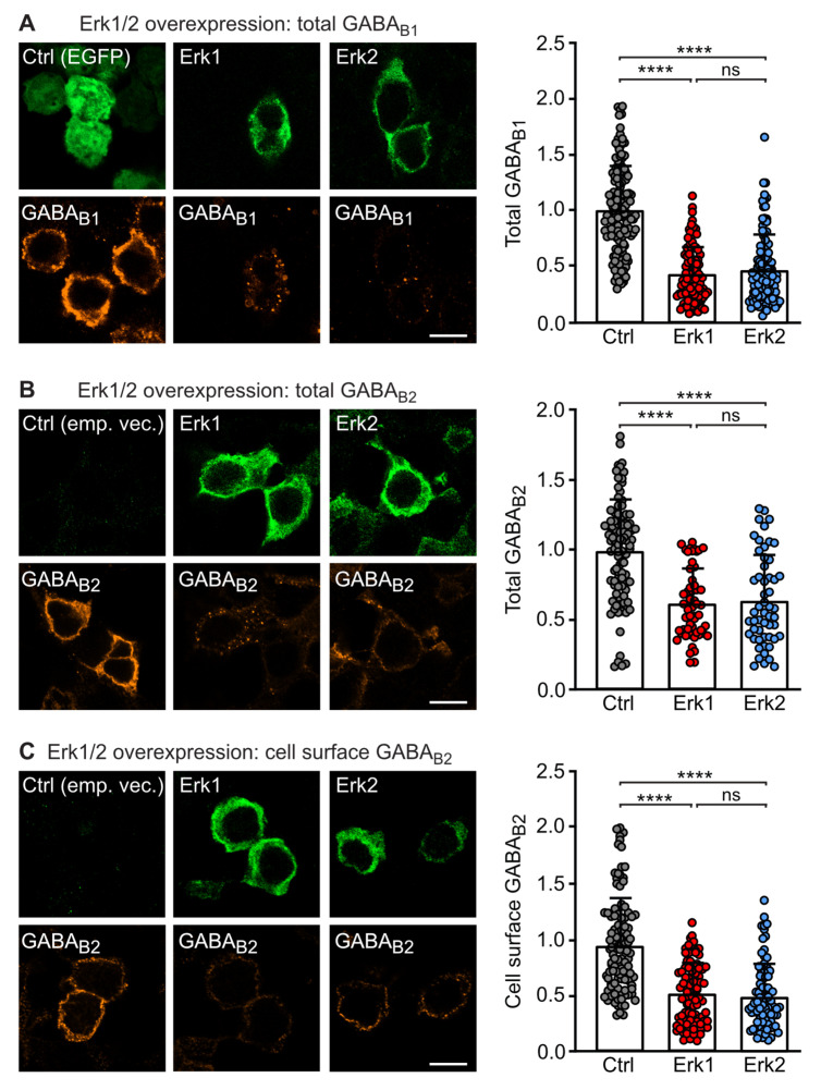
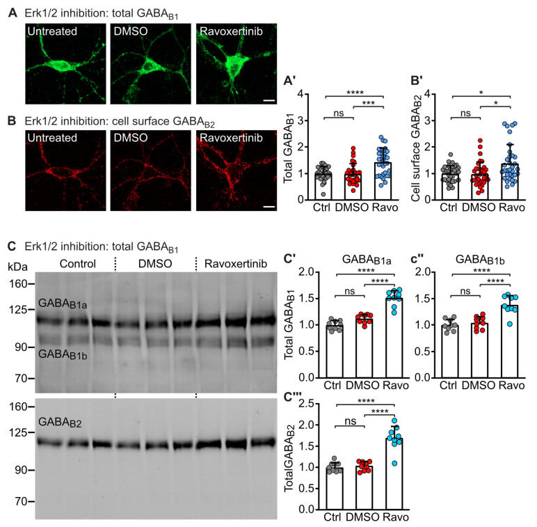
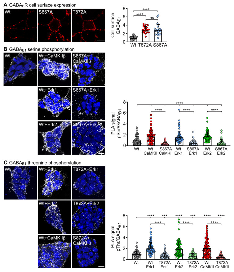
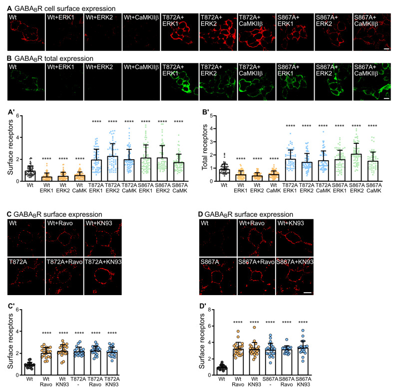
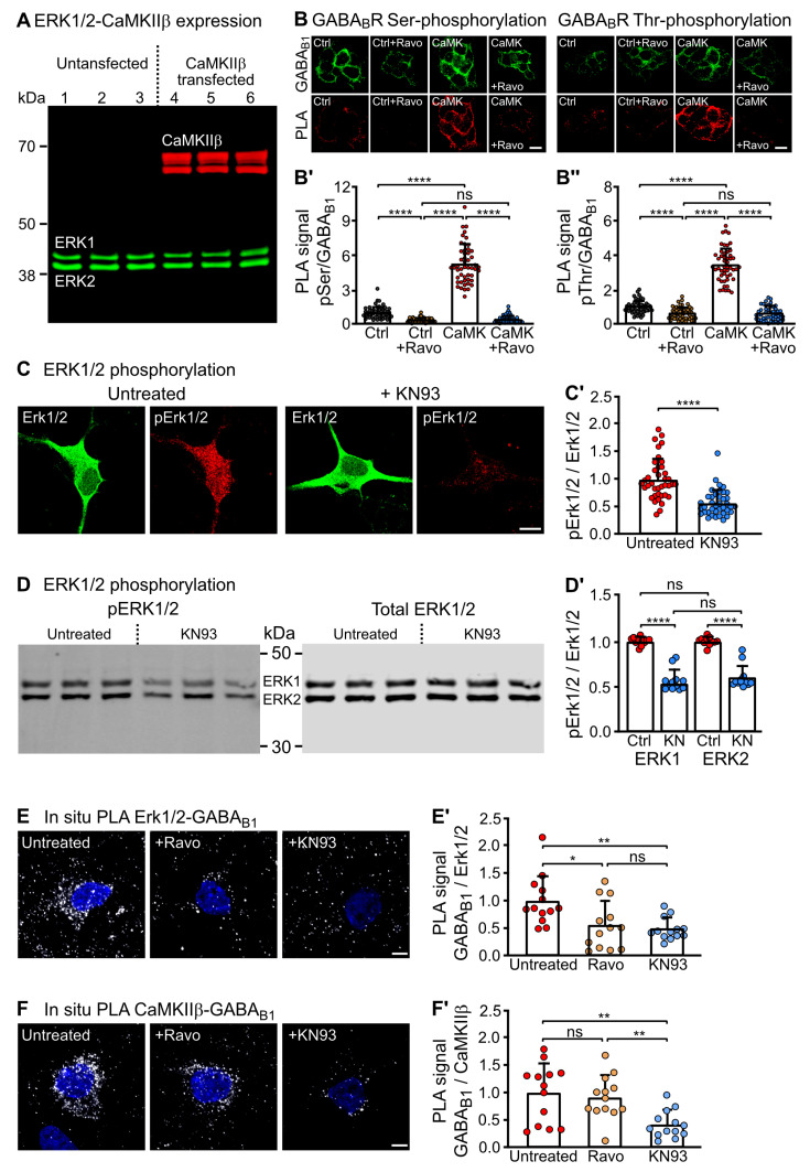
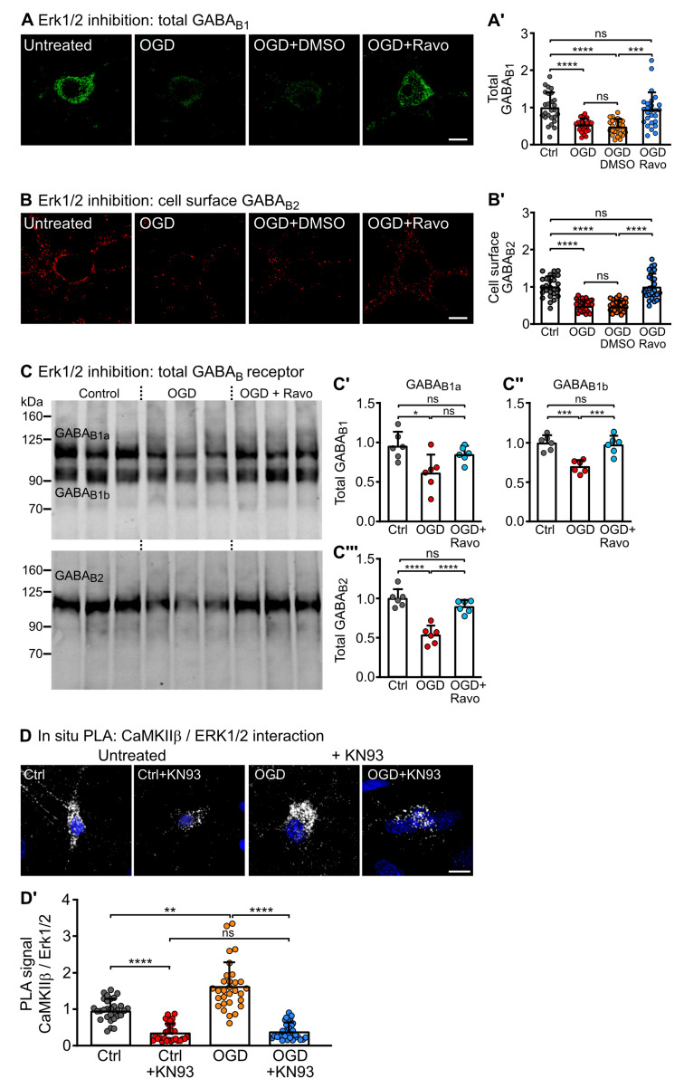
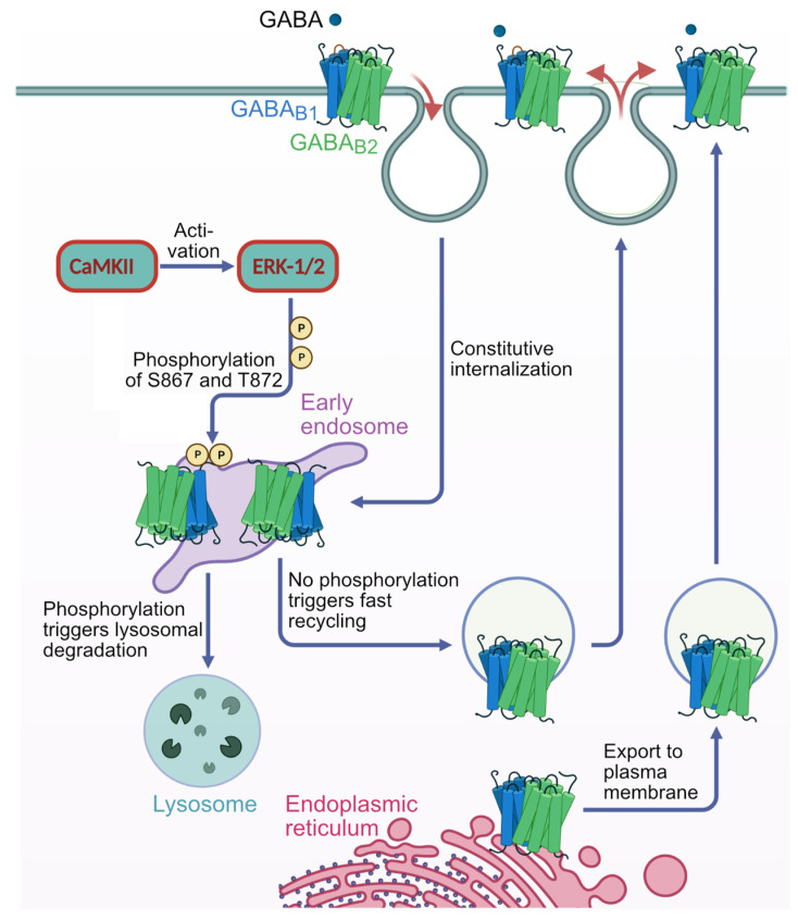
GABAB receptor-mediated inhibition is indispensable for maintaining a healthy neuronal excitation/inhibition balance. Many neurological diseases are associated with a disturbed excitation/inhibition balance and downregulation of GABAB receptors due to enhanced sorting of the receptors to lysosomal degradation. A key event triggering the downregulation of the receptors is the phosphorylation of S867 in the GABAB1 subunit mediated by CaMKIIβ. Interestingly, close to S867 in GABAB1 exists another phosphorylation site, T872. Therefore, the question arose as to whether phosphorylation of T872 is involved in downregulating the receptors and whether phosphorylation of this site is also mediated by CaMKIIβ or by another protein kinase. Here, we show that mutational inactivation of T872 in GABAB1 prevented the degradation of the receptors in cultured neurons. We found that, in addition to CaMKIIβ, also ERK1/2 is involved in the degradation pathway of GABAB receptors under physiological and ischemic conditions. In contrast to our previous view, CaMKIIβ does not appear to directly phosphorylate S867. Instead, the data support a mechanism in which CaMKIIβ activates ERK1/2, which then phosphorylates S867 and T872 in GABAB1. Blocking ERK activity after subjecting neurons to ischemic stress completely restored downregulated GABAB receptor expression to normal levels. Thus, preventing ERK1/2-mediated phosphorylation of S867/T872 in GABAB1 is an opportunity to inhibit the pathological downregulation of the receptors after ischemic stress and is expected to restore a healthy neuronal excitation/inhibition balance.

Keywords: CaMKII; ERK1/2; GABAB receptors; cerebral ischemia; degradation; phosphorylation.


Conflict of interest statement

The authors declare no conflict of interest.

Figures

Figure 1
Figure 1
Overexpression of ERK1/2 downregulates GABAB receptors. HEK 293 cells were transfected with GABAB1, GABAB2 and empty vector or EGFP as a control (Ctrl) or with GABAB1, GABAB2 and either ERK1 or ERK2. After 2 days, the cells were tested for ERK1/2 and GABAB receptor expression by immunofluorescence staining using antibodies directed against ERK1/2 and GABAB1 or GABAB2. (A) Transfection with ERK1 or ERK2 downregulated the expression of total GABAB1. Left: representative images (scale bar: 10 µm). Right: quantification of fluorescence intensities. The data represent the mean ± SD of 101–148 cells per condition from four independent experiments. Brown–Forsythe and Welch’s ANOVA with Games-Howell’s post-test (ns, p > 0.05; ****, p < 0.0001). (B) Transfection with ERK1 or ERK2 downregulated the expression of total GABAB2. Left: representative images (scale bar: 10 µm). Right: quantification of fluorescence intensities. The data represent the mean ± SD of 54–95 cells per condition from four independent experiments. One Way ANOVA with Tukey’s post-test (ns, p > 0.05; ****, p < 0.0001). (C) Transfection with ERK1 or ERK2 downregulated the cell surface expression of GABAB receptors as probed with antibodies directed against an extracellular located epitope in the N-terminal domain of GABAB2. Left: representative images (scale bar: 10 µm). Right: quantification of fluorescence intensities. The data represent the mean ± SD of 84–113 cells per condition from four independent experiments. Brown–Forsythe and Welch’s ANOVA with Games-Howell’s post-test (ns, p > 0.05; ****, p < 0.0001).
Figure 2
Figure 2
ERK1/2 regulates GABAB receptor expression in cultured neurons. Neurons were treated or not for 10 min with the ERK1/2 inhibitor Ravoxertinib (10 nM) or with DMSO (1% final concentration, as a control for the effect of the solvent used for dissolving Ravoxertinib) and then tested for GABAB receptor expression. (A) Treatment with Ravoxertinib upregulated the expression of total GABAB receptors as determined using antibodies directed against GABAB1. Left (A): representative images (scale bar: 10 μm). Right (A’): quantification of fluorescence intensities. Signals were normalized to control (cultures not treated with Ravoxertinib). The data represent the mean ± SD of 33 neurons per condition from three independent experiments. Brown–Forsythe and Welch’s ANOVA with Dunnett’s T3 post test (ns, p > 0.05; ***, p < 0.0005; ****, p < 0.0001). (B) Treatment with Ravoxertinib upregulated the cell surface expression of GABAB receptors as determined using antibodies directed against an extracellular located epitope in the N-terminal domain of GABAB2. Left (B): representative images (scale bar: 10 μm). Right (B’): quantification of fluorescence intensities. Signals were normalized to control (cultures not treated with Ravoxertinib). The data represent the mean ± SD of 33 neurons per condition from three independent experiments. Brown–Forsythe and Welch’s ANOVA with Dunnett’s T3 post-test (ns, p > 0.05; *, p < 0.05). (C) Treatment with Ravoxertinib upregulated total GABAB receptor expression as tested by Western blotting using antibodies directed against GABAB1 and GABAB2. (C’C’’’): quantification of Western blot signals. Signals were normalized to control (cultures not treated with Ravoxertinib). Cultures treated with DMSO (1% final concentration) served as controls for the effect of the solvent used for dissolving Ravoxertinib. The data represent the mean ± SD of 9 cultures per condition from three independent neuron preparations. One-way ANOVA with Tukey’s post-test (ns, p > 0.05; ****, p < 0.0001).
Figure 3
Figure 3
ERK1/2 and CaMKIIβ mediated phosphorylation of GABAB1 at serine 867 (S867) and threonine 872 (T872). (A) Transfection of neurons with mutant GABAB1 containing inactivated phosphorylation sites T872A or S867A upregulated the cell surface expression of GABAB receptors as determined by immunofluorescence staining using antibodies directed against the HA-tagged GABAB1 phospho-mutants. Left: representative images (scale bar: 10 µm). Right: quantification of fluorescent intensities. The data represent the mean ± SD of 24 cells per condition from three independent experiments. Brown–Forsythe and Welch’s ANOVA with Games-Howell’s post-test (ns, p > 0.05; ****, p < 0.0001). (B) ERK1/2 and CaMKII mediated phosphorylation of S867 in GABAB1. HEK-293 cells were transfected with wild-type GABAB1/GABAB2 (wt) or with the phospho-mutant GABAB1(S867A)/GABAB2 with or without CaMKIIβ, ERK1 or ERK2 and tested for GABAB receptor phosphorylation by in situ PLA using antibodies directed against phospho-serine and HA-tagged GABAB1. Signals were normalized to the in situ PLA signals in HEK-cells transfected with wild-type GABAB1/GABAB2, which served as control. Left: representative images; in situ PLA signals (white dots) represent serine phosphorylated GABAB receptors (scale bar: 10 µm). Right: quantification of in situ PLA signals. The data represent the mean ± SD of 78 cells per condition from three independent preparations. Brown–Forsythe and Welch’s ANOVA with Dunnett’s T3 multiple comparison test (****, p < 0.0001). The upper line depicts the statistical evaluation between the wild-type control and all other conditions. (C) ERK1/2 and CaMKII mediated phosphorylation of T872 in GABAB1. HEK-293 cells were transfected with wild-type GABAB1/GABAB2 (wt) or with the phospho-mutant GABAB1(T872A)/GABAB2 with or without CaMKIIβ, ERK1 or ERK2 and tested for GABAB receptor phosphorylation by in situ PLA using antibodies directed against phospho-threonine and HA-tagged GABAB1. Signals were normalized to the in situ PLA signals in HEK cells transfected with wild-type GABAB1/GABAB2, which served as control. Left: representative images; in situ PLA signals (white dots) represent threonine phosphorylated GABAB receptors (scale bar: 10 µm). Right: quantification of in situ PLA signals. The data represent the mean ± SD of 86 cells per condition from three independent preparations. Brown–Forsythe and Welch’s ANOVA with Dunnett’s T3 multiple comparison test (***, p < 0.0005; ****, p < 0.0001). The upper line depicts the statistical evaluation between the wild-type control and all other conditions. PLA, proximity ligation assay.
Figure 4
Figure 4
Effect of ERK1/2 and CaMKIIβ mediated phosphorylation of GABAB1 T872 and S867 on GABAB receptor expression. (A,B) HEK 293 cells were transfected with wild-type GABAB1/GABAB2 (wt) or with the phospho-mutant GABAB1(T872A) plus GABAB2 or GABAB1(S867A) plus GABAB2 with or without CaMKIIβ, ERK1 or ERK2 and tested for total and cell surface expression of GABAB receptors by fluorescence staining using antibodies directed against GABAB1 for total receptor staining and antibodies directed against an extracellular located epitope in the N-terminal domain of GABAB2 for cell surface receptor staining. Co-transfection with GABAB1(S867A) or GABAB1(T872A) upregulated the cell surface (A) and total (B) expression of GABAB receptors. Top: representative images (scale bar: 10 µm). Bottom (A’,B’): quantification of fluorescence intensities. The data represent the mean ± SD of 62–68 cells per condition from three independent experiments. Brown–Forsythe and Welch’s ANOVA with Games-Howell’s post-test (****, p < 0.0001). (C) The cell surface expression of GABAB receptors containing the phospho-mutant GABAB1(T872A) was unaffected by CaMKII and ERK1/2 inhibitors. Neurons were transfected with wild-type HA-tagged GABAB1 or the phospho-mutant HA-tagged GABAB1(T872A), treated or not for 10 min with the CaMKII inhibitor KN93 or ERK1/2 inhibitor Ravoxertinib and tested for cell surface expression of GABAB receptors by immunofluorescence staining using antibodies directed against the extracellularly located HA-tag. Top (C): representative images (scale bar: 10 µm). Bottom (C’): quantification of fluorescence intensities. The data represent the mean ± SD of 19 cells per condition from three independent experiments. Brown–Forsythe and Welch’s ANOVA with Games-Howell’s post-test (****, p < 0.0001). (D) The cell surface expression of GABAB receptors containing the phospho-mutant GABAB1(T867A) was unaffected by CaMKII and ERK1/2 inhibitors. Neurons were transfected with wild-type HA-tagged GABAB1 or the phospho-mutant HA-tagged GABAB1(T867A), treated or not for 10 min with the CaMKII inhibitor KN93 or ERK1/2 inhibitor Ravoxertinib and tested for cell surface expression of GABAB receptors by immunofluorescence staining using antibodies directed against the extracellularly located HA-tag. Top (D): representative images (scale bar: 10 µm). Bottom (D’): quantification of fluorescence intensities. The data represent the mean ± SD of 22 cells per condition from three independent experiments. Brown–Forsythe and Welch’s ANOVA with Games-Howell’s post-test (****, p < 0.0001).
Figure 5
Figure 5
ERK1/2 was activated by CaMKII and interacted activity-dependently with GABAB receptors. (A) HEK-293 cells endogenously express ERK1/2 but not CaMKIIβ. Homogenates of untransfected HEK-293 cells were probed for the presence of ERK1/2 and CaMKIIβ by Western blot analysis (lanes 1–3 depict three homogenate preparations). As a control, HEK-293 cells were transfected with CaMKIIβ plasmid (lanes 4–6). (B) ERK1/2 but not CaMKIIβ directly phosphorylates GABAB receptors. HEK-293 cells were transfected with GABAB1 and GABAB2 (Ctrl, Ctrl + Ravo) or with GABAB1 and GABAB2 plus CaMKIIβ (CaM, CaMK + Ravo) and tested for Ser and Thr phosphorylation by in situ PLA after blocking ERK1/2 activity with 10 nM Ravoxertinib for 10 min (+Ravo) or not. Top (B): representative images (in situ PLA signals: red dots, scale bar: 10 µm). Bottom (B’,B’’): quantification of in situ PLA signals. The data represent the mean ± SD of 50 cells per condition from two independent experiments. Brown–Forsythe and Welch’s ANOVA with Dunnets’s T3 post-test (ns, p > 0.05; ****, p < 0.0001). (C,D) CaMKII activated ERK1/2. Neurons were treated for 10 min with the CaMKII inhibitor KN93 and then tested for activated ERK1/2 (pERK1/2, phosphorylated at Thr183 and Tyr185) and total ERK1/2 expression by immunofluorescence staining (C) or Western blotting (D) using phospho-specific and pan ERK1/2 antibodies. Inhibition of CaMKII by KN93 also inhibited the activity of ERK1/2. Left (C,D): representative images (scale bar: 10 µm). Right (C’,D’): quantification of fluorescence intensities. (C) The data represent the mean ± SD of 39 neurons per condition from three independent experiments. Unpaired two-tailed t-test (****, p < 0.0001). (D) The data represent the mean ± SD of 12 cultures per condition from four independent neuron preparations. One-way ANOVA with Tukey’s post-test (ns, p > 0.05; ****, p < 0.0001). (E) The interaction between GABAB receptors and ERK1/2 in neurons was reduced by inhibition of CaMKII (KN93) or ERK1/2 (Ravoxertinib) as determined by in situ PLA using antibodies directed against GABAB1 and ERK1/2. PLA signals were normalized to the untreated control. Left (C): representative images of in situ PLA signals (white dots, scale bar: 5 µm). Right (E’): quantification of in situ PLA signals. The data represent the mean ± SD of 13 neurons per condition from two independent experiments. Brown–Forsythe and Welch’s ANOVA with Games-Howell’s post-test (ns, p > 0.05; *, p < 0.05; **, p < 0.01). (F) The interaction of CaMKIIβ with GABAB receptors is independent of ERK1/2 activity. Neurons were either untreated or treated with the CaMKII inhibitor KN93 or the ERK1/2 inhibitor Ravoxertinib for 10 min and subsequently analyzed for the interaction of CaMKIIβ with GABAB receptors using antibodies directed against CaMKIIβ and GABAB1. Signals were normalized to the untreated control. Left (F): representative images of in situ PLA signals (white dots, scale bar: 5 µm). Right (F’): quantification of in situ PLA signals. The data represent the mean ± SD of 13 neurons per condition from two independent experiments. Brown–Forsythe and Welch’s ANOVA with Games-Howell’s post-test (ns, p > 0.05; **, p < 0.01).
Figure 6
Figure 6
ERK1/2 is involved in ischemic stress-induced downregulation of GABAB receptors. (A,B) Inhibition of ERK1/2 normalized GABAB receptor total (A) and cell surface (B) expression after oxygen and glucose deprivation (OGD) induced stress. Neurons were subjected to 1 h of OGD, then treated for 10 min with the ERK1/2 inhibitor Ravoxertinib (10 nM) and analyzed for GABAB receptor expression using antibodies directed against GABAB1 (A) and GABAB2 (B) by immunofluorescence staining. OGD downregulated GABAB receptors, and treatment with Ravoxertinib restored normal expression levels. Right (A,B): representative images (scale bar: 10 µm). Left (A’,B’): quantification of fluorescence intensities. The data represent the mean ± SD of 26 neurons per condition from three independent experiments. Brown–Forsythe and Welch’s ANOVA with Dunnett’s T3 post test (ns, p > 0.05; ***, p < 0.0005; ****, p < 0.0001). (C) OGD-induced downregulation of GABAB receptor expression was recovered by inhibition of ERK1/2 with Ravoxertinib as verified by Western Blotting. (C’–C’’’): quantification of Western blot signals. Signals were normalized to untreated control cultures (control). The data represent the mean ± SD of 3 independent neuron preparations per condition and one technical replicate. One-way ANOVA with Tukey’s post-test (ns, p > 0.05; ***, * p < 0.05; p < 0.0005; ****, p < 0.0001). (D) The interaction between CaMKIIβ and ERK1/2 in neurons was increased after OGD and blocked by inhibition of CaMKII activity. Neurons were subjected to 1 h of OGD, then treated for 10 min with the CaMKII inhibitor KN93 (10 µM) and analyzed for the interaction of CaMKIIβ and ERK1/2 by in situ PLA. Signals were normalized to untreated cultures (control). Top (D): representative images of in situ PLA signals (white dots, scale bar: 10 µm). Bottom (D’): quantification of in situ PLA signals. The data represent the mean ± SD of 30 neurons per condition from three independent experiments. Two-way ANOVA with Tukey’s multiple comparison test (ns, p > 0.05; **, p < 0.01; ****, p < 0.0001).
Figure 7
Figure 7
Proposed mechanism of CaMKIIβ and ERK1/2 mediated downregulation/degradation of GABAB receptors. GABAB receptors are constitutively internalized to early endosomes. From early endosomes, receptors are recycled back to the plasma membrane or sorted into lysosomes for degradation. Degraded receptors are replaced by newly synthesized receptors exported from the ER to ensure a constant number of cell surface receptors. The initial signal that tags GABAB receptors for degradation appears to be phosphorylating GABAB1 by CaMKIIβ and ERK1/2 at the level of early endosomes. Our data support a mechanism in which CaMKIIβ activates ERK1/2, which phosphorylates GABAB1 at S867 and T872 for further sorting of the receptors to lysosomes. As CaMKIIβ most likely does not directly phosphorylate ERK1/2, activated CaMKIIβ might recruit the components of the ERK1/2 activation cascade to GABAB receptors and induce activation of ERK1/2 by phosphorylation of Ref [24,26,28]. This figure was created using BioRender (www.biorender.com, accessed on 12 April 2023).

Similar articles

Cited by

References

    1. Chalifoux J.R., Carter A.G. GABAB receptor modulation of synaptic function. Curr. Opin. Neurobiol. 2011;21:339–344. doi: 10.1016/j.conb.2011.02.004. - DOI - PMC - PubMed
    1. Jones K.A., Borowsky B., Tamm J.A., Craig D.A., Durkin M.M., Dai M., Yao W.-J., Johnson M., Gunwaldsen C., Huang L.-Y., et al. GABAB receptor function as a heteromeric assembly of the subunits GABABR1 and GABABR2. Nature. 1998;396:674–679. doi: 10.1038/25348. - DOI - PubMed
    1. Kaupmann K., Malitschek B., Schuler V., Heid J., Froestl W., Beck P., Mosbacher J., Bischoff S., Kulik A., Shigemoto R., et al. GABAB receptor subtypes assemble into functional heteromeric complexes. Nature. 1998;396:683–687. doi: 10.1038/25360. - DOI - PubMed
    1. White J.H., Wise A., Main M.J., Green A., Fraser N.J., Disney G.H., Barnes A.A., Emson P., Foord S.M., Marshall F.H. Heterodimerization is required for the formation of functional GABAB receptors. Nature. 1998;396:679–682. doi: 10.1038/25354. - DOI - PubMed
    1. Gassmann M., Bettler B. Regulation of neuronal GABAB receptor functions by subunit composition. Nat. Rev. Neurosci. 2012;13:380–394. doi: 10.1038/nrn3249. - DOI - PubMed

LinkOut - more resources