. 2023 Aug 30;24(17):13439.
doi: 10.3390/ijms241713439.

TRIM67 Implicates in Regulating the Homeostasis and Synaptic Development of Mitral Cells in the Olfactory Bulb

Affiliations

TRIM67 Implicates in Regulating the Homeostasis and Synaptic Development of Mitral Cells in the Olfactory Bulb

Chunyu Cai et al. Int J Mol Sci. .

Abstract

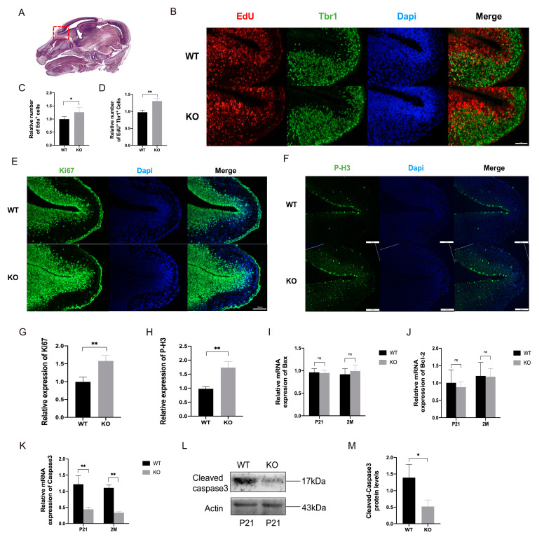
In recent years, olfactory dysfunction has attracted increasingly more attention as a hallmark symptom of neurodegenerative diseases (ND). Deeply understanding the molecular basis underlying the development of the olfactory bulb (OB) will provide important insights for ND studies and treatments. Now, with a genetic knockout mouse model, we show that TRIM67, a new member of the tripartite motif (TRIM) protein family, plays an important role in regulating the proliferation and development of mitral cells in the OB. TRIM67 is abundantly expressed in the mitral cell layer of the OB. The genetic deletion of TRIM67 in mice leads to excessive proliferation of mitral cells in the OB and defects in its synaptic development, resulting in reduced olfactory function in mice. Finally, we show that TRIM67 may achieve its effect on mitral cells by regulating the Semaphorin 7A/Plexin C1 (Sema7A/PlxnC1) signaling pathway.

Keywords: TRIM67; development; mitral cells; olfactory bulb.


Conflict of interest statement

The authors declare no conflict of interest.

Figures

Figure 1
Figure 1
TRIM67 highly concentrates in the mitral cell layer of the olfactory bulb. (AC) RT-qPCR and Western blots show the temporal expression pattern of TRIM67 in the postnatal mouse OB (n = 3). (D) In situ hybridization shows the spatial expression of TRIM67 in the mouse OB. Error bars represent SEM. Red arrows indicate mitral cells.
Figure 2
Figure 2
TRIM67 KO affects the homeostasis of mitral cells in the olfactory bulb. (A) TRIM67 knockout strategy. (B) Genotyping PCR for the validation of TRIM67 knockout mice. (C) The picture shows that deletion of the TRIM67 gene does not affect OB size. (D) Quantification shows normal organ indices of the OB in TRIM67 KO mice (n = 4). (E) HE staining show the basic structure in the OB of TRIM67 KO mice. (F,G) Representative images and quantification of NISSL staining show increased number of mitral cells in the OB of TRIM67 KO mice. (HJ) RT-qPCR and Western blot show increased expression of mitral cell markers Tbx21 and Tbr1 in the OB of TRIM67 KO mice (n = 3). (K,L) Quantifications reveal that deletion of the TRIM67 gene does not affect the number of granule cells and periglomerular cells in the mouse OB. Error bars represent SEM. * p-value < 0.05; ** p-value < 0.01. Red arrows indicate mitral cells.
Figure 3
Figure 3
TRIM67 affects the proliferation of mitral cells in an embryonic mouse. (A) Schematic diagram of the OB in E14 mice [23]. The red dotted area represents the OB. (BD) Immunostaining and quantifications of Edu and Tbr1 reveal an increased number of newly proliferating mitral cells in the OB of E14 TRIM67 KO mice (n = 3). (EH) Immunostaining and quantifications of Ki67 and P-H3 reveal that the proliferation in OB cells of E14 TRIM67 KO mice was enhanced (n = 3). (IM) RT-qPCR and Western blot show reduced apoptosis in the OB of TRIM67 KO mice (n = 4). Error bars represent SEM. * p-value < 0.05; ** p-value < 0.01.
Figure 4
Figure 4
Loss of TRIM67 causes olfactory dysfunction in mice. (AC) Schematic diagrams showing the food burial, olfactory discrimination, and odor exploration experiments. (D) Statistical analysis of the time for WT and KO mice to find hidden or exposed food particles in the food burial and the food exposure tests (n = 8). (E) Statistical analysis of time spent in familiar and unfamiliar odor environments by WT and KO mice (n = 8). (F) Statistical analysis of odor exploration time for H2O, PA, PEA and TMT in WT and KO mice (n = 8). Error bars represent SEM. * p-value < 0.05; ** p-value < 0.01.
Figure 5
Figure 5
Impaired synapse formation in mitral cells of TRIM67 KO mice. (AC) Representative images of Golgi-stained mitral cells and statistical analysis of dendrites show normal development of mitral cell dendrites in the OB of TRIM67 KO mice (n = 3). (D,E) Representative images of Golgi-stained dendritic spines of mitral cells and schematic diagrams of various types of dendritic spines. (FH) Statistical analysis of mature, immature, and total dendritic spine numbers show decreased dendritic spines in mitral cells in TRIM67 KO mice (n = 3). (IK) Electron microscopy and quantification of representative images of synapses in the OB show a decrease in the number of synapses in the OB of TRIM67 KO mice (n = 3). Red arrows indicate asymmetric synapses, yellow arrows indicate symmetric synapses. (L,M) RT-qPCR reveal decreased expression of synapse-associated markers in the olfactory bulb of KO mice compared with WT mice (n = 4). (N,O) RT-qPCR reveal that the expressions of factors that promote synapse development were decreased in the olfactory bulb of KO mice compared with WT mice (n = 4). (PS) Immunostaining, Western blot, and quantification show decreased protein expression of VGLUT2 and Synaptophysin in the olfactory bulb of KO mice compared with WT mice (n = 4). Error bars represent SEM. * p-value < 0.05; ** p-value < 0.01.
Figure 6
Figure 6
Sema7A/PlxnC1 signaling implicates in TRIM67-regulated synapse development of mitral cells. (A) RT-qPCR reveals that the expression level of Sema7A mRNA was decreased in the OB of KO mice compared with WT mice (n = 4). (B) RT-qPCR reveals that the expression level of PlxnC1 mRNA was decreased in the OB of KO mice compared with WT mice (n = 4). (C) RT-qPCR reveals that the expression level of CDC42 mRNA was reduced in the OB of KO mice compared with WT mice (n = 4). (D) RT-qPCR reveals that the expression level of NMDA-R mRNA in the OB of KO mice was reduced compared with that of WT mice (n = 4). (E,F) Western blot and quantification show reduced protein expression of CDC42 in the OB of TRIM67 KO mice (n = 3). (GI) RT-qPCR quantifications show increased expression of genes related with the Sema7A/PlxnC1 signaling pathway in TRIM67 overexpressed 293T cells (n = 4). Error bars represent SEM. * p-value < 0.05; ** p-value < 0.01.

Similar articles

References

    1. Brunert D., Rothermel M. Extrinsic neuromodulation in the rodent olfactory bulb. Cell Tissue Res. 2021;383:507–524. doi: 10.1007/s00441-020-03365-9. - DOI - PMC - PubMed
    1. Graven S.N., Browne J.V. Sensory development in the fetus, neonate, and infant: Introduction and overview. Newborn Infant Nurs. Rev. 2008;8:169–172. doi: 10.1053/j.nainr.2008.10.007. - DOI
    1. Hinds J.W. Autoradiographic study of histogenesis in the mouse olfactory bulb. I. Time of origin of neurons and neuroglia. J. Comp. Neurol. 1968;134:287–304. doi: 10.1002/cne.901340304. - DOI - PubMed
    1. Sabiniewicz A., Hoffmann L., Haehner A., Hummel T. Symptoms of depression change with olfactory function. Sci. Rep. 2022;12:5656. doi: 10.1038/s41598-022-09650-7. - DOI - PMC - PubMed
    1. Son G., Jahanshahi A., Yoo S.J., Boonstra J.T., Hopkins D.A., Steinbusch H.W.M., Moon C. Olfactory neuropathology in Alzheimer’s disease: A sign of ongoing neurodegeneration. BMB Rep. 2021;54:295–304. doi: 10.5483/BMBRep.2021.54.6.055. - DOI - PMC - PubMed