Review
. 2023 Aug 30;24(17):13447.
doi: 10.3390/ijms241713447.

CRISPR/Cas-Based Techniques for Live-Cell Imaging and Bioanalysis

Affiliations
Review

CRISPR/Cas-Based Techniques for Live-Cell Imaging and Bioanalysis

Shuo Huang et al. Int J Mol Sci. .

Abstract

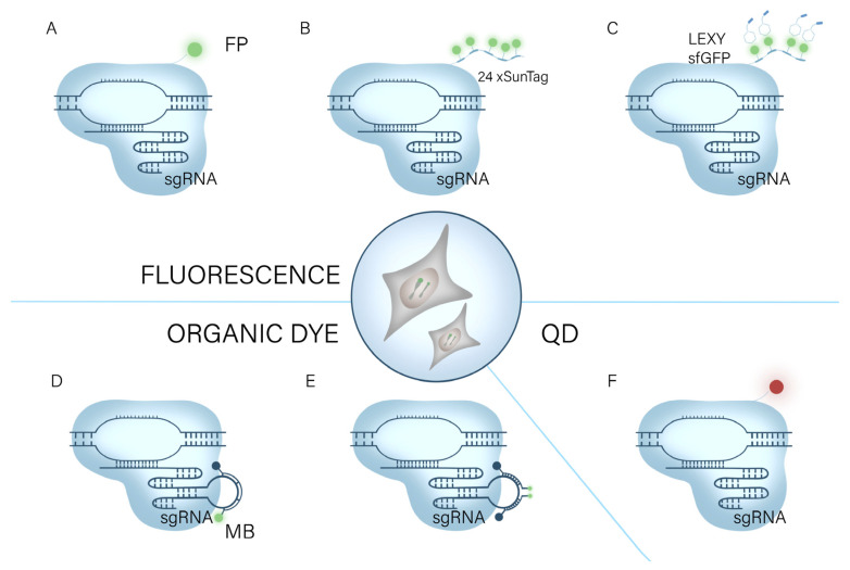
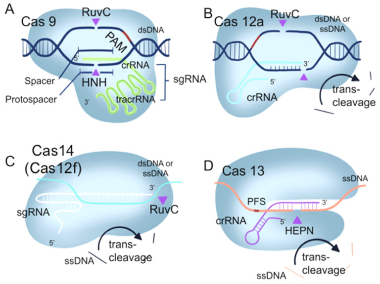
CRISPR/Cas systems have found widespread applications in gene editing due to their high accuracy, high programmability, ease of use, and affordability. Benefiting from the cleavage properties (trans- or cis-) of Cas enzymes, the scope of CRISPR/Cas systems has expanded beyond gene editing and they have been utilized in various fields, particularly in live-cell imaging and bioanalysis. In this review, we summarize some fundamental working mechanisms and concepts of the CRISPR/Cas systems, describe the recent advances and design principles of CRISPR/Cas mediated techniques employed in live-cell imaging and bioanalysis, highlight the main applications in the imaging and biosensing of a wide range of molecular targets, and discuss the challenges and prospects of CRISPR/Cas systems in live-cell imaging and biosensing. By illustrating the imaging and bio-sensing processes, we hope this review will guide the best use of the CRISPR/Cas in imaging and quantifying biological and clinical elements and inspire new ideas for better tool design in live-cell imaging and bioanalysis.

Keywords: CRISPR/Cas; bioanalysis; bioimaging; nucleic acid analysis; protein analysis.


Conflict of interest statement

The authors declare no conflict of interest.

Figures

Figure 2
Figure 2
The CRISPR imaging system for genomic imaging. It relies on dCas9 or gRNA-bound fluorescent substances to visualize genomic loci in living cells. (A) dCas9 binds to a fluorescent protein (FP) [31]. (B) The supernova labeling (SunTag) system binds multiple fluorescent proteins [36]. (C) The SunTag system binds to light-induced nuclear output tags (LEXY) [37]. (D) RNA aptamers bind to molecular beacons MB [39]. (E) sgRNA carries two different molecular beacons (MBs). (F) dCas9 combined with DQ for imaging [40].
Figure 1
Figure 1
Fundamental components of four CRISPR/Cas systems. (A) CRISPR/Cas9. (B) CRISPR/Cas12a. (C) CRISPR/Cas14(Cas12f). (D) CRISPR/Cas13a. Purple arrowheads indicate cis-cleavage sites, and black arrows indicate trans-cleavag.
Figure 3
Figure 3
Using RCas9 to target mRNA in living cells. (A) Schematic diagram of RNA imaging system using CRISPR/dCas9 [43]. The cross symbol represents sequences that do not contain the PAM sequence. (B) Schematic diagram of the CRISPR-Sunspot imaging strategy, depicting the utilization of three target sites within a single mRNA for visualization purposes [45]. (C) Schematic representation of the Pepper530 complex [46].
Figure 4
Figure 4
Targeting mRNA in living cells. (A) Strategy for imaging NEAT 1, SatIII, MUC 4, and GCN 4 RNAs in live cells using the CRISPR/dCas13 system [47]. (B) Development of RNA imaging strategy by a combination of Tat peptide with fluorescent RNA aptamers (TRAP-tag) [49]. (C) Schematic diagram of applying CRISPR-TRAP-tag to track SatIII lncRNA under SA treatment [49]. (D) Imaging demonstrating the localization and expression patterns of each mammalian construct [48]. Scale bars, 10 μm. (E) Schematic of CRISPR/dCas13 system binding with sgRNA containing [66].
Figure 5
Figure 5
Nucleic acid detection based on CRISPR/Cas9 system. (A) Nucleic acid detection based on NASBACC [92]. (B) CAS-EXPAR nucleic acid analysis system [102]. (C) CRISPR/Cas9 nucleic acid analysis system in conjunction with E-DNA sensors [104].
Figure 6
Figure 6
Nucleic acid analysis system based on Cas12. (A) Schematic of direct nucleic detection acid using HOLMES system [90]. (B) Schematic of direct nucleic acid using DETECTR system [24]. (C) Schematic of direct nucleic acid using HOLMESv2 system [105]. (D) Double-amplification sensing strategy based on terminal deoxynucleotide transferase (TdT) and CRISPR/Cas12a [98]. Scissors represent nicking endonuclease (Cas12a) cleaving the reporters.
Figure 7
Figure 7
Schematic diagram of nucleic acid detection system based on Cas13. (A) Schematic of ZIKV RNA detection by SHERLOCK [30]. (B) Schematic of direct viral detection using HUDSON and SHERLOCK [91]. (C) Overview of Cas13-based CREST modifications [89]. (D) Schematic of complementary Cas13 and Cas12a enzymes in 4-channel in-sample multiplexing [93].
Figure 8
Figure 8
CRISPR protein analysis system with fluorescence sensor. (A) Schematic of the TdT-combined CRISPR/Cas12a method for UDG activity assay [108]. (B) Schematic of a Cas13a/crRNA-mediated, RNA-triggered signal amplification system following DNA transcription [134]. (C) Schematic of a DNA-regulated CRISPR/Cas12a biosensor designed to detect non-nucleic acid targets. This biosensor combines enhanced upconversion LRET with luminescence amplification facilitated by a biomimetic PC chip [139].

Similar articles

Cited by

References

    1. Hsu P.D., Lander E.S., Zhang F. Development and applications of CRISPR-Cas9 for genome engineering. Cell. 2014;157:1262–1278. doi: 10.1016/j.cell.2014.05.010. - DOI - PMC - PubMed
    1. Jinek M., Chylinski K., Fonfara I., Hauer M., Doudna J.A., Charpentier E. A programmable dual-RNA-guided DNA endonuclease in adaptive bacterial immunity. Science. 2012;337:816–821. doi: 10.1126/science.1225829. - DOI - PMC - PubMed
    1. Makarova K.S., Wolf Y.I., Koonin E.V. The basic building blocks and evolution of CRISPR-CAS systems. Biochem. Soc. Trans. 2013;41:1392–1400. doi: 10.1042/BST20130038. - DOI - PMC - PubMed
    1. Xu Y., Li Z. CRISPR-Cas systems: Overview, innovations and applications in human disease research and gene therapy. Comput. Struct. Biotechnol. J. 2020;18:2401–2415. doi: 10.1016/j.csbj.2020.08.031. - DOI - PMC - PubMed
    1. Glotzer J.B., Saffrich R., Glotzer M., Ephrussi A. Cytoplasmic flows localize injected oskar RNA in Drosophila oocytes. Curr. Biol. 1997;7:326–337. doi: 10.1016/S0960-9822(06)00156-4. - DOI - PubMed