. 2023 Aug 30;24(17):13476.
doi: 10.3390/ijms241713476.

BAP31 Knockout in Macrophages Affects CD4+T Cell Activation through Upregulation of MHC Class II Molecule

Affiliations

BAP31 Knockout in Macrophages Affects CD4+T Cell Activation through Upregulation of MHC Class II Molecule

Bo Zhao et al. Int J Mol Sci. .

Abstract

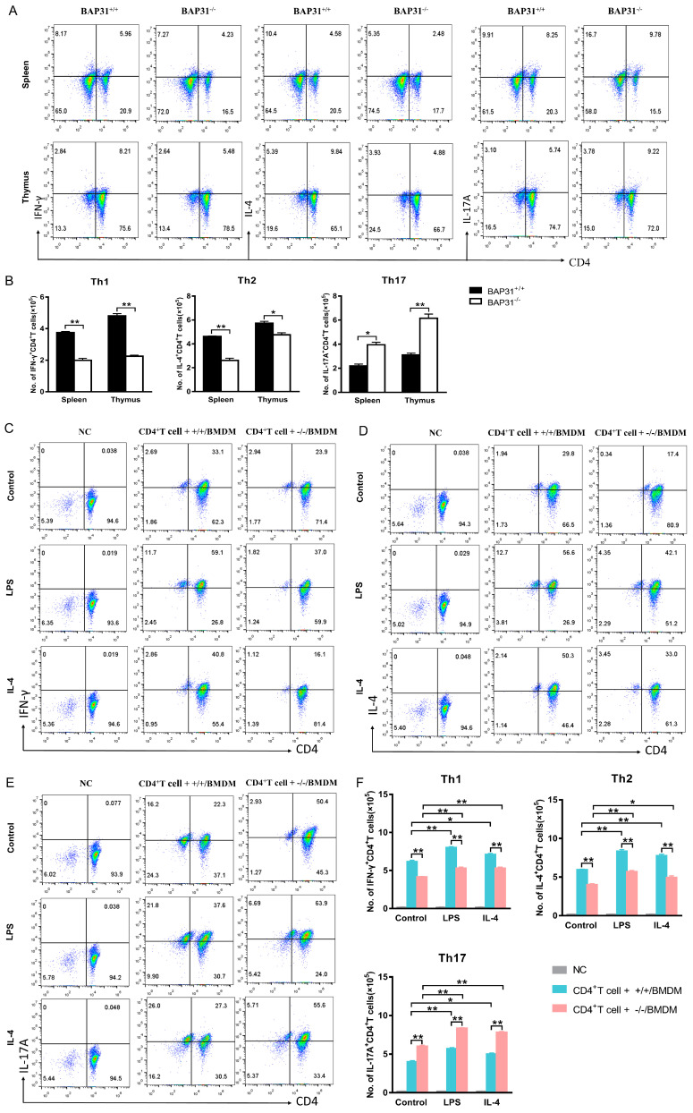
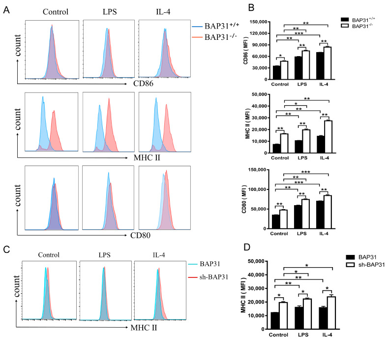
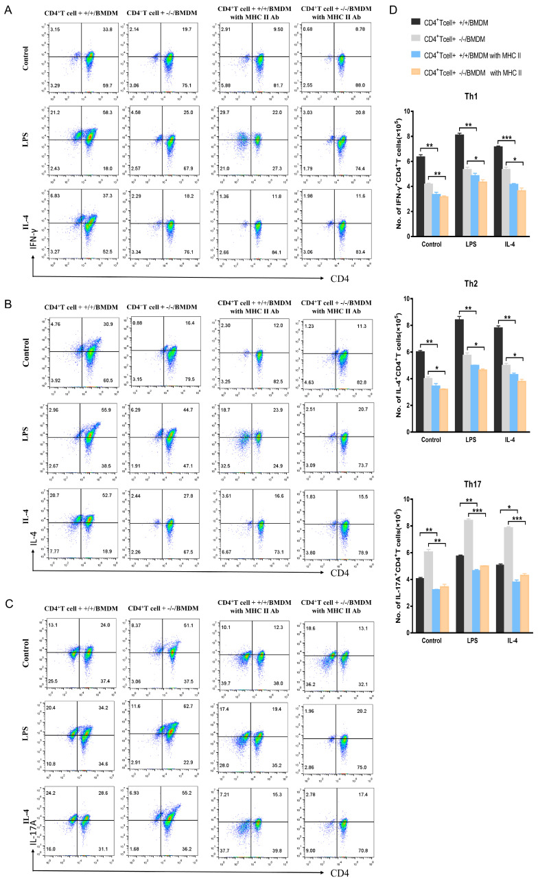
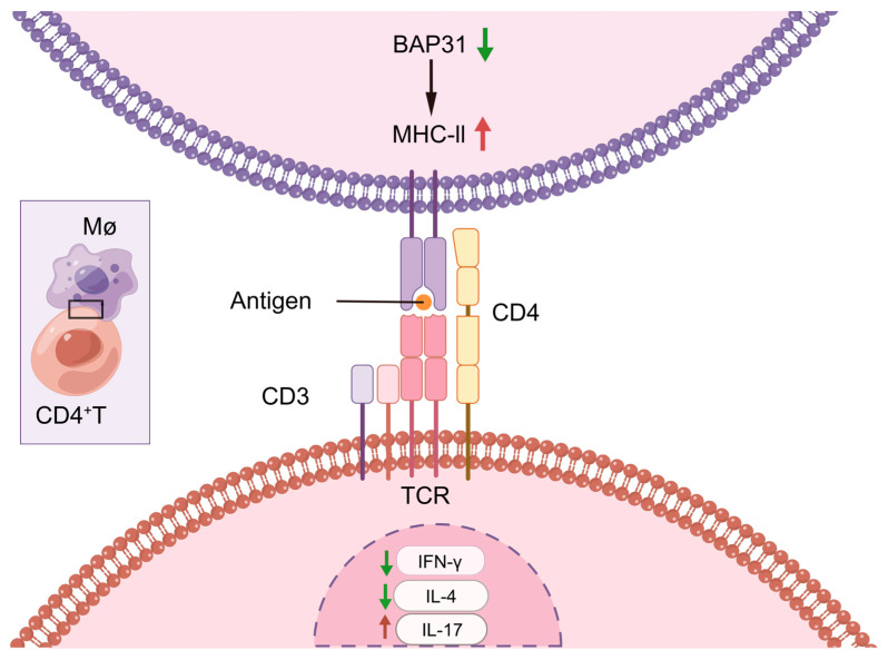
The differentiation of CD4+T cells is a crucial component of the immune response. The spleen and thymus, as immune organs, are closely associated with the differentiation and development of T cells. Previous studies have suggested that BAP31 may play a role in modulating T cell activation, but the specific impact of BAP31 on T cells through macrophages remains uncertain. In this study, we present evidence that BAP31 macrophage conditional knockout (BAP31-MCKO) mice display an enlarged spleen and thymus, accompanied by activated clustering and disrupted differentiation of CD4+T cells. In vitro co-culture studies were conducted to investigate the impact of BAP31-MCKO on the activation and differentiation of CD4+T cells. The examination of costimulatory molecule expression in BMDMs and RAW 264.7 cells, based on the endoplasmic reticulum function of BAP31, revealed an increase in the expression of antigen presenting molecules, particularly MHC-II molecule, in the absence of BAP31 in BMDMs or RAW264.7 cells. These findings suggest that BAP31 plays a role in the activation and differentiation of CD4+T cells by regulating the MHC class II molecule on macrophages. These results provide further support for the importance of BAP31 in developing interaction between macrophages and CD4+T cells.

Keywords: BAP31; CD4+T cell; MHC-II; activation; differentiation; macrophages.


Conflict of interest statement

The authors declare no conflict of interest.

Figures

Figure 1
Figure 1
BAP31 is involved in the development of spleen and thymus in BAP31-MCKO mice. (A) Flow cytometry-detected BMDM (CD11b+F4/80+) purity of BAP31flox/flox (BAP31+/+) and BAP31flox/floxLyz2-cre (BAP31−/−) mice. (B) Western blotting analysis of knockout efficiency of BAP31 from BMDMs (n = 3). Relative protein expression is expressed as the ratio of BAP31 to β-actin. (C) Real-time PCR analysis of knockout efficiency of BAP31 from BMDMs (n = 3). Relative BAP31 expression was normalized by GAPDH expression. (D) Statistics of the weight of spleen and thymus from mice (n = 5). (E) RT-qPCR analysis of genomic RNA of T cell activation molecules (CD25, CD69) from spleen and thymus. Total splenocyte and thymocyte RNA from BAP31flox/flox mice (BAP31+/+) and BAP31flox/floxLyz2-cre mice (BAP31−/−) (n = 3). (F) RT-qPCR analysis of Th1 cell transcription factor (T-bet) and cytokine (IFN-γ) from spleen and thymus (n = 3). (G) RT-qPCR analysis of Th2 cell transcription factor (GATA3) and cytokine (IL-4) from spleen and thymus (n = 3). (H) RT-qPCR analysis of Th17 cell transcription factor (RORγt) and cytokine (IL-17) from spleen and thymus (n = 3). Relative gene expression was measured by qRT-PCR and normalized by GAPDH. * p < 0.05. ** p < 0.01.
Figure 2
Figure 2
BAP31-MCKO facilitates CD4+T cell activation. (A) Flow cytometry detected T cell activation markers (CD25, CD69) of naïve CD4+T cells co-cultured for 72 h with bone marrow-derived macrophages divided into control, LPS and IL-4 groups. Naïve CD4+T cells without co-culture served as the negative control (NC). (B) Statistical bar charts showing the cell numbers of CD4+T cell + +/+/BMDM and CD4+T cell + −/−/BMDM group (n = 3). (C) Flow cytometry detected T cell activation markers (CD25, CD69) of EL4 cells co-cultured for 72 h with RAW264.7 cells divided into control, LPS and IL-4 groups. EL4 cells without co-culture served as the negative control (NC). (D) Statistical bar charts showing the cell numbers of EL4 + RAW264.7 and EL4 + sh-BAP31 RAW264.7 group (n = 3). (E) RT-qPCR analysis of T cell activation markers (CD25, CD69) of EL4 cells co-cultured for 72 h with RAW264.7 cells divided into control, LPS and IL-4 groups (n = 3). Relative gene expression was measured by qRT-PCR and normalized by GAPDH. * p < 0.05. ** p < 0.01. *** p < 0.001.
Figure 3
Figure 3
Macrophage BAP31 knockout influences CD4+T cell differentiation. (A) Flow cytometry detected differentiation of CD4+T cell subsets Th1 (IFN-γ), Th2 (IL-4), Th17 (IL-17A) in splenocytes and thymocytes from BAP31flox/flox mice (BAP31+/+) and BAP31flox/floxfLyz2-cre mice (BAP31−/−). (B) Statistical bar charts showing the cell numbers of BAP31+/+ and BAP31−/−groups (n = 3). (CE) Flow cytometry detected differentiation of CD4+T cell subsets Th1 (IFN-γ), Th2 (IL-4), and Th17 (IL-17A). Naïve CD4+T cell co-cultured with bone marrow-derived macrophages for 72 h compared with control, LPS and IL-4 groups. Naïve CD4+T cells without co-culture served as the negative control (NC) (F) Statistical bar charts showing the cell numbers of CD4+T cell + +/+/BMDM and CD4+T cell + −/−/BMDM group (n = 3). * p < 0.05. ** p < 0.01.
Figure 4
Figure 4
BAP31 influences macrophage antigen. (A) Flow cytometry detected antigen-presenting molecules (CD86, MHC-II, CD80) of bone marrow-derived macrophages divided into control, LPS and IL-4 groups. (B) Quantification of the mean fluorescence intensity (MFI) of the antigen-presenting molecules (CD86, MHC-II, CD80) by flow cytometric analysis (n = 3). (C) Flow cytometry detected antigen-presenting molecules (MHC-II) of RAW264.7 cells divided into control, LPS and IL-4 groups. (D) Quantification of the mean fluorescence intensity (MFI) of the antigen-presenting molecule MHC-II by flow cytometric analysis (n = 3). * p < 0.05. ** p < 0.01. *** p < 0.001.
Figure 5
Figure 5
BAP31-MCKO influences T cell differentiation by regulating MHC-II expression levels. (AC) Flow cytometry detected CD4+T cell differentiation of Th1 (IFN-γ), Th2 (IL-4) and Th17 (IL-17A) under four different conditions: CD4+T cells + +/+/BMDMs, CD4+T cells + −/−/BMDMs, CD4+T cells + +/+/BMDMs with MHC-II Abs, CD4+T cells + −/−/BMDMs with MHC-II Abs. (D) Statistical bar charts showing the cell numbers of CD4+T cells + +/+/BMDMs, CD4+T cells + −/−/BMDMs, CD4+T cells + +/+/BMDMs with MHC-II Abs, CD4+T cells + −/−/BMDMs with MHC-II Abs. * p < 0.05. ** p < 0.01. *** p < 0.001. n = 3 for each group.
Figure 6
Figure 6
Schematic diagram. BAP31 knockdown led to an increase in MHC-II expression in macrophages, which promoted an increase in CD4+T cell activation levels. Ultimately, this series of events impacted the differentiation of CD4+T cells, resulting in a decrease in Th1 and Th2 cells and an increase in Th17 cells.

Similar articles

Cited by

References

    1. Roche P.A., Furuta K. The ins and outs of MHC class II-mediated antigen processing and presentation. Nat. Rev. Immunol. 2015;15:203–216. doi: 10.1038/nri3818. - DOI - PMC - PubMed
    1. Shapouri-Moghaddam A., Mohammadian S., Vazini H., Taghadosi M., Esmaeili S.-A., Mardani F., Seifi B., Mohammadi A., Afshari J.T., Sahebkar A. Macrophage plasticity, polarization, and function in health and disease. J. Cell. Physiol. 2018;233:6425–6440. doi: 10.1002/jcp.26429. - DOI - PubMed
    1. Chow A., Brown B.D., Merad M. Studying the mononuclear phagocyte system in the molecular age. Nat. Rev. Immunol. 2011;11:788–798. doi: 10.1038/nri3087. - DOI - PubMed
    1. Guerriero J.L. Macrophages: Their Untold Story in T Cell Activation and Function. Int. Rev. Cell Mol. Biol. 2019;342:73–93. doi: 10.1016/bs.ircmb.2018.07.001. - DOI - PubMed
    1. Szeto C., Bloom J.I., Sloane H., Lobos C.A., Fodor J., Jayasinghe D., Chatzileontiadou D.S.M., Grant E.J., Buckle A.M., Gras S. Impact of HLA-DR Antigen Binding Cleft Rigidity on T Cell Recognition. Int. J. Mol. Sci. 2020;21:7081. doi: 10.3390/ijms21197081. - DOI - PMC - PubMed

Substances

LinkOut - more resources