. 2023 Aug 24;15(17):4240.
doi: 10.3390/cancers15174240.

Expression of Stem Cell Markers in High-LET Space Radiation-Induced Intestinal Tumors in Apc1638N/+ Mouse Intestine

Affiliations

Expression of Stem Cell Markers in High-LET Space Radiation-Induced Intestinal Tumors in Apc1638N/+ Mouse Intestine

Elaina Kwiatkowski et al. Cancers (Basel). .

Abstract

Estimation of cancer risk among astronauts planning to undertake future deep-space missions requires understanding the quantitative and qualitative differences in radiogenic cancers after low- and high-LET radiation exposures. Previously, we reported a multifold higher RBE for high-LET radiation-induced gastrointestinal (GI) tumorigenesis in Apc1638N/+ mice. Using the same model system, i.e., Apc1638N/+ mice, here, we report qualitative differences in the cellular phenotype of low- and high-LET radiation-induced GI tumors. Stem cell (SC) phenotypes were identified using BMI1, ALDH1, CD133, DCLK1, MSI1, and LGR5 markers in low (γ-rays)- and high (56Fe)-LET radiation-induced and spontaneous tumors. We also assessed the expression of these markers in the adjacent normal mucosa. All six of these putative SC markers were shown to be overexpressed in tumors compared to the adjacent normal intestinal tissue. A differential SC phenotype for spontaneous and radiogenic intestinal tumors in Apc1638N/+ mice was observed, where the ALDH1, BMI1, CD133, MSI1, and DCLK1 expressing cells were increased, while LGR5 expressing cells were decreased in 56Fe-induced tumors compared to γ-ray-induced and spontaneous tumors. Furthermore, higher β-catenin activation (marked by nuclear localization) was observed in 56Fe-induced tumors compared to γ and spontaneous tumors. Since differential tumor cell phenotype along with activated β-catenin may very well affect malignant progression, our findings are relevant to understanding the higher carcinogenic risk of high-LET radiation. This study has implications for the assessment of GI-cancer risk among astronauts, as well as for the estimation of secondary cancer risk among patients receiving hadron therapy, considering that our results indicate increased stemness properties after radiation.

Keywords: Apc1638N/+; heavy-ion radiation; intestinal tumor; space radiation; stem cells; β-catenin.


Conflict of interest statement

The authors have no potential conflict of interest relevant to this article. The funders had no role in the in the writing of the manuscript; or in the decision to publish the results.

Figures

Figure 1
Figure 1
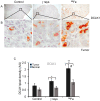
Detection of LGR5 expression in intestinal tumors after irradiation. Sample images of spontaneous, γ-rays, and 56Fe-irradiated intestinal tumor cells (A,B) stained for LGR5. (C) The bar graph represents the average DAB pixel density of LGR5 in all images taken of spontaneous, γ-rays, and 56Fe-irradiated tumor and normal tissue. The nuclei were counterstained with hematoxylin in blue. Scale bar tumor: 100 μm. *, significant relative to normal tissue, **, significant relative to spontaneous tumor sample. Statistical significance is set at p < 0.05 error bars represent mean ± SEM.
Figure 2
Figure 2
Differential response of CD133 and MSI1 after low- and high-LET irradiation in intestinal tumor. Sample images (A,B) of spontaneous, γ-rays, and 56Fe irradiated intestinal tumor cells stained for CD133 in dark brown DAB. (C) DAB pixel density was quantified from at least 20 different fields of view, statistically analyzed, and graphically represented. Bar graph represents the average DAB pixel density of CD133 in all images taken of control, γ-rays, and 56Fe-irradiated tumor and normal tissue. Sample images (D,E) of spontaneous, γ-rays, and 56Fe irradiated tumor intestinal cells stained for MSI1 in dark brown DAB. (F) DAB pixel density was quantified from at least 10 different fields of view, statistically analyzed, and graphically represented. Bar graph represents the average DAB pixel density of MSI1 in all images taken of unirradiated control, γ-rays, and 56Fe-irradiated tumor and normal tissue. The nuclei were counterstained with hematoxylin in blue. Scale bar tumor: 100 μm. *, significant relative to normal tissue, #, significant relative to γ-ray tumor sample. Statistical significance is set at p < 0.05 error bars represent mean ± SEM.
Figure 3
Figure 3
Response of DCLK1 after low- and high-LET irradiation in intestinal tumor. (A,B) Sample images of spontaneous, γ-rays, and 56Fe-irradiated tumor stained with DCLK1 in dark brown DAB. DAB-positive cells were counted from at least 20 different fields of view, statistically analyzed, and graphically represented. (C) Bar graph represents an average number of DCLK1-positive cells per 10× field. Higher DCLK1 positive cells were observed in radiation-induced tumors compared to the control. The nuclei are stained with hematoxylin in blue. Scale bar 100 μm. *, significant relative to normal tissue, **, significant relative to spontaneous tumor sample, #, significant relative to γ-ray tumor sample. Statistical significance is set at p < 0.05; error bars represent mean ± SEM.
Figure 4
Figure 4
Enhanced ALDH1 and BMI1 expression in intestinal tumor cells after 56Fe-ions. (A,B) Representative images of spontaneous, γ-rays, and 56Fe irradiated intestinal tumor cells stained for ALDH1 in dark brown DAB. (C) Bar graph represents the average DAB pixel expression of ALDH1 in images taken of control, γ-rays, and 56Fe-irradiated tumor and normal tissue. (D,E) Sample images of spontaneous, γ-rays, and 56Fe-irradiated tumor stained with BMI1 in dark brown DAB after brightfield immunohistochemistry. (F) Average DAB expression of BMI1 staining in all images taken of spontaneous, γ-rays, and 56Fe-irradiated tumor and normal tissue. The nuclei are counterstained with hematoxylin in blue. Scale bar 100 μm. DAB pixel density was quantified from at least 25 different fields of view and statistically analyzed *, significant relative to normal tissue, **, significant relative to control sample, #, significant relative to γ-ray tumor sample. Statistical significance is set at p < 0.05; error bars represent mean ± SEM.
Figure 5
Figure 5
Enhanced β-catenin expression and nuclear localization coincide with higher BMI1 expression in 56Fe-exposed mice. (A) Sample images of spontaneous, γ-rays, and 56Fe-irradiated intestinal tumor after ALDH1 and β-catenin fluorescent immunostaining. The images show β-catenin (red), ALDH1 (green); nuclei with DAPI (blue). (B) Fluorescent intensity of β-catenin and ALDH1 in tumor cells was quantified from at least ten different FOV, and mean fluorescent intensity was calculated and graphically represented. (C) Representative images of BMI1 and β-catenin fluorescent immunostaining in tumors across the treatment group. Arrow marks show nuclear β-catenin expression in the area of BMI1 expression. (D) Bar graph showing the mean fluorescent intensity of BMI1 or β-catenin in intestinal tumors. Scale bar: 20 μm. **, significant relative to spontaneous tumor; #, significant relative to γ-ray tumor sample. Statistical significance is set at p < 0.05; error bars represent mean ± SEM.

References

    1. Rahmanifard F., de Wet W.C., Schwadron N.A., Owens M.J., Jordan A.P., Wilson J.K., Joyce C.J., Spence H.E., Smith C.W., Townsend L.W. Galactic Cosmic Radiation in the Interplanetary Space Through a Modern Secular Minimum. Space Weather. 2020;18:e2019SW002428. doi: 10.1029/2019SW002428. - DOI
    1. Asaithamby A., Uematsu N., Chatterjee A., Story M.D., Burma S., Chen D.J. Repair of HZE-particle-induced DNA double-strand breaks in normal human fibroblasts. Radiat. Res. 2008;169:437–446. doi: 10.1667/RR1165.1. - DOI - PubMed
    1. Mishra B., Lawson G.W., Ripperdan R., Ortiz L., Luderer U. Charged-Iron-Particles Found in Galactic Cosmic Rays are Potent Inducers of Epithelial Ovarian Tumors. Radiat. Res. 2018;190:142–150. doi: 10.1667/RR15028.1. - DOI - PMC - PubMed
    1. Datta K., Suman S., Kallakury B.V., Fornace A.J. Heavy ion radiation exposure triggered higher intestinal tumor frequency and greater β-catenin activation than γ radiation in APC(Min/+) mice. PLoS ONE. 2013;8:e59295. doi: 10.1371/journal.pone.0059295. - DOI - PMC - PubMed
    1. Kumar S., Suman S., Fornace A.J., Jr., Datta K. Space radiation triggers persistent stress response, increases senescent signaling, and decreases cell migration in mouse intestine. Proc. Natl. Acad. Sci. USA. 2018;115:E9832–E9841. doi: 10.1073/pnas.1807522115. - DOI - PMC - PubMed

LinkOut - more resources