. 2023 Sep 2;15(17):4399.
doi: 10.3390/cancers15174399.

Development and Validation of the Oxidative Stress Related lncRNAs for Prognosis in Esophageal Squamous Cell Carcinoma

Affiliations

Development and Validation of the Oxidative Stress Related lncRNAs for Prognosis in Esophageal Squamous Cell Carcinoma

Xuan Zheng et al. Cancers (Basel). .

Abstract

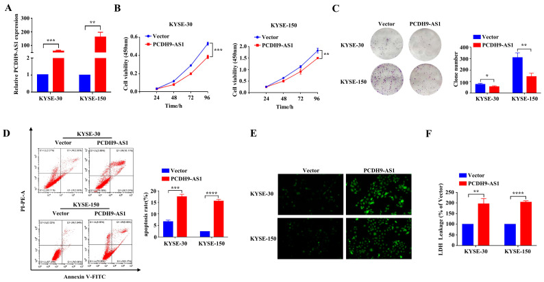
Esophageal squamous cell cancer (ESCC) is an aggressive disease associated with a poor prognosis. Long non-coding RNAs (lncRNAs) and oxidative stress play crucial roles in tumor progression. We aimed to identify an oxidative stress-related lncRNA signature that could predict the prognosis in ESCC. In the GSE53625 dataset, we identified 332 differentially expressed lncRNAs (DElncRNAs) between ESCC and control samples, out of which 174 were oxidative stress-related DElncRNAs. Subsequently, seven oxidative stress-related DElncRNAs (CCR5AS, LINC01749, PCDH9-AS1, TMEM220-AS1, KCNMA1-AS1, SNHG1, LINC01672) were selected based on univariate and LASSO Cox to build a prognostic risk model, and their expression was detected by RT-qPCR. The model exhibited an excellent ability for the prediction of overall survival (OS) and other clinicopathological traits using Kaplan-Meier (K-M) survival curves, receiver operating characteristic (ROC) curves, and the Wilcoxon test. Additionally, analysis of infiltrated immune cells and immune checkpoints indicated differences in immune status between the two risk groups. Finally, the in vitro experiments showed that PCDH9-AS1 overexpression inhibited proliferation ability and promoted apoptosis and oxidative stress levels in ESCC cells. In conclusion, our study demonstrated that a novel oxidative stress-related DElncRNA prognostic model performed favorably in predicting ESCC patient prognosis and benefits personalized clinical applications.

Keywords: ESCC; lncRNA; overall survival; oxidative stress; prognosis.


Conflict of interest statement

The authors declare no conflict of interest.

Figures

Figure 1
Figure 1
Study flowchart.
Figure 2
Figure 2
Identification of differentially expressed lncRNAs (DElncRNAs) in esophageal squamous cell carcinoma (ESCC). (A) Volcano plot illustrating the DElncRNAs in ESCC tissues compared with paired paracancerous tissues from the GSE53625 dataset. Red dots denote upregulated genes, blue dots represent downregulated genes, and grey dots indicate genes with no significant difference. (B) Heat map displaying the top 10 DElncRNAs in the GSE53625 dataset. Red represents high expression, while blue indicates low expression.
Figure 3
Figure 3
Construction and validation of a prognostic oxidative stress-related risk model composed of seven DElncRNAs. (A) Forest plots presenting the results of Cox univariate regression analysis of prognostic oxidative stress-related DElncRNAs. (B,C) cvfit and lambda curves illustrate the application of the least absolute shrinkage and selection operator (LASSO) regression using the minimum criteria. Each line represents one oxidative stress-related DElncRNAs (CCR5AS, LINC01749, PCDH9-AS1, TMEM220-AS1, KCNMA1-AS1, SNHG1, LINC01672) in subfigure B. (DG) Distribution of the risk scores, overall survival status (OS), and risk score in the training and validation datasets. (H,I) Kaplan–Meier curves demonstrating the survival status and survival time in the training dataset and validation dataset. (J,K) Receiver operating characteristic (ROC) curve showcasing the potential of the prognostic oxidative stress-related DElncRNA signature in predicting 1-year, 2-year, and 3-year OS in the training dataset and validation dataset.
Figure 4
Figure 4
Construction of a nomogram for OS prediction in ESCC. (A,B) Univariate (A) and multivariate (B) Cox regression analysis of prognostic clinical indicators. (C) Nomogram to predict the 1-year, 3-year, and 5-year OS rates in ESCC patients. (D) Calibration curve used to evaluate the accuracy of the nomogram model at 1-year, 3-year, and 5-year time points.
Figure 5
Figure 5
Association between clinicopathological data and risk score in ESCC. (AD) The boxplot shows the association between the risk score and clinical stage (A), T stage (B), N stage (C), and gender (D). The “•” represents outliers. NS p > 0.05 and * p < 0.05.
Figure 6
Figure 6
Gene Set Variation Analysis (GSVA) of high-risk and low-risk groups based on the prognostic signature of oxidative stress-related DElncRNAs. (AC) Enriched categories of biological process (A), cellular component (B), and molecular function (C) in the high-risk and low-risk groups. (D) Significant Kyoto Encyclopedia of Genes and Genomes (KEGG) pathways in the high-risk and low-risk groups.
Figure 7
Figure 7
Immune-related analysis using the prognostic signature in ESCC. (A) Stacked column chart showing the proportions of tumor-infiltrating cells in the high-risk and low-risk groups. (B) Boxplots comparing the immune cell levels between the high-risk and low-risk groups. (C) Boxplots comparing the expression levels of immune checkpoint genes between the high-risk and low-risk groups. ns p > 0.05, * p < 0.05, ** p < 0.01, and *** p < 0.001.
Figure 8
Figure 8
Verification of the expression levels of the seven oxidative stress-related DElncRNAs in cell lines and tissues. (A) Relative expression of the seven oxidative stress-related DElncRNAs in the normal human esophageal epithelial line Het-1A and ESCC cells (KYSE-30, KYSE-150, KYSE-410, TE-1, Eca-109). (B) Relative expression of the seven oxidative stress-related DElncRNAs in 10 pairs of carcinoma tissue and paracancerous tissue of ESCC. NS p > 0.05, * p < 0.05, ** p < 0.01, *** p < 0.001, and **** p < 0.0001.
Figure 9
Figure 9
Immunofluorescence analysis of PCDH9-AS1 expression in carcinoma tissues and paracancerous tissues of ESCC. (A) Representative images (magnification 200×). DAPI was used for nuclear staining. (B) Quantitative map of the relative fluorescence intensity for 80 pairs of carcinoma tissue and paracancerous tissue of ESCC. **** p < 0.0001.
Figure 10
Figure 10
The effect of PCDH9-AS1 overexpression on ESCC cells. (A) Relative expression level of PCDH9-AS1 after transfection with the corresponding pcDNA 3.1 (+) vector. (BD) Cell proliferation ability (B), colony formation ability (C), and apoptosis level (D) in the PCDH9-AS1 overexpression and control groups. (E,F) Oxidative stress level evaluated by detecting ROS (E) (green, magnification 200×) and LDH (F) levels in the PCDH9-AS1 overexpression and control groups. * p < 0.05, ** p < 0.01, *** p < 0.001, and **** p < 0.0001.

References

    1. Sung H., Ferlay J., Siegel R.L., Laversanne M., Soerjomataram I., Jemal A., Bray F. Global Cancer Statistics 2020: GLOBOCAN Estimates of Incidence and Mortality Worldwide for 36 Cancers in 185 Countries. CA Cancer J. Clin. 2021;71:209–249. doi: 10.3322/caac.21660. - DOI - PubMed
    1. Sheikh M., Roshandel G., McCormack V., Malekzadeh R. Current Status and Future Prospects for Esophageal Cancer. Cancers. 2023;15:765. doi: 10.3390/cancers15030765. - DOI - PMC - PubMed
    1. Waters J.K., Reznik S.I. Update on Management of Squamous Cell Esophageal Cancer. Curr. Oncol. Rep. 2022;24:375–385. doi: 10.1007/s11912-021-01153-4. - DOI - PubMed
    1. Rogers J.E., Sewastjanow-Silva M., Waters R.E., Ajani J.A. Esophageal cancer: Emerging therapeutics. Expert Opin. Ther. Targets. 2022;26:107–117. doi: 10.1080/14728222.2022.2036718. - DOI - PubMed
    1. Dimitrova N., Zamudio J.R., Jong R.M., Soukup D., Resnick R., Sarma K., Ward A.J., Raj A., Lee J.T., Sharp P.A., et al. LincRNA-p21 activates p21 in cis to promote Polycomb target gene expression and to enforce the G1/S checkpoint. Mol. Cell. 2014;54:777–790. doi: 10.1016/j.molcel.2014.04.025. - DOI - PMC - PubMed

LinkOut - more resources