Review
. 2023 Sep 4;15(17):4419.
doi: 10.3390/cancers15174419.

Human Aldehyde Dehydrogenases: A Superfamily of Similar Yet Different Proteins Highly Related to Cancer

Affiliations
Review

Human Aldehyde Dehydrogenases: A Superfamily of Similar Yet Different Proteins Highly Related to Cancer

Vasileios Xanthis et al. Cancers (Basel). .

Abstract

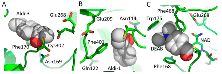
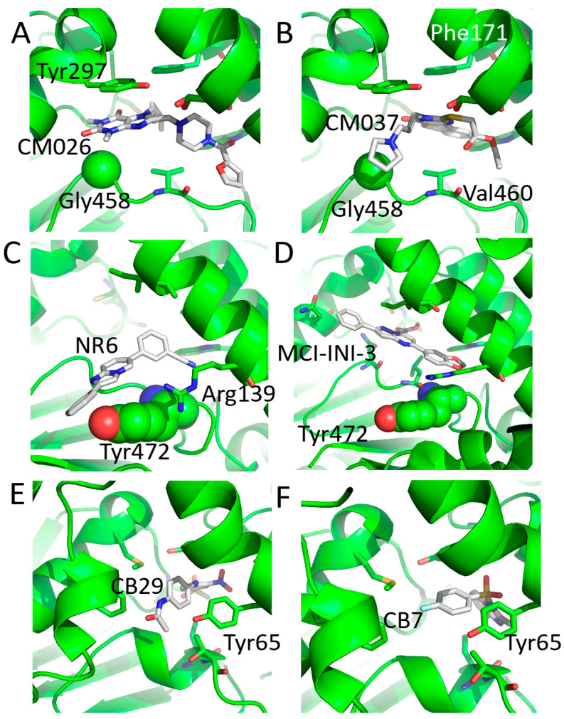
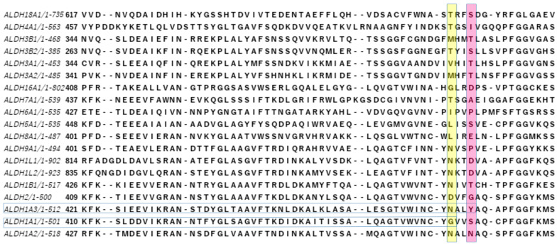
The superfamily of human aldehyde dehydrogenases (hALDHs) consists of 19 isoenzymes which are critical for several physiological and biosynthetic processes and play a major role in the organism's detoxification via the NAD(P) dependent oxidation of numerous endogenous and exogenous aldehyde substrates to their corresponding carboxylic acids. Over the last decades, ALDHs have been the subject of several studies as it was revealed that their differential expression patterns in various cancer types are associated either with carcinogenesis or promotion of cell survival. Here, we attempt to provide a thorough review of hALDHs' diverse functions and 3D structures with particular emphasis on their role in cancer pathology and resistance to chemotherapy. We are especially interested in findings regarding the association of structural features and their changes with effects on enzymes' functionalities. Moreover, we provide an updated outline of the hALDHs inhibitors utilized in experimental or clinical settings for cancer therapy. Overall, this review aims to provide a better understanding of the impact of ALDHs in cancer pathology and therapy from a structural perspective.

Keywords: 3D structure; aldehyde dehydrogenases (ALDHs); cancer; chemotherapy resistance; crystallins; inhibitors; post-translational modifications; quaternary association; topology.


Conflict of interest statement

The authors declare no conflict of interest. The funders had no role in the design of the study; in the collection, analyses, or interpretation of data; in the writing of the manuscript; or in the decision to publish the results.

Figures

Figure 1
Figure 1
The structure of mitochondrial hALDH1B1 monomer in complex with the NAD (Nicotinamide Adenine Dinucleotide) cofactor and the ZGG (8-(2-methoxyphenyl)-10-(4-phenylphenyl)-1,8-diazabicyclo[5.3.0]deca-1(7),9-diene) inhibitor (PDBid:7MJD). The protein is shown in cartoon representation. Cofactor and inhibitor are shown as space-filling models and indicate the substrate and cofactor binding sites on the protein, respectively. The three catalytic residues, i.e., Cys 302, Glu 268, and Asn 169, are shown with spheres and indicate the protein’s active site. The monomer’s structure consists of a catalytic domain (pink), a cofactor binding domain (blue), and an oligomerization domain (green). (AC) Three views of the monomer related with 90-degree rotations around the indicated axis. The zoom-in view of (B) focuses on the NAD and ZGG binding sites and thus highlights the spatial proximity of cofactor and substrate binding sites on the protein’s structure. The catalytic (D) and cofactor (E) binding domains fold as Rossmann motifs. In the interface of catalytic and cofactor binding domains, the active site of the enzyme is located.
Figure 2
Figure 2
Multiple sequence alignment of the 19 hALDHs. The part of the alignment which includes the catalytic triad (highlighted in blue) is shown. The alignment shows that all hALDHs but ALDH16A1, ALDH6A1, and ALDH18A1 have in common a catalytic triad consisting of a cysteine (catalytic thiol), a glutamic acid (general base), and an asparagine (residue important for stabilizing the reaction’s intermediate). ALDH6A1 follows a slightly different mechanism compared with the other members of the superfamily (see text) and possesses a slightly modified catalytic triad where glutamic acid has been substituted by an asparagine, while the other two catalytic residues (cysteine and asparagine) are conserved. ALDH18A1 is the most distant member of the superfamily (see text and Figure 3) which is also evident from the fact that its active site incorporates only the catalytic cysteine. Last, ALDH16A1 is a pseudoenzyme without enzymatic activity and includes none of the catalytic residues.
Figure 3
Figure 3
All against all structure analysis of hALDHs with determined 3D structures through DALI server [30]. Protein names and the PDB codes of the structures used as representatives of each protein for the analysis are shown together. (A) Heatmap of structural similarity matrix based on Dali Z-scores. (B) Structural similarity dendrogram. The dendrogram is derived by average linkage clustering of the structural similarity matrix (Dali Z-scores).The dendrogram figure was prepared with the iTOL tool [31].
Figure 4
Figure 4
Topology diagrams of the three domains for each of the 12 hALDHs of known structure. All three domains of hALDH18A1 are distinct, while all the other members have in common at least the topology of catalytic domain. hALDH3A1 and 3A2 have in addition a common topology of their cofactor binding domains. ALDH4A1 shares with the hALDH3Ai pair a quite common oligomerization domain. hALDH1/2, 5A1, and 9A1 have common topologies in all three domains, while hALDH4A1 and 7A1 might be the bridge between the 3Ai subgroup and the 1/2, 5A1, 9A1 subgroup.
Figure 5
Figure 5
Dimer organization—from three different views, 90 degrees apart—for three representative structures of hALDH superfamily. The structures are shown with surface representation and the colours indicate the catalytic (pink), cofactor binding (blue), and oligomerization (green) domains. (A) hALDH3A1 (PDBid: 3SZB). (B) hALDH1A1 (PDBid: 4WB9). (C) hALDH18A1 (PDBid: 2H5G).
Figure 6
Figure 6
Representative examples of hALDH quaternary assemblies. Each structure is shown from three different views, as indicated. Space-filling models are used, and the different colours represent different monomers. (A) hALDH3A1 dimer (PDBid: 3SZB). (B) hALDH1A1 tetramer (PDBid: 4WB9). (C) hALDH5A1 dodecamer of reduced protein (PDBid: 2W8O).
Figure 7
Figure 7
Diagram presenting the multiple roles of ALDHs.
Figure 8
Figure 8
Location of Lysine 353 on the surface of ALDH1A1 tetramer and how its acetylation imposes steric hindrance and affects its catalytic ability. (A,B) Cartoon representation of hALDH1A1. Each monomer is shown with a different colour. Lysine 353 (gray space-filling model) is found on the rim of NAD(H) (yellow space-filling model) binding pocket. (C,D) Zoom in on the Lysine 353/NADH binding area. Lysine is shown in a gray sticks model superimposed with van der Waals dots of non-hydrogen atoms. In (C), the unmodified Lys makes optimum interactions with the bound NADH. (D) A model of acetylated Lys353 shows how the extra acetyl-group restricts the available space and makes unfavourable the NADH binding.
Figure 9
Figure 9
Non-specific inhibitors in complex with hALDHs. (A) hALDH2/Aldi-3 complex (PDBid: 3SZ9). The inhibitor is inside the active site and forms a covalent bond with the catalytic cysteine. (B) hALDH3A1/Aldi-1 complex (PDBid: 3SZB). (C) hALDH7A1/DEAB complex (PDBid: 4X0T).
Figure 10
Figure 10
Specific inhibitors in complex with hALDHs. Proteins are shown in cartoon representation and residues significant for the inhibitor-protein interactions are shown with sticks. Especially, non-conserved driving-specificity residues are shown in spacefill. Protein elements are all in green. Inhibitors are represented with gray-carbon stick models. (A) hALDH1A1/CM026 complex (PDBid:4WP7). (B) hALDH1A1/CM037 complex (PDBid:4X4L). (C) hALDH1A3/NR6 complex (PDBid:7A6Q). (D) hALDH1A3/MCI-INI-3 complex (PDBid:6TGW). (E) hALDH3A1/CB29 complex (PDBid:4H80). (F) hALDH3A1/CB7 complex (PDBid:4L2O).
Figure 11
Figure 11
A small window from the sequence alignment of hALDHs, which highlights two residues experimentally characterized as driving-specificity residues for designing inhibitors. Gly458 for ALDH1A1 (last residue of the horizontal yellow highlight) and Tyr472 for ALDH1A3 (last residue of the horizontal pink highlight). For more details, see text and Figure 10.

References

    1. Marchitti S.A., Brocker C., Stagos D., Vasiliou V. Non-P450 Aldehyde Oxidizing Enzymes: The Aldehyde Dehydrogenase Superfamily. Expert Opin. Drug Metab. Toxicol. 2008;4:697. doi: 10.1517/17425255.4.6.697. - DOI - PMC - PubMed
    1. Rodríguez-Zavala J.S., Calleja L.F., Moreno-Sánchez R., Yoval-Sánchez B. Role of Aldehyde Dehydrogenases in Physiopathological Processes. Chem. Res. Toxicol. 2019;32:405–420. doi: 10.1021/acs.chemrestox.8b00256. - DOI - PubMed
    1. Ursini F., Maiorino M. Lipid Peroxidation and Ferroptosis: The Role of GSH and GPx4. Free Radic. Biol. Med. 2020;152:175–185. doi: 10.1016/j.freeradbiomed.2020.02.027. - DOI - PubMed
    1. Kayani M.A., Parry J.M. The in Vitro Genotoxicity of Ethanol and Acetaldehyde. Toxicol. Vitr. Int. J. Publ. Assoc. BIBRA. 2010;24:56–60. doi: 10.1016/j.tiv.2009.09.003. - DOI - PubMed
    1. O’Brien P., Siraki A., Shangari N. Aldehyde Sources, Metabolism, Molecular Toxicity Mechanisms, and Possible Effects on Human Health. Crit. Rev. Toxicol. 2008;35:609–662. doi: 10.1080/10408440591002183. - DOI - PubMed

LinkOut - more resources