. 2023 Aug 24;28(17):6236.
doi: 10.3390/molecules28176236.

Molecular Docking Assessment of Cathinones as 5-HT2AR Ligands: Developing of Predictive Structure-Based Bioactive Conformations and Three-Dimensional Structure-Activity Relationships Models for Future Recognition of Abuse Drugs

Affiliations

Molecular Docking Assessment of Cathinones as 5-HT2AR Ligands: Developing of Predictive Structure-Based Bioactive Conformations and Three-Dimensional Structure-Activity Relationships Models for Future Recognition of Abuse Drugs

Nevena Tomašević et al. Molecules. .

Abstract

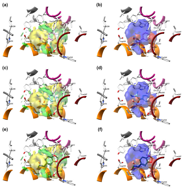
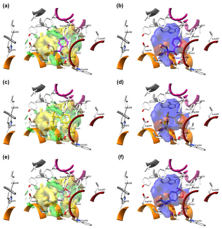
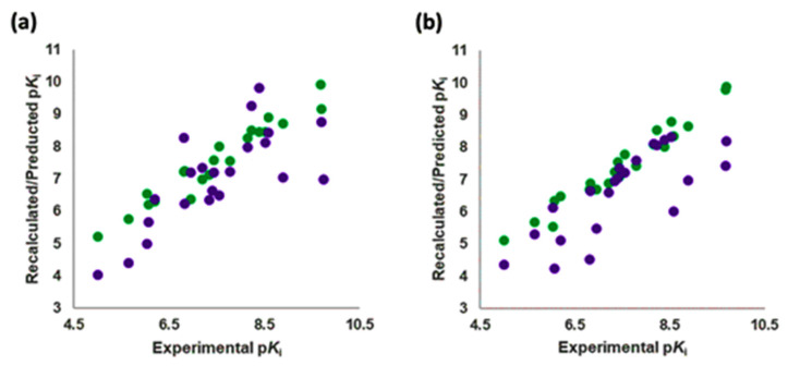
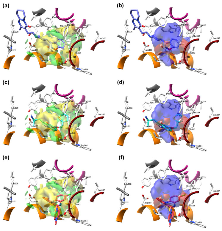
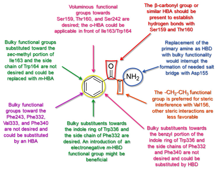
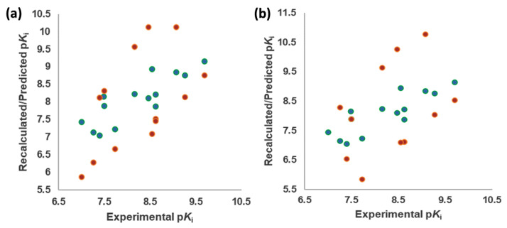
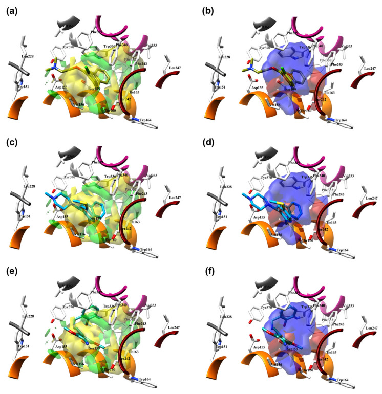
Commercially available cathinones are drugs of long-term abuse drugs whose pharmacology is fairly well understood. While their psychedelic effects are associated with 5-HT2AR, the enclosed study summarizes efforts to shed light on the pharmacodynamic profiles, not yet known at the receptor level, using molecular docking and three-dimensional quantitative structure-activity relationship (3-D QSAR) studies. The bioactive conformations of cathinones were modeled by AutoDock Vina and were used to build structure-based (SB) 3-D QSAR models using the Open3DQSAR engine. Graphical inspection of the results led to the depiction of a 3-D structure analysis-activity relationship (SAR) scheme that could be used as a guideline for molecular determinants by which any untested cathinone molecule can be predicted as a potential 5-HT2AR binder prior to experimental evaluation. The obtained models, which showed a good agreement with the chemical properties of co-crystallized 5-HT2AR ligands, proved to be valuable for future virtual screening campaigns to recognize unused cathinones and similar compounds, such as 5-HT2AR ligands, minimizing both time and financial resources for the characterization of their psychedelic effects.

Keywords: 3-D QSAR; 5-HT2AR; cathinones; molecular docking.


Conflict of interest statement

The authors declare no conflict of interest.

Figures

Figure 1
Figure 1
Left: The crystal structure of 5-HT2AR in complex with LSD (as deposited at PDB, https://www.rcsb.org/, PDB ID: 6HWT [16], accessed on 1 December 2022). LSD is depicted in blue encircled by the transparent sphere, TM1 (residues 69–101) is colored in blue, TM2 (residues 111–137) is depicted in plum, TM3 (residues 114–179) is painted in orange, TM4 (residues 188–217) is colored in lime green, TM5 (residues 231–265) is depicted in dark red, TM6 (residues 315–349) is painted in violet-red, TM7 (residues 353–383) is colored in dark grey, intracellular amphipathic helix H8 (residues 384–399) is depicted in yellow, two intracellular loops ICL1 and ICL2 (residues 102–111 and 179–187) are illustrated in cyan and hot pink, respectively, three extracellular loops ECL1, ECL2, and ECL3 (residues 134–138, 218–230, and 350–352) are labeled in gold, dim gray, and dark slate gray, respectively; Right: The crystal structure of 5-HT2AR immersed into dipalmitoylphosphatidylcholine (DPPC) lipid bilayer (as deposited at OPM, https://opm.phar.umich.edu/, entry: 4282, accessed on 2 March 2023): the DPPCs are illustrated with transparent gray spheres, oxygen atoms from water molecules are colored in red, sodium atoms are presented as pink spheres, and chlorine atoms are described as green spheres. For the clarity of the presentation, hydrogen atoms were omitted.
Figure 2
Figure 2
Sequence alignment between human 5-HT2AR (Uniprot entry P28223) and 5-HT2BR (UniProt entry P41596) performed by Clustal Omega [66]. Special characters meanings: * (Asterix) positions with a single, fully conserved residue; “:” (colon) positions with conservation between amino acid groups of similar properties; “.” (period) positions with conservation between amino acid groups of weakly similar properties.
Figure 3
Figure 3
The docking assessment of LSD co-crystallized into 5-HT2AR (the 6WGT crystal [16]), EC conformation in pink, ECRD conformation in blue, RCRD conformation in green, ECCD conformation in red, and RCCD conformation in yellow (a); LSD co-crystallized into 5-HT2BR (the 5VNT crystal [31]), EC conformation in pink, ECRD conformation in blue, RCRD conformation in green, ECCD conformation in red, and RCCD conformation in yellow (b).
Figure 4
Figure 4
Experimental versus recalculated (blue circles) and predicted (red circles) pKis for TR using OH2 probe BOTH (STE + ELE) LOO model at 5 PCs (a); OH2 probe BOTH (STE + ELE) LSO model at 5 PCs (b).
Figure 5
Figure 5
The structure-based alignment within 5-HT2AR-DPPC and OH2 probe-based PLS-coefficients (positive steric coefficients presented in green, negative steric coefficients depicted in yellow, positive electrostatic coefficients portrayed in red, negative electrostatic coefficients displayed in blue) for 1 (a,b); 2 (c,d); 17 (e,f). For the clarity of presentation, DPPC was omitted.
Figure 6
Figure 6
The structure-based alignment within 5-HT2AR-DPPC and OH2 probe-based PLS-coefficients (positive steric coefficients presented in green, negative steric coefficients depicted in yellow, positive electrostatic coefficients portrayed in red, negative electrostatic coefficients displayed in blue) for 4 (a,b), 9 (c,d), and 18 (e,f). For the clarity of presentation, DPPC was omitted.
Figure 7
Figure 7
Experimental versus recalculated (green circles) and predicted (purple circles) pKis for TS using OH2 probe BOTH (STE + ELE) LOO model at 5 PCs (a); OH2 probe BOTH (STE + ELE) LSO model at 5 PCs (b).
Figure 8
Figure 8
The structure-based alignment within 5-HT2AR-DPPC and OH2 probe-based PLS-coefficients (positive steric coefficients presented in green, negative steric coefficients depicted in yellow, positive electrostatic coefficients portrayed in red, negative electrostatic coefficients displayed in blue) for 25 (a,b); 26 (c,d); 27 (e,f). For the clarity of presentation, DPPC was omitted.
Figure 9
Figure 9
The SAR model for CSs as 5-HT2AR ligands and agonists is seen through 1.
Figure 10
Figure 10
Experimental versus recalculated (green circles) and predicted (orange circles) pKis for TSCRY using OH2 probe BOTH (STE + ELE) LOO model at 5 PCs (a); OH2 probe BOTH (STE + ELE) LSO model at 5 PCs (b).
Figure 11
Figure 11
The structure-based alignment within 5-HT2AR-DPPC and OH2 probe-based PLS-coefficients (positive steric coefficients presented in green, negative steric coefficients depicted in yellow, positive electrostatic coefficients portrayed in red, negative electrostatic coefficients displayed in blue) for 6A94 (a,b); 6WH4 (c,d); 6WHA (e,f). For the clarity of presentation, DPPC was omitted.

References

    1. Nichols D.E., Nichols C.D. Serotonin Receptors. Chem. Rev. 2008;108:1614–1641. doi: 10.1021/cr078224o. - DOI - PubMed
    1. Hauser A.S., Attwood M.M., Rask-Andersen M., Schiöth H.B., Gloriam D.E. Trends in GPCR Drug Discovery: New Agents, Targets and Indications. Nat. Rev. Drug Discov. 2017;16:829–842. doi: 10.1038/nrd.2017.178. - DOI - PMC - PubMed
    1. Welsh D.J., Harnett M., MacLean M., Peacock A.J. Proliferation and Signaling in Fibroblasts: Role of 5-hydroxytryptamine2A Receptor and Transporter. Am. J. Respir. Crit. Care Med. 2004;170:252–259. doi: 10.1164/rccm.200302-264OC. - DOI - PubMed
    1. El-Merahbi R., Loffler M., Mayer A., Sumara G. The Roles of Peripheral Serotonin in Metabolic Homeostasis. FEBS Lett. 2015;589:1728–1734. doi: 10.1016/j.febslet.2015.05.054. - DOI - PubMed
    1. Matsumoto T., Kobayashi T., Ishida K., Taguchi K., Kamata K. Enhancement of Mesenteric Artery Contraction to 5-HT Depends on Rho Kinase and Src Kinase Pathways in the Ob/Ob Mouse Model of Type 2 Diabetes. Br. J. Pharmacol. 2010;160:1092–1104. doi: 10.1111/j.1476-5381.2010.00753.x. - DOI - PMC - PubMed

LinkOut - more resources