. 2023 Aug 28;28(17):6278.
doi: 10.3390/molecules28176278.

Membrane-Targeting Perylenylethynylphenols Inactivate Medically Important Coronaviruses via the Singlet Oxygen Photogeneration Mechanism

Affiliations

Membrane-Targeting Perylenylethynylphenols Inactivate Medically Important Coronaviruses via the Singlet Oxygen Photogeneration Mechanism

Kseniya A Mariewskaya et al. Molecules. .

Abstract

Perylenylethynyl derivatives have been recognized as broad-spectrum antivirals that target the lipid envelope of enveloped viruses. In this study, we present novel perylenylethynylphenols that exhibit nanomolar or submicromolar antiviral activity against Severe Acute Respiratory Syndrome Coronavirus-2 (SARS-CoV-2) and feline infectious peritonitis virus (FIPV) in vitro. Perylenylethynylphenols incorporate into viral and cellular membranes and block the entry of the virus into the host cell. Furthermore, these compounds demonstrate an ability to generate singlet oxygen when exposed to visible light. The rate of singlet oxygen production is positively correlated with antiviral activity, confirming that the inhibition of fusion is primarily due to singlet-oxygen-induced damage to the viral envelope. The unique combination of a shape that affords affinity to the lipid bilayer and the capacity to generate singlet oxygen makes perylenylethynylphenols highly effective scaffolds against enveloped viruses. The anticoronaviral activity of perylenylethynylphenols is strictly light-dependent and disappears in the absence of daylight (under red light). Moreover, these compounds exhibit negligible cytotoxicity, highlighting their significant potential for further exploration of the precise antiviral mechanism and the broader scope and limitations of this compound class.

Keywords: SARS-CoV-2; antivirals; perylene; photosensitizers; singlet oxygen.


Conflict of interest statement

The authors declare no conflict of interest.

Figures

Figure 1
Figure 1
Recently studied perylene antivirals and the parent compound for this study (HOPY11).
Figure 2
Figure 2
Structural design of phenol-conjugated perylenylethynyl derivatives.
Scheme 1
Scheme 1
Synthesis of perylenylethynylphenols.
Figure 3
Figure 3
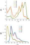
Normalized UV-Vis (A) and fluorescence (B) spectra of compounds 3af in 96% EtOH.
Figure 4
Figure 4
Normalized absorbance spectra of perylene 3e and SOSG in methanol solution. Region of LED irradiation for singlet oxygen generation by perylene compounds is shown in blue, wavelength of SOSG excitation is shown in green. Inset: time course of SOSG fluorescence intensity in a reaction with 1O2 generated by perylene compound 3e under blue light irradiation.
Figure 5
Figure 5
Cytotoxicity and anti-coronaviral activity of perylenylethynylphenols in vitro. (A) Schematic representation of the cytotoxicity assay using Cell Counting Kit-8 (Dojindo Molecular Technologies, Munich, Germany). (B) Schematic representation of the viral titer reduction assay. (C) Cytotoxicity of perylenylethynylphenols at the indicated concentrations for Vero cells. (D) Anti-SARS-CoV-2 activity of perylenylethynylphenols at the indicated concentrations in Vero cells. (E) Anti-FIPV activity of compound 3a in CRFK cells. Data are expressed as the mean ± SD of two independent experiments, each performed in triplicate. The horizontal dashed line indicates the minimum detectable threshold of 1.44 log10 PFU/mL.
Figure 6
Figure 6
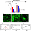
Interaction of compound 3a with viral envelopes, cellular membranes and liposomal mebranes. (A) Demonstration of direct (virucidal) activity of 3a against SARS-CoV-2 and its interaction with the viral envelope. Schematic representation of the experiment (virucidal plaque assay). (B) Quantification of the virucidal activity of 3a using Vero E6 cells. The virus at the indicated titers was incubated with the compounds (10 μM) for 120 min. Viral titers were then quantified by plaque assays. (C) Penetration of 3a into PS cells. Cells were seeded on slides for 24 h, then treated with 3a (10 µM) and incubated for 1 h. Photomicrographs were taken using confocal microscopy. (D) Excitation and emission spectra of 3a (10 µM) in DMSO. (E) Fluorescence spectra of free compound 3a in PBS (10 µM, dashed line) and a mixture of 3a and LPS in PBS. (F) Kinetics of the penetration of 3a (10 µM) into liposomes, measured at 520 nm. Data are expressed as the mean ± SD of two independent experiments, each performed in triplicate.
Figure 7
Figure 7
Light-dependent cytotoxicity and antiviral activity of perylenylethynylphenols. (A) Determination of photocytotoxicity (schematic representation of experiments). (B) Cytotoxicity of perylenylethynylphenols under normal light conditions (sample preparation in daylight, incubation of compounds with CRFK cells in the dark). (C) Cytotoxicity of perylenylethynylphenols after irradiation with blue light for 10 min at RT with LEDs (465–480 nm, 30 mW/cm2). (D) Light-dependent anti-FIPV activity of compound 3a (schematic representation of the experiments). (E) FIPV was treated with 3a, as described in (D), and the viability of the compound-treated virus was determined by plaque assays. Data are expressed as the mean ± SD of two independent experiments, each performed in triplicate. The horizontal dashed line indicates the minimum detectable threshold of 1.44 log10 PFU/mL.

References

    1. Murakami N., Hayden R., Hills T., Al-Samkari H., Casey J., Del Sorbo L., Lawler P.R., Sise M.E., Leaf D.E. Therapeutic advances in COVID-19. Nat. Rev. Nephrol. 2023;19:38–52. doi: 10.1038/s41581-022-00642-4. - DOI - PMC - PubMed
    1. Li G., Hilgenfeld R., Whitley R., De Clercq E. Therapeutic strategies for COVID-19: Progress and lessons learned. Nat. Rev. Drug Discov. 2023;22:449–475. doi: 10.1038/s41573-023-00672-y. - DOI - PMC - PubMed
    1. Von Delft A., Hall M.D., Kwong A.D., Purcell L.A., Saikatendu K.S., Schmitz U., Tallarico J.A., Lee A.A. Accelerating antiviral drug discovery: Lessons from COVID-19. Nat. Rev. Drug Discov. 2023;22:585–603. doi: 10.1038/s41573-023-00692-8. - DOI - PMC - PubMed
    1. Mohammed Ibrahim O., Bara Allawe A., Ali Kadhim H. Isolation and molecular detection of Feline infectious peritonitis virus. Arch. Razi Inst. 2022;77:1709–1714. doi: 10.22092/ari.2022.357997.2135. - DOI - PMC - PubMed
    1. Hudson J.B., Zhou J., Chen J., Harris L., Yip L., Towers G.H.N. Hypocrellin, from Hypocrella bambuase, is phototoxic to human immunodeficiency virus. Photochem. Photobiol. 1994;60:253–255. doi: 10.1111/j.1751-1097.1994.tb05100.x. - DOI - PubMed