. 2023 Aug 25;16(17):5821.
doi: 10.3390/ma16175821.

The Morphology Dependent Interaction between Silver Nanoparticles and Bovine Serum Albumin

Affiliations

The Morphology Dependent Interaction between Silver Nanoparticles and Bovine Serum Albumin

Jingyi Zhang et al. Materials (Basel). .

Abstract

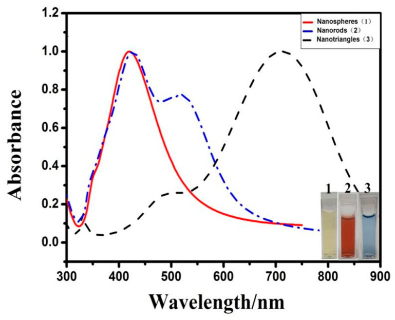
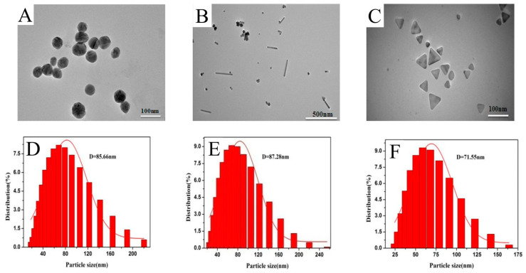
Biological applications of silver nanoparticles (AgNPs) depend on the covalently attached or adsorbed proteins. A series of biological effects of AgNPs within cells are determined by the size, shape, aspect ratio, surface charge, and modifiers. Herein, the morphology dependent interaction between AgNPs and protein was investigated. AgNPs with three different morphologies, such as silver nanospheres, silver nanorods, and silver nanotriangles, were employed to investigate the morphological effect on the interaction with a model protein: bovine serum albumin (BSA). The adsorptive interactions between BSA and the AgNPs were probed by UV-Vis spectroscopy, fluorescence spectroscopy, dynamic light scattering (DLS), Fourier transform infrared spectrometry (FTIR), transmission electron microscopy (TEM), and circular dichroism (CD) techniques. The results revealed that the particle size, shape, and dispersion of the three types of AgNPs markedly influence the interaction with BSA. Silver nanospheres and nanorods were capsulated by protein coronas, which led to slightly enlarged outer size. The silver nanotriangles evolved gradually into nanodisks in the presence of BSA. Fluorescence spectroscopy confirmed the static quenching the fluorescence emission of BSA by the three AgNPs. The FTIR and CD results suggested that the AgNPs with different morphologies had different effects on the secondary structure of BSA. The silver nanospheres and silver nanorods induced more pronounced structural changes than silver nanotriangles. These results suggest that the formation of a protein corona and the aggregation behaviors of AgNPs are markedly determined by their inherent morphologies.

Keywords: bovine serum albumin; morphology; protein corona; silver nanoparticle.


Conflict of interest statement

The authors declare no conflict interests.

Figures

Figure 1
Figure 1
UV-Vis spectra of as-synthesized silver nanospheres, silver nanorods, and silver nanotriangles.
Figure 2
Figure 2
TEM of (A) silver nanospheres, (B) silver nanorods, and (C) silver nanotriangles. Histograms of the particle size distribution of (D) silver nanospheres, (E) silver nanorods, and (F) silver nanotriangles. The solid curve is the Gaussian fit to the histogram.
Figure 3
Figure 3
Fluorescence spectra of the interaction of BSA with (A) silver nanospheres: (1–11: 0, 0.01, 0.05, 0.10, 0.20, 0.30, 0.40, 0.60, 0.80, 0.90, 1.04) × 10−10 M, (B) silver nanorods: (1–8: 0, 1.0, 2.0, 3.0, 4.0, 5.0, 6.51, 7.45) × 10−10 M, and (C) silver nanotriangles: (1–11: 0, 0.02, 0.04, 0.08, 0.16, 0.32, 0.64, 0.80, 0.90, 1.09, 1.177) × 10−4 M.
Figure 4
Figure 4
CD spectra of the interaction of BSA with (A) silver nanospheres (1–4: 0, 1 × 10⁻¹² M, 2 × 10⁻¹² M, 3.9 × 10⁻¹² M), (B) silver nanorods (1–4: 0, 0.33 × 10⁻¹⁰ M, 0.615 × 10⁻¹⁰ M, 1.104 × 10⁻¹⁰ M), and (C) silver nanotriangles (1–4: 0, 0.336 × 10⁻⁶ M, 0.811 × 10⁻⁶ M, 1.801 × 10⁻⁶ M).
Figure 5
Figure 5
FTIR of the interaction of BSA with (A) silver nanospheres (0.052 nM), (B) silver nanorods (0.184 nM), and (C) silver nanotriangles (0.24 μM) in aqueous solution.
Figure 6
Figure 6
Representative TEM images of the interactions of BSA with (A) silver nanospheres, (B) silver nanorods, and (C) silver nanotriangles. Histograms of the particle size distribution in the interaction of BSA with (D) silver nanospheres, (E) silver nanorods, and (F) silver nanotriangles. The solid curve is a Gaussian fit to the histogram.
Figure 6
Figure 6
Representative TEM images of the interactions of BSA with (A) silver nanospheres, (B) silver nanorods, and (C) silver nanotriangles. Histograms of the particle size distribution in the interaction of BSA with (D) silver nanospheres, (E) silver nanorods, and (F) silver nanotriangles. The solid curve is a Gaussian fit to the histogram.

Similar articles

Cited by

References

    1. Wu W., Wu M., Sun Z., Li G., Ma Y., Liu X., Wang X., Chen X. Morphology controllable synthesis of silver nanoparticles: Optical properties study and SERS application. J. Alloys Compd. 2013;579:117–123. doi: 10.1016/j.jallcom.2013.05.044. - DOI
    1. Ravindran A., Singh A., Raichur A.M., Chandrasekaran N., Mukherjee A. Studies on interaction of colloidal Ag nanoparticles with bovine serum albumin (BSA) Colloids Surf. B Biointerfaces. 2010;76:32–37. doi: 10.1016/j.colsurfb.2009.10.005. - DOI - PubMed
    1. Sivolella S., Stellini E., Brunello G., Gardin C., Ferroni L., Bressan E., Zavan B. Silver nanoparticles in alveolar bone surgery devices. J. Nanomater. 2012;2012:15–26. doi: 10.1155/2012/975842. - DOI
    1. Chaloupka K., Malam Y., Seifalian A.M. Nanosilver as a new generation of nanoproduct in biomedical applications. Trends Biotechnol. 2010;28:580–588. doi: 10.1016/j.tibtech.2010.07.006. - DOI - PubMed
    1. Wei L., Lu J., Xu H., Patel A., Chen Z.S., Chen G. Silver nanoparticles: Synthesis, properties, and therapeutic applications. Drug Discov. Today. 2015;20:595–601. doi: 10.1016/j.drudis.2014.11.014. - DOI - PMC - PubMed

LinkOut - more resources