Links between Neuroanatomy and Neurophysiology with Turning Performance in People with Multiple Sclerosis
- PMID: 37688084
- PMCID: PMC10490793
- DOI: 10.3390/s23177629
Links between Neuroanatomy and Neurophysiology with Turning Performance in People with Multiple Sclerosis
Abstract
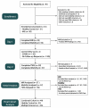
Multiple sclerosis is accompanied by decreased mobility and various adaptations affecting neural structure and function. Therefore, the purpose of this project was to understand how motor cortex thickness and corticospinal excitation and inhibition contribute to turning performance in healthy controls and people with multiple sclerosis. In total, 49 participants (23 controls, 26 multiple sclerosis) were included in the final analysis of this study. All participants were instructed to complete a series of turns while wearing wireless inertial sensors. Motor cortex gray matter thickness was measured via magnetic resonance imaging. Corticospinal excitation and inhibition were assessed via transcranial magnetic stimulation and electromyography place on the tibialis anterior muscles bilaterally. People with multiple sclerosis demonstrated reduced turning performance for a variety of turning variables. Further, we observed significant cortical thinning of the motor cortex in the multiple sclerosis group. People with multiple sclerosis demonstrated no significant reductions in excitatory neurotransmission, whereas a reduction in inhibitory activity was observed. Significant correlations were primarily observed in the multiple sclerosis group, demonstrating lateralization to the left hemisphere. The results showed that both cortical thickness and inhibitory activity were associated with turning performance in people with multiple sclerosis and may indicate that people with multiple sclerosis rely on different neural resources to perform dynamic movements typically associated with fall risk.
Keywords: MRI; TMS; inhibition; motor cortex; multiple sclerosis; turning.
Conflict of interest statement
The authors declare no conflict of interest.
Figures


References
-
- Bendfeldt K., Kuster P., Traud S., Egger H., Winklhofer S., Mueller-Lenke N., Naegelin Y., Gass A., Kappos L., Matthews P.M., et al. Association of regional gray matter volume loss and progression of white matter lesions in multiple sclerosis—A longitudinal voxel-based morphometry study. Neuroimage. 2009;45:60–67. doi: 10.1016/j.neuroimage.2008.10.006. - DOI - PubMed
-
- Bergsland N., Horakova D., Dwyer M.G., Uher T., Vaneckova M., Tyblova M., Seidl Z., Krasensky J., Havrdova E., Zivadinov R. Gray matter atrophy patterns in multiple sclerosis: A 10-year source-based morphometry study. Neuroimage Clin. 2018;17:444–451. doi: 10.1016/j.nicl.2017.11.002. - DOI - PMC - PubMed
MeSH terms
Grants and funding
LinkOut - more resources
Full Text Sources
Medical

