

Microstates of the cortical brain-heart axis
- PMID: 37688575
- PMCID: PMC10619395
- DOI: 10.1002/hbm.26480
Microstates of the cortical brain-heart axis
Abstract
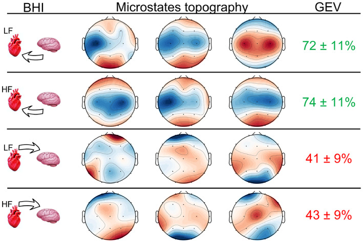
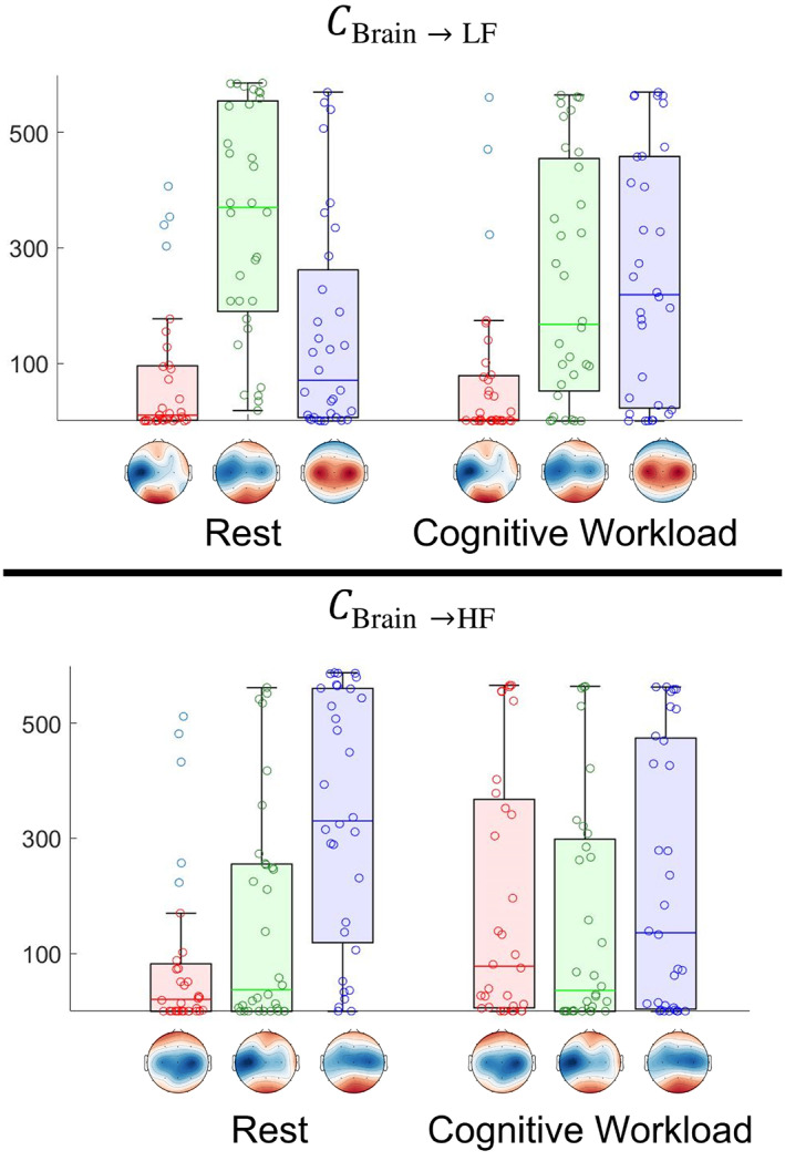
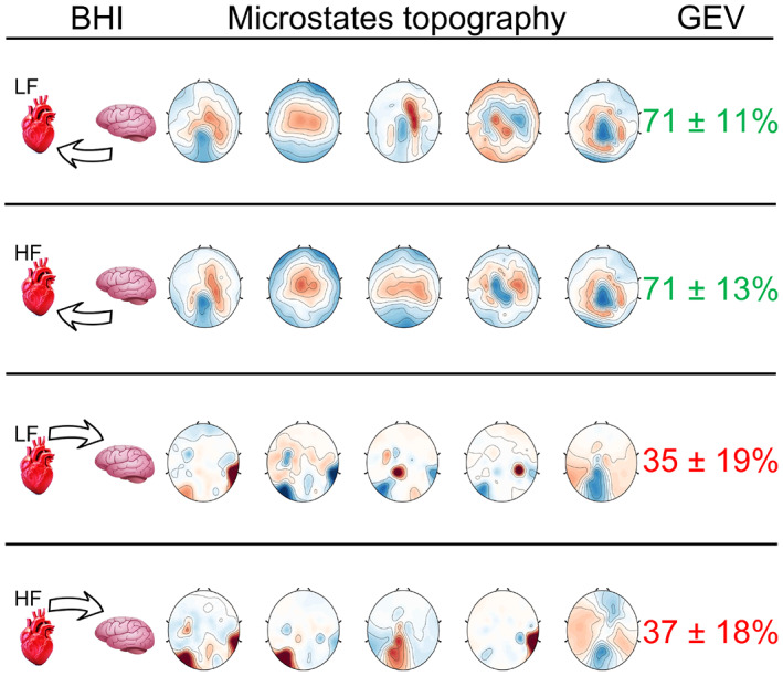
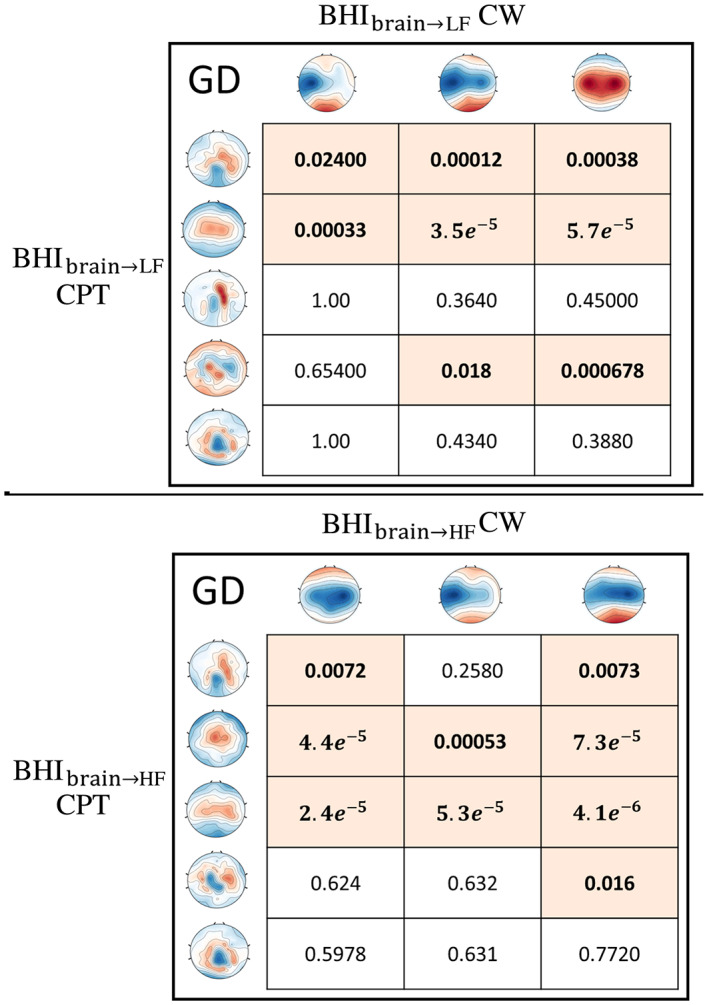
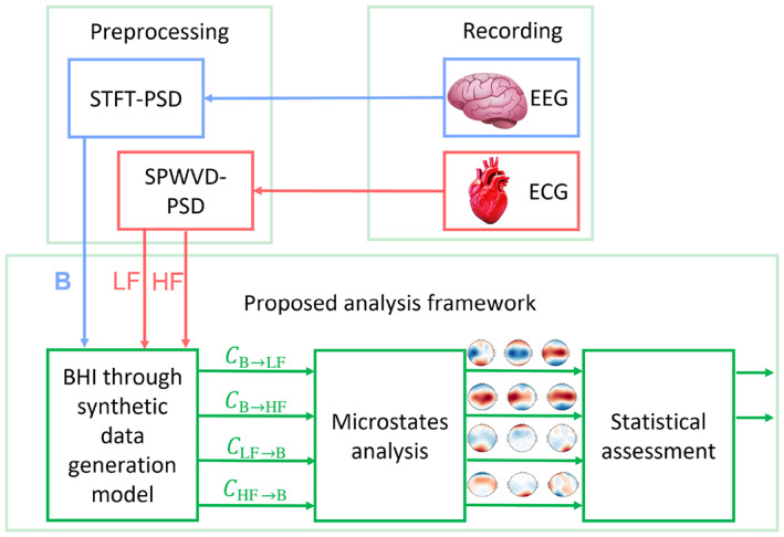
Electroencephalographic (EEG) microstates are brain states with quasi-stable scalp topography. Whether such states extend to the body level, that is, the peripheral autonomic nerves, remains unknown. We hypothesized that microstates extend at the brain-heart axis level as a functional state of the central autonomic network. Thus, we combined the EEG and heartbeat dynamics series to estimate the directional information transfer originating in the cortex targeting the sympathovagal and parasympathetic activity oscillations and vice versa for the afferent functional direction. Data were from two groups of participants: 36 healthy volunteers who were subjected to cognitive workload induced by mental arithmetic, and 26 participants who underwent physical stress induced by a cold pressure test. All participants were healthy at the time of the study. Based on statistical testing and goodness-of-fit evaluations, we demonstrated the existence of microstates of the functional brain-heart axis, with emphasis on the cerebral cortex, since the microstates are derived from EEG. Such nervous-system microstates are spatio-temporal quasi-stable states that exclusively refer to the efferent brain-to-heart direction. We demonstrated brain-heart microstates that could be associated with specific experimental conditions as well as brain-heart microstates that are non-specific to tasks.
Keywords: EEG; HRV; brain-heart; microstates.
© 2023 The Authors. Human Brain Mapping published by Wiley Periodicals LLC.
Conflict of interest statement
All authors declare that they have no conflicts of interest.
Figures
References
-
- Al‐Nashash, H. , Al‐Assaf, Y. , Paul, J. , & Thakor, N. (2004). EEG signal modeling using adaptive Markov process amplitude. IEEE Transactions on Biomedical Engineering, 51(5), 744–9751. - PubMed
-
- Benarroch, E. E. (1993). The central autonomic network: Functional organization, dysfunction, and perspective. Mayo Clinic Proceedings, 68–10, 988–1001. - PubMed
-
- Bernardi, L. , Wdowczyk‐Szulc, J. , Valenti, C. , Castoldi, S. , Passino, C. , Spadacini, G. , & Sleight, P. (2000). Effects of controlled breathing, mental activity and mental stress with or without verbalization on heart rate variability. Journal of the American College of Cardiology, 35(6), 1462–1469. - PubMed
-
- Brennan, M. , Palaniswami, M. , & Kamen, P. (2002). Poincare plot interpretation using a physiological model of HRV based on a network of oscillators. American Journal of Physiology‐Heart and Circulatory Physiology, 283(5), H1873–H1886. - PubMed
-
- Bressler, S. L. , & Menon, V. (2010). Large‐scale brain networks in cognition: Emerging methods and principles. Trends in Cognitive Sciences, 14(6), 277–290. - PubMed
Publication types
MeSH terms
LinkOut - more resources
Full Text Sources
